. 2003 Aug;77(15):8462-9.
doi: 10.1128/jvi.77.15.8462-8469.2003.

Evaluation of quinacrine treatment for prion diseases

Affiliations

Evaluation of quinacrine treatment for prion diseases

A Barret et al. J Virol. 2003 Aug.

Abstract

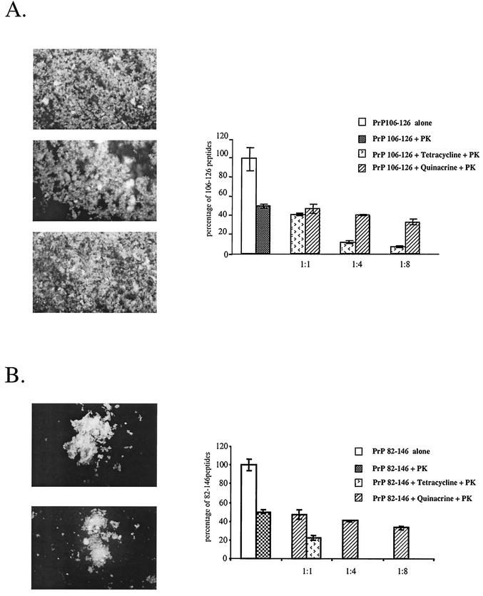
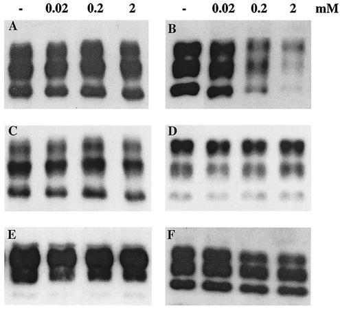
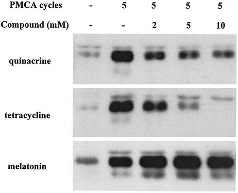
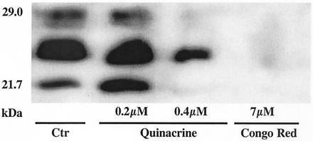
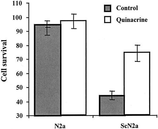
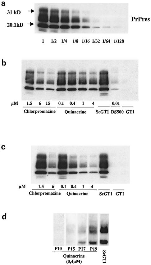
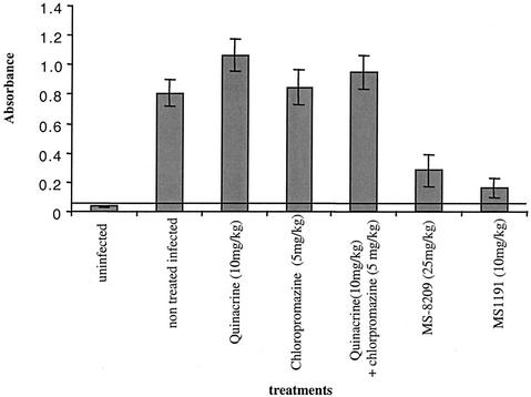
Based on in vitro observations in scrapie-infected neuroblastoma cells, quinacrine has recently been proposed as a treatment for Creutzfeldt-Jakob disease (CJD), including a new variant CJD which is linked to contamination of food by the bovine spongiform encephalopathy (BSE) agent. The present study investigated possible mechanisms of action of quinacrine on prions. The ability of quinacrine to interact with and to reduce the protease resistance of PrP peptide aggregates and PrPres of human and animal origin were analyzed, together with its ability to inhibit the in vitro conversion of the normal prion protein (PrPc) to the abnormal form (PrPres). Furthermore, the efficiencies of quinacrine and chlorpromazine, another tricyclic compound, were examined in different in vitro models and in an experimental murine model of BSE. Quinacrine efficiently hampered de novo generation of fibrillogenic prion protein and PrPres accumulation in ScN2a cells. However, it was unable to affect the protease resistance of preexisting PrP fibrils and PrPres from brain homogenates, and a "curing" effect was obtained in ScGT1 cells only after lengthy treatment. In vivo, no detectable effect was observed in the animal model used, consistent with other recent studies and preliminary observations in humans. Despite its ability to cross the blood-brain barrier, the use of quinacrine for the treatment of CJD is questionable, at least as a monotherapy. The multistep experimental approach employed here could be used to test new therapeutic regimes before their use in human trials.


Figures

FIG. 1.
FIG. 1.
Binding of quinacrine, tetracycline, and thioflavin T to PrP106-126 and PrP82-146 peptides and their effects on proteinase K (PK) resistance of these peptides. (A) Fluorescence microscopy of PrP106-126 aggregates incubated with quinacrine (top), tetracycline (middle), or thioflavin T (bottom) and effects of quinacrine and tetracycline on proteinase K resistance of the peptide (graph). (B) Fluorescence microscopy of PrP82-146 aggregates incubated with quinacrine (top) or tetracyline (bottom) and effects of quinacrine and tetracycline on proteinase K resistance of the peptide (graph). The extents of proteolysis of PrP106-126 (A) and PrP82-146 (B) in the absence or presence of quinacrine or tetracycline were calculated as the percentage of peptide present in the pellet compared to the total amount originally present.
FIG. 2.
FIG. 2.
Effects of quinacrine (A and C to F) and tetracycline (B) on the protease resistance of a variety of PrPres isoforms. Partially purified PrPres was incubated with increasing concentrations of the compounds (indicated at the top; −, absence of treatment) and then digested with proteinase K. (A and B) Sporadic CJD with type 1 PrPres; (C) sporadic CJD with type 2 PrPres; (D) variant CJD; (E) 263K-infected hamster; (F) 139A-infected mouse. The immunoblot analysis was carried out using the MAb 3F4 (A to E), and the polyclonal antibody PrP95-108 (F).
FIG. 3.
FIG. 3.
Effects of quinacrine, tetracycline, and melatonin on cyclic amplification of PrPres from scrapie-infected hamster brain. The number of cycles of incubation-sonication and the concentrations of the compounds are indicated. −, none; PMCA, protein misfolding cyclic amplification.
FIG. 4.
FIG. 4.
Quinacrine inhibits PrPres accumulation in N2a58/22L cells. Anti-PrP immunoblots with SAF75 of N2a58/22L cells that were incubated with quinacrine and Congo red (three administrations) for 3 days are shown. Control (Ctr) corresponds to the absence of treatment.
FIG. 5.
FIG. 5.
Quinacrine protects ScN2a cells against microglia-mediated killing. The survival of prion-infected (ScN2a) and uninfected (N2a) neuroblastoma cells cocultured with microglia in the presence (open bars) or absence (shaded bars) of 0.2 μM quinacrine is illustrated. The values given are the means ± standard deviations of triplicate experiments repeated twice (six observations).
FIG. 6.
FIG. 6.
In vitro evaluation by Western blotting (with SAF83) of the efficacy of treatment with quinacrine, chlorpromazine, and DS500 on PrPres accumulation in ScGT1 cells. (a) Dilution scale of ScGT1 cells (lane 1 corresponds to 300 μg of total protein). (b) Effects of a range of quinacrine and chlorpromazine concentrations and of DS500 (0.01 μM) after a unique 4-day treatment. (c) Same doses of chlorpromazine, quinacrine, and DS500 applied three times in 6 days. (d) Effects of 0.4 μM quinacrine treatment every day for 3 weeks and at several passages (P) after treatment was stopped.
FIG. 7.
FIG. 7.
Evaluation by enzyme immunoassay of the efficacies of different drugs in reducing PrPres accumulation in the spleens of scrapie-infected mice. Mice infected intraperitoneally with the 6PB1 strain were treated intraperitoneally each day for 3 weeks with quinacrine alone (10 mg/kg), chlorpromazine alone (5 mg/kg), both drugs, and MS-8209 (25 mg/kg) or MS1191 (10 mg/kg). The PrPres concentrations in the spleens of the animals after 30 days were evaluated by optical densitometry. Day 30 is the time when the scrapie PrP concentration reaches a plateau in this organ (3). The horizontal line at an absorbance of 0.08 corresponds to the cutoff, calculated as 2.5 times the mean absorbance of the negative controls.

References

    1. Adjou, K. T., N. Privat, S. Demart, J. P. Deslys, M. Seman, J. J. Hauw, and D. Dormont. 2000. MS-8209, an amphotericin B analogue, delays the appearance of spongiosis, astrogliosis and PrPres accumulation in the brain of scrapie-infected hamsters. J. Comp. Pathol. 122:3-8. - PubMed
    1. Bate, C., S. Reid, and A. Williams. 2001. Killing of prion-damaged neurones by microglia. Neuroreport 12:2589-2594. - PubMed
    1. Beringue, V., F. Lamoury, K. T. Adjou, T. Maignien, M. Demoy, P. Couvreur, and D. Dormont. 2000. Pharmacological manipulation of early PrPres accumulation in the spleen of scrapie-infected mice. Arch. Virol. Suppl. 16:39-56. - PubMed
    1. Bolton, D. C., M. P. McKinley, and S. B. Prusiner. 1982. Identification of a protein that purifies with the scrapie prion. Science 218:1309-1311. - PubMed
    1. Caughey, B., D. Ernst, and R. E. Race. 1993. Congo red inhibition of scrapie agent replication. J. Virol. 67:6270-6272. - PMC - PubMed

Publication types

MeSH terms