. 2003 Jul 22;100(15):8981-6.
doi: 10.1073/pnas.1531172100. Epub 2003 Jul 11.

Solitary chemoreceptor cells in the nasal cavity serve as sentinels of respiration

Affiliations

Solitary chemoreceptor cells in the nasal cavity serve as sentinels of respiration

Thomas E Finger et al. Proc Natl Acad Sci U S A. .

Abstract

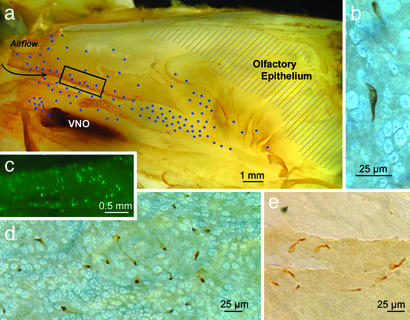
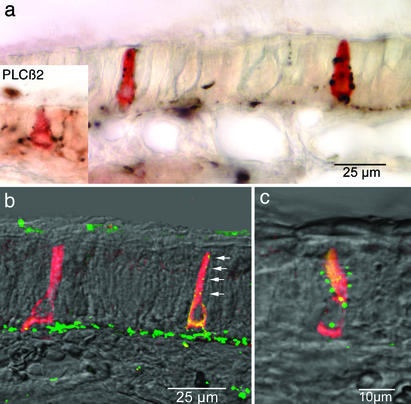
Inhalation of irritating substances leads to activation of the trigeminal nerve, triggering protective reflexes that include apnea or sneezing. Receptors for trigeminal irritants are generally assumed to be located exclusively on free nerve endings within the nasal epithelium, requiring that trigeminal irritants diffuse through the junctional barrier at the epithelial surface to activate receptors. We find, in both rats and mice, an extensive population of chemosensory cells that reach the surface of the nasal epithelium and form synaptic contacts with trigeminal afferent nerve fibers. These chemosensory cells express T2R "bitter-taste" receptors and alpha-gustducin, a G protein involved in chemosensory transduction. Functional studies indicate that bitter substances applied to the nasal epithelium activate the trigeminal nerve and evoke changes in respiratory rate. By extending to the surface of the nasal epithelium, these chemosensory cells serve to expand the repertoire of compounds that can activate trigeminal protective reflexes. The trigeminal chemoreceptor cells are likely to be remnants of the phylogenetically ancient population of solitary chemoreceptor cells found in the epithelium of all anamniote aquatic vertebrates.


Figures

Fig. 1.
Fig. 1.
Gustducin-expressing cells in the nasal epithelium. (a) The distribution of gustducin-immunoreactive cells on the turbinates and lateral nasal wall of a 6-week-old rat plotted on a lateral view of the nasal cavity split along the sagittal plane. The blue dots indicate the relative density and location of reactive cells. The region of olfactory epithelium is indicated by shading. The dark region at the left side of the figure is the vomeronasal organ (VNO). (Scale bar, 1.0 mm.) (b) Micrograph of an immunoreactive cell in the epithelium of a whole mount is shown. The elongate cell runs obliquely through the thin epithelium. (c) Fluorescent cells in the anterior portions of the nasal cavity, just caudal to the vestibule (boxed region in a), from an adult transgenic mouse in which GFP is driven by the gustducin promoter. The distribution of these fluorescent cells is similar to that revealed by gustducin immunocytochemistry. (d) Low-power micrograph of a field of gustducin-immunoreactive cells of a rat as revealed by whole-mount immunocytochemistry. The packing density of the scattered gustducin-containing cells varies according to the region of the nose in which they lie. In rats, the maximum density is ≈300 cells per mm2. More often, the density is one order of magnitude less. Because of the convoluted nature of the nasal passageways and the variable density of gustducin-containing cells, it is difficult to give a reliable estimate of the total number of cells involved; however, in rat, the number on one side of the nose is >1,000. (e) Immunoreactive cells along the torn edge of the respiratory epithelium from a rat reacted as a whole mount. The apical portion of the cell is generally more immunoreactive (darker) than lower portions.
Fig. 2.
Fig. 2.
Innervation of gustducin-immunoreactive epithelial cells as seen by double-label immunocytochemistry. In a, two elongate immunoreactive cells are apparent. Each is contacted repeatedly by PGP 9.5-immunoreactive nerve fibers (black). The gustducin-immunoreactive cells are not reactive for PGP 9.5, which reacts with other types of paraneurons. Inset, at the same magnification, shows a similar situation for a cell reacted with antisera to PLC β2, a downstream component in the T2R-gustducin transduction cascade. (b and c) Gustducin-immunoreactive cells (red-yellow) being contacted by CGRP-immunoreactive nerve fibers (green). Arrows in b indicate a series of neuronal varicosities in close proximity to the apical part of a gustducin-immunoreactive epithelial cell. The yellow color of the gustducin-immunoreactive cells is due to the double-labeling method employing primary antibodies raised in the same species. When CGRP antiserum is used without gustducin antiserum, the cells are not labeled (5).
Fig. 3.
Fig. 3.
Electron micrographs of a gustducin-immunoreactive epithelial cell. The dark flocculent reaction product marks the cytoplasm of the immunoreactive cell, which has been artificially tinted yellow for visibility. (a) The apical half of a gustducin-immunoreactive cell showing repeated contacts with nerve processes (N). (b) The contact between a nerve process and the apex of the cell. (c) From an adjacent section, the apex of the immunoreactive cell appears much narrower than the surrounding ciliated respiratory cells (crc). The microvilli (mv) of the immunoreactive cell are intermediate in size between the microvilli and cilia (c) of the ciliated respiratory cells. The microvilli of the gustducin-immunoreactive cell are wavy and have no subsurface filamentous web, unlike microvilli of respiratory brush cells. Scale as in d. (d, e1, and e2) Sites of specialized contact between the immunoreactive cells and nerve fibers display several features typical of synapses, including small vesicles (arrowheads) and a slight presynaptic thickening, are shown. The fine processes of a multiple branched nerve fiber (black N with arrows) are visible at the lower right in d. (e1 and e2) Shown are nearby sections through a synaptic contact between an immunoreactive cell and a nerve fiber. Vesicles (arrowheads) are apparent within the chemoreceptor cell (SCC), whereas a modest postsynaptic density (psd) is visible in the nerve fiber. [Scale bars, 0.5 μm (a and d) and 0.25 μm (b and e).]
Fig. 4.
Fig. 4.
Trigeminal chemoreceptors express T2R bitter-receptor mRNA and respond to stimulation with bitter-tasting ligands applied to the nasal passages. (a) Coexpression of mT2R8 (red, in situ hybridization) and gustducin (green, immunocytochemistry) in nasal SCCs. Although the gustducin immunoreactivity is evident throughout the cell, the in situ hybridization signal, which reveals the location of T2R8 mRNA, is perinuclear, where rough ER is typically located. (Scale bar, 10 μm.) (b) Relative chemosensory component (mean response to stimulus - mean response to saline) of the integrated neural activity in the ethmoid branch of the trigeminal nerve in anesthetized rats. The graph shows the percentage increase in neural activity during chemical stimulation compared with stimulation of the system with saline only. Responses to all compounds were significantly greater than responses to control (saline) solutions (*, paired t test, P < 0.05; six animals). Denat, denatonium (0.01 M); quinine (0.01 M); and Cyclo., cycloheximide (0.01 M). Error bars indicate SEM. (c) Mean percentage depression in respiratory rate when saline or bitter-tasting substances are applied to the nasal epithelium. The rate decreases significantly (*, paired t test, P < 0.05; nine animals) when the nasal epithelium is bathed in 0.01 M denatonium, quinine, or cycloheximide. Error bars indicate SEM. (d) Respiratory record in an anaesthetized rat is shown. The respiratory cycle is indicated by the sinusoidal purple line showing a basal respiration rate of approximately six breaths in 5 sec. A profound respiratory depression is evoked by 0.01 M cycloheximide. In this example, on application of cycloheximide, the steady prestimulus rate is drastically reduced to a near apnea (rate is less than one breath per 10 sec.). Shallow, but relatively normally paced respiration returns ≈10 sec after washout of the strong trigeminal stimulant.

References

    1. Bryant, B. P. & Silver, W. L. (2000) in The Neurobiology of Taste and Smell, eds. Finger, T. E., Silver, W. L. & Restrepo, D. (Wiley, New York), pp. 73-100.
    1. Liu, L. & Simon, S. A. (2000) Physiol. Behav. 69, 363-378. - PubMed
    1. McKemy, D. D., Neuhausser, W. M. & Julius, D. (2002) Nature 416, 52-58. - PubMed
    1. Silverman, J. D. & Kruger, L. (1989) J. Comp. Neurol. 280, 303-330. - PubMed
    1. Finger, T. E., St. Jeor, V. L., Kinnamon, J. C. & Silver, W. L. (1990) J. Comp. Neurol. 294, 293-305. - PubMed

Publication types

LinkOut - more resources