

Experimental infection of piglets with a korean strain of porcine epidemic diarrhoea virus
- PMID: 12859908
- PMCID: PMC7127761
- DOI: 10.1016/s0021-9975(02)00170-6
Experimental infection of piglets with a korean strain of porcine epidemic diarrhoea virus
Abstract
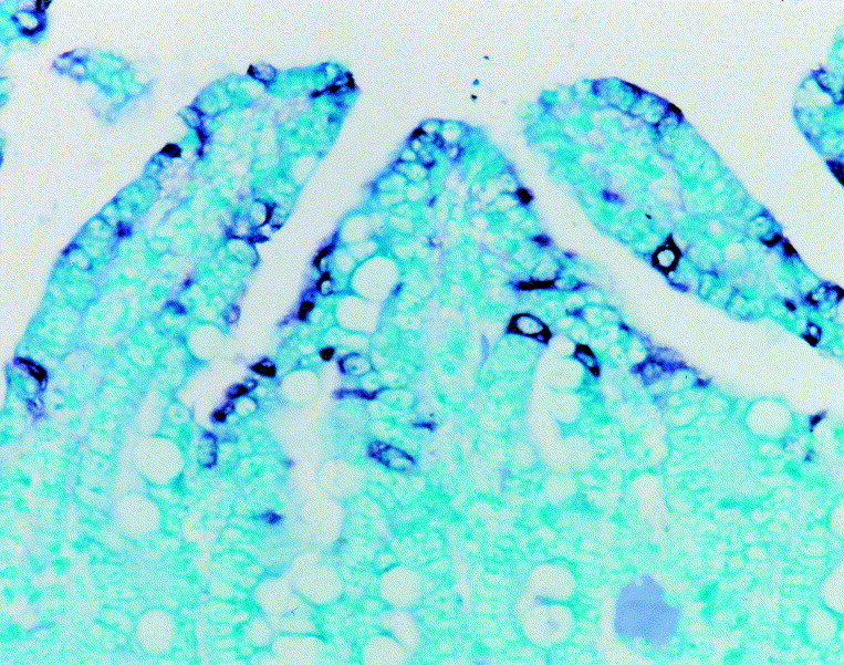
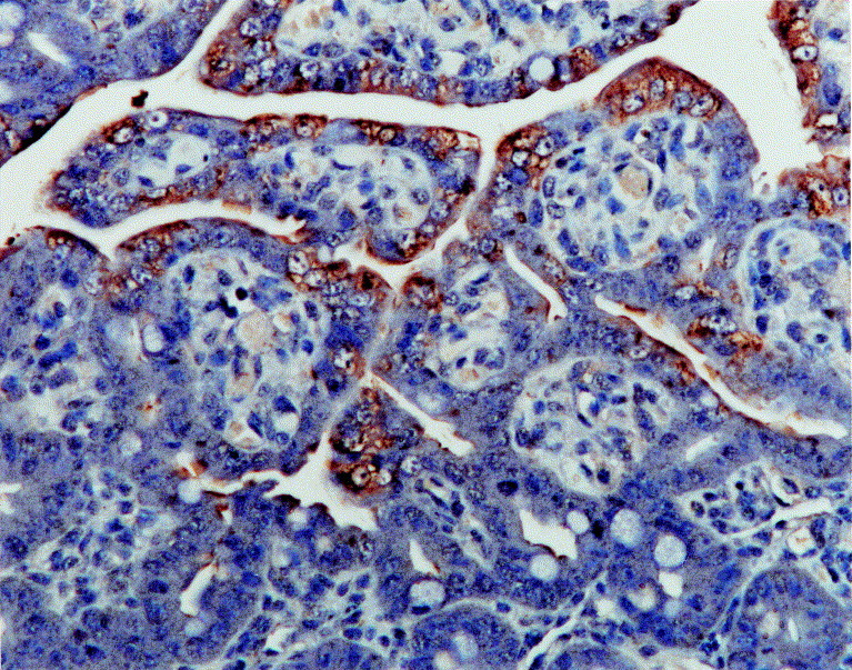
Thirty colostrum-deprived piglets aged 1 day were inoculated with a Korean strain of porcine epidemic diarrhoea virus (PEDV). The purpose was to elucidate the pathogenicity and viral distribution in PEDV-infected piglets over a period of 60 h, by morphometric analysis, in-situ hybridization and immunohistochemistry. At 24-60 h post-inoculation (hpi), the villous height/crypt depth (VH/CD) ratio of infected pigs was significantly less than that of control pigs. Positive cells typically exhibited a dark black reaction product (in-situ hybridization) or brown reaction product (immunohistochemistry) in the cytoplasm, without background staining. PEDV nucleic acid and antigen were detected in the duodenum, jejunum and ileum of experimentally infected pigs. The results suggested that the Korean strain was virulent and caused severe villous atrophy in the small intestine.
Figures
References
-
- Anonymous Information supplement. Veterinary Record. 1972;95:49.
-
- Cavanagh D. Nidovirales: a new order comprising Coronaviridae and Arteriviridae. Archives of Virology. 1997;142:629–633. - PubMed
-
- Chae C., Kim O., Choi C., Min K., Cho W.-S., Kim J., Tai J.H. Prevalence of porcine epidemic diarrhoea virus and transmissible gastroenteritis virus infection in Korean pigs. Veterinary Record. 2000;147:606–608. - PubMed
-
- Coussement W., Ducatelle R., Debouck P., Hoorens J. Pathology of experimental CV777 coronavirus enteritis in piglets. I. Histological and histochemical study. Veterinary Pathology. 1982;19:46–56. - PubMed
Publication types
MeSH terms
Substances
LinkOut - more resources
Full Text Sources
Medical
