

Tissue Doppler imaging predicts the development of hypertrophic cardiomyopathy in subjects with subclinical disease
- PMID: 12860897
- PMCID: PMC2908312
- DOI: 10.1161/01.CIR.0000084500.72232.8D
Tissue Doppler imaging predicts the development of hypertrophic cardiomyopathy in subjects with subclinical disease
Abstract
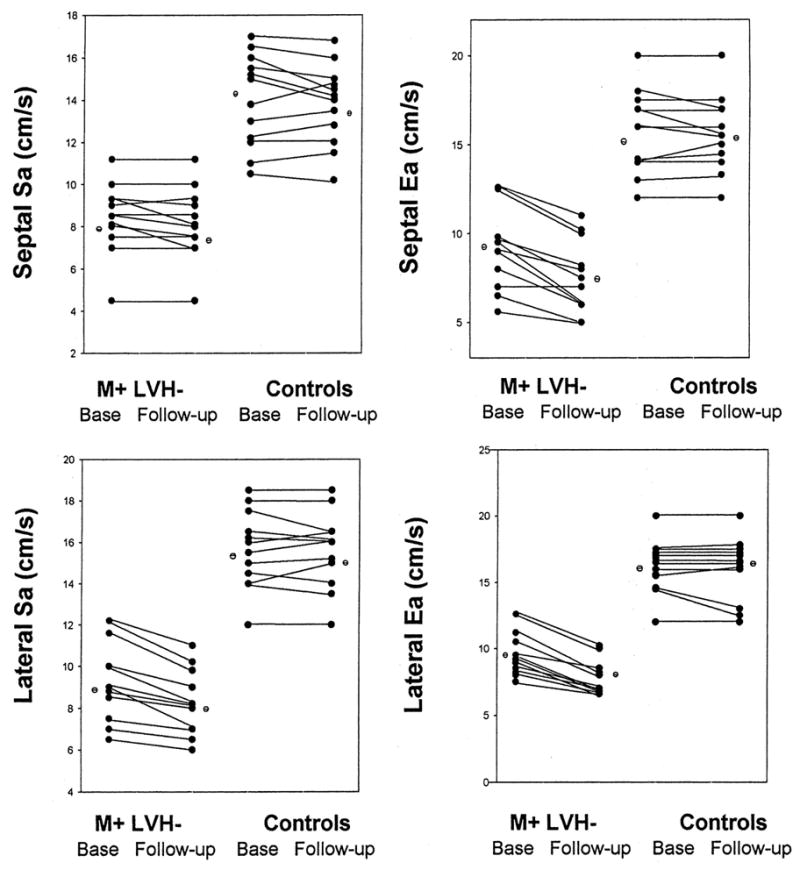
Background: Systolic (Sa) and diastolic (Ea) myocardial velocities measured by tissue Doppler (TD) imaging (TDI) recently were shown to be decreased in subjects who have mutations causing hypertrophic cardiomyopathy (HCM) but who do not have left ventricular (LV) hypertrophy. By studying these subjects at a later date, we sought to determine whether TDI predicts the subsequent evolution of the HCM phenotype.
Methods and results: Serial 2D and Doppler echocardiography were performed in 12 subjects (age range, 17 to 51 years) with HCM-causing mutations on 2 occasions: before development of hypertrophy and 2 years later. Twelve age- and gender-matched family members without mutations were included as control subjects. In those with mutations, mean septal thickness and LV mass were 1.07+/-0.14 cm and 103.0+/-11 g at baseline, respectively, and increased to 1.30+/-0.36 cm and 193.0+/-78 g at follow-up (P<0.01), with 6 subjects satisfying HCM diagnostic criteria. Sa and Ea velocities in those with mutations were lower compared with control subjects at baseline and follow-up (lateral Sa, 15+/-1.2 versus 8.2+/-2.1 cm/s; lateral Ea, 16.5+/-2.8 versus 8.1+/-2.3 cm/s; P<0.01). At 2 years, left atrial volume and pulmonary venous flow indices of LV filling pressures increased, whereas TD early and late diastolic velocities decreased (all P<0.05) in those with the mutations. Control subjects had no significant interval changes of the above parameters.
Conclusions: Subsequent development of HCM in subjects with initially reduced TD velocities establishes TDI as a reliable method for early identification of HCM mutation carriers.
Figures
References
-
- Ho CY, Sweitzer NK, McDonough B, et al. Assessment of diastolic function with Doppler tissue imaging to predict genotype in preclinical hypertrophic cardiomyopathy. Circulation. 2002;105:2992–2997. - PubMed
Publication types
MeSH terms
Substances
Grants and funding
LinkOut - more resources
Full Text Sources
