. 2003 Oct 1;552(Pt 1):23-34.
doi: 10.1113/jphysiol.2003.048843. Epub 2003 Jul 14.

Molecular mechanism for ATP-dependent closure of the K+ channel Kir6.2

Affiliations

Molecular mechanism for ATP-dependent closure of the K+ channel Kir6.2

Scott A John et al. J Physiol. .

Abstract

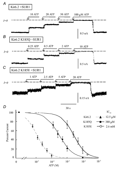
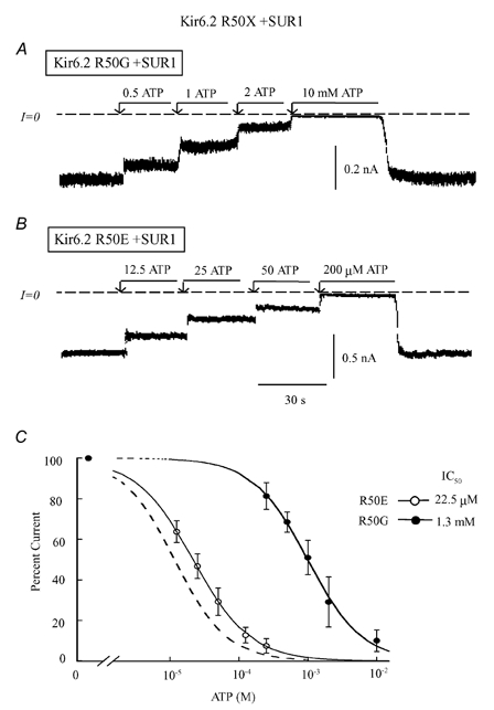
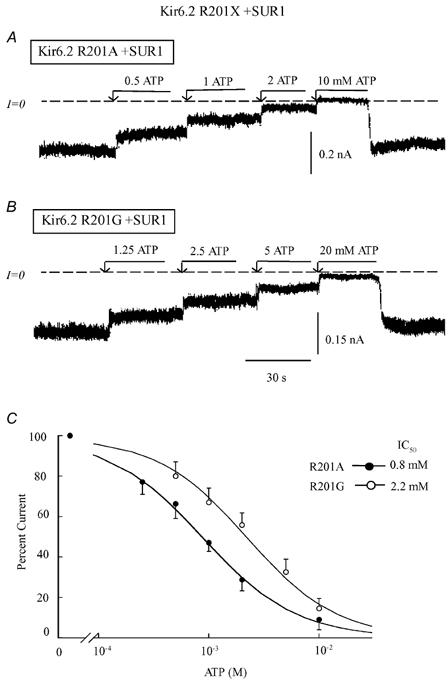
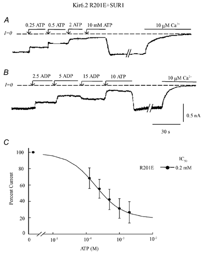
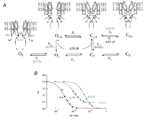
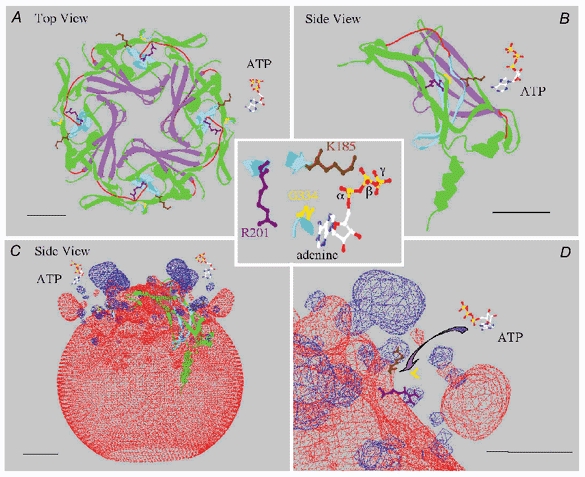
In the ATP-dependent K+ (KATP) channel pore-forming protein Kir6.2, mutation of three positively charged residues, R50, K185 and R201, impairs the ability of ATP to close the channel. The mutations do not change the channel open probability (Po) in the absence of ATP, supporting the involvement of these residues in ATP binding. We recently proposed that at least two of these positively charged residues, K185 and R201, interact with ATP phosphate groups to cause channel closure: the beta phosphate group of ATP interacts with K185 to initiate closure, while the alpha phosphate interacts with R201 to stabilize the channel's closed state. In the present study we replaced these three positive residues with residues of different charge, size and hydropathy. For K185 and R201, we found that charge, more than any other property, controls the interaction of ATP with Kir6.2. At these positions, replacement with another positive residue had minor effects on ATP sensitivity. In contrast, replacement of K185 with a negative residue (K185D/E) decreased ATP sensitivity much more than neutral substitutions, suggesting that an electrostatic interaction between the beta phosphate group of ATP and K185 destabilizes the open state of the channel. At R201, replacement with a negative charge (R201E) had multiple effects, decreasing ATP sensitivity and preventing full channel closure at high concentrations. In contrast, the R50E mutation had a modest effect on ATP sensitivity, and only residues such as proline and glycine that affect protein structure caused major decreases in ATP sensitivity at the R50 position. Based on these results and the recently published structure of Kir3.1 cytoplasmic domain, we propose a scheme where binding of the beta phosphate group of ATP to K185 induces a motion of the surrounding region, which destabilizes the open state, favouring closure of the M2 gate. Binding of the alpha phosphate group of ATP to R201 then stabilizes the closed state. R50 on the N-terminus controls ATP binding by facilitating the interaction of the beta phosphate group of ATP with K185 to destabilize the open state.


Figures

Figure 1
Figure 1. ATP sensitivity of K185X mutants co-expressed with SUR1
Current traces in the three upper panels depict inward currents measured in the absence of Mg2+, for wild-type Kir6.2 channels co-expressed with SUR1 (A) and for two Kir6.2 mutants, K185Q (B) and K185E (C) also co-expressed with SUR1. Currents were progressively suppressed by increasing ATP concentration, with the IC50 near 12 μM, 0.4 mM and 2.5 mM, respectively. On average K185 replacement with neutral residues had a modest effect on ATP-induced inhibition, while replacement with negatively charged residues dramatically decreased ATP sensitivity. C, plot of channel ATP sensitivity for wild-type Kir6.2 (▴), K185Q (•) and K185E (○), all co-expressed with SUR1. A fit of the data points to the Hill equation yielded IC50 values of 12.5 μM (n = 5) for wild-type channels, 0.38 mM (n = 5) for K185Q and 2.6 mM (n = 3) for K185E. The three Hill coefficients were close to unity.
Figure 2
Figure 2. ATP sensitivity of R50X mutants co-expressed with SUR1
Current traces in A and B illustrate inward currents measured in the absence of Mg2+, for Kir6.2 mutants R50G (A) and R50E (B), both co-expressed with SUR1. These panels depict progressive suppression of inward current by increasing ATP concentration, with the IC50 values near 22 μM and 1.3 mM, respectively. R50G had major effects on ATP sensitivity, while R50E had little effect. C, plot of channel ATP sensitivity for R50G (•) and R50E (○) co-expressed with SUR1, with the wild-type curve indicated by the dashed line for comparison. A fit of the data points to the Hill equation yielded IC50 values of 1.3 mM (n = 5) for R50G channels and 22.5 μM (n = 5) for R50E. The two Hill coefficients were close to unity.
Figure 3
Figure 3. ATP sensitivity of R201X mutants co-expressed with SUR1
Current traces in A and B depict inward currents measured in the absence of Mg2+ with Kir6.2 mutants, for R201A (A) and R201G (B) co-expressed with SUR. These panels illustrate progressive suppression of inward current by increasing ATP concentrations with the IC50 values near 0.8 mM and 2.2 mM, respectively. R201A had substantial effects on ATP sensitivity, while R201G, which may modify the protein structure, had an even larger effect. C, plot of channel ATP sensitivity for R201A (•) and R201G (○) co-expressed with SUR1. A fit of the data points to the Hill equation yielded IC50 values of 0.8 mM (n = 5) for R201A channels and 2.2 mM (n = 3) for R201G. The two Hill coefficients were close to unity.
Figure 4
Figure 4. ATP and ADP sensitivity of R201E co-expressed with SUR1
Current traces in A and B depict inward currents measured in the absence of Mg2+ with the Kir6.2 mutant R201E + SUR1. These panels illustrate progressive suppression of inward current by increasing concentrations of ATP (A) and ADP (B). At the end of each trace, full current inhibition by Ca2+ is shown to illustrate that ATP or ADP failed to fully suppress channel activity. With high ADP there was even channel re-opening (B). C, plot of channel ATP sensitivity for R201E (•) co-expressed with SUR1. For this plot three patches were selected to illustrate the wide difference in ATP sensitivity among patches. A fit of the data points to the Hill equation was poor unless a pedestal of non-suppressible current was included, with a maximum inhibition by ATP of 81 %, IC50 of 197 μM (n = 3) and Hill coefficient close to unity.
Figure 5
Figure 5. Plasma membrane insertion of Kir6.2 wild-type, R50E and K185E mutants linked to GFP and co-expressed with SUR1
Three images obtained from different transfection experiments are representative of the distribution of GFP linked to the Kir6.2 C-terminus of Kir6.2 + SUR1, R50E + SUR1 and K185E + SUR1. A and B, data obtained with wild-type Kir6.2 and R50E showed bright uniform fluorescence associated with the plasma membrane and minimal labelling of intracellular lamellar structures. These patterns, together with the high level of channel activity recorded in both cases, support our hypothesis that R50E mutant channels behave like wild-type channels. Similar results were obtained with R201E, but not R201D. C, with K185E and K185D (not shown) fluorescence labelling of an intracellular lamellar structure tentatively identified as the endoplasmic reticulum was observed together with some plasma membrane labelling. The reduced currents recorded in these cases suggest that channel insertion into the cell membrane is partly impaired. Scale bar is 10 μm.
Figure 6
Figure 6
A, allosteric model for the interaction of the α and β phosphate groups of adenine nucleotides with positively charged residues in Kir6.2. In the stable channel open configuration (OS), the lack of interaction between the R50 region in the N-terminus and the C-terminus does not permit binding of the ATP β phosphate with K185. Thus, R50 mutations with structure-breaking residues proline or glycine prevent this transition from OS to the unstable open state (OU), but otherwise the R50 site is relatively insensitive to charge, hydropathy or side chain size. The interaction between the R50 and K185 regions destabilizes the open state causing a transition to OU, in which interaction of the β phosphate group with K185 is permissive (OUA). Destabilization of the open state leads to unstable closure of the channel gate within M2 (CU), and binding of the ATP β phosphate to K185 allosterically promotes the transition from CU to CUA. Thus, in this model binding of the ATP β phosphate to K185 is state independent. Charge reversal at K185, however, prevents the ATP β phosphate from binding to K185, and therefore inhibits the latter transition, markedly suppressing ATP-dependent inhibition. Finally, once the channel is in the CUA state, the ATP α phosphate group interacts with R201 to stabilize the channel in a stable closed state (CSA). Insertion of a negatively charged residue at position 201 repels the approaching α phosphate group during channel closure, accounting for incomplete suppression of channel activity by ATP, since the channel can never reach the stable closed state CS. B, fitting results of the model in A to the steady-state ATP sensitivity curves for wild-type Kir6.2 (black), K185E (blue) and R201E (green). The Po in the absence of ATP was assumed to be 0.4 in each case. Fitting of this Po value in the absence of ATP yielded K1 = 2.7, K2 = 1.92 and K3 = 0.07. Fitting of the ATP dose-response curves yielded the following parameter values. For wild-type channel K5 = 16, Ka1 = 2000 and Ka2 = 11 000. For K185E mutant channels ATP binding to K185 varied yielding Ka1 = 5 and Ka2 = 40. To fit the R201E mutant data we only removed the last transition to CSA (K5 = 0).
Figure 7
Figure 7. Structure of mouse Kir6.2 ATP-binding region
A, view of the predicted ribbon structure of Kir6.2 cytoplasmic ‘pore’ region formed by the four subunits, viewed from the top down (towards the cytoplasm), along with an ATP molecule to the right. The purple structure delineates the pore-lining residues, and the pale blue structure corresponds to two anti-parallel sets of β strands containing residues K185 and R201, whose side chains are coloured brown and purple, respectively. The remainder of the protein is coloured green and red. Red identifies regions that differ structurally from the Kir3.1 channel or have insufficient electron density, as determined by Nishida & MacKinnon (2002), to place in a defined secondary structure. These regions are part of the linker region between the N-terminus and the C-terminus (see Nishida & MacKinnon, 2002). The cytoplasmic pore is formed by four identical subunits. B, side view of a single subunit, with an ATP molecule at the upper right, showing a detailed view of the β strands that bind ATP. C, side view of the electrostatic fields around the Kir6.2 tetramer. The negative fields are red and positive fields are blue. At the top (near the membrane), two small blue regions are formed by K185 and R201, and the larger blue region corresponds to a positive field generated by the juxtaposition of R206 and R177, which have been shown to interact with PIP2 in the plasma membrane to stabilize the channel structure. D, enlargement of the ATP-binding region, which shows the two positive residues K185 and R201 as well as G334 in yellow, on a set of β strands that face the ATP-binding region. The G334D mutation causes a 1000-fold decrease in ATP sensitivity (Drain et al. 1998). These observations suggest that ATP may bind in a pocket with the phosphate groups interacting with the positive charges at K185 and R201, while the adenine ring interacts via hydrogen bonds with G334. The estimated distance between G334 and K185 (≈8 Å) is consistent with this possibility, but the distance between the K185 and R201 side chains (≈16 Å), is very likely to be too large for the β and α phosphate groups of ATP to bind simultaneously to K185 and R201, respectively, in the purported open channel configuration shown here. We tentatively speculate that a movement (rotation) of K185 bound to the β phosphate, possibly together with G334 bound to adenine, may allow simultaneous binding of the two phosphate groups to K185 and R201. This scheme puts torsion in the side chain of 185 that may affect the backbone and hence the position of M2 to enable gating of the channel in the presence of ATP. In each panel the bar represents a distance of 10 Å.

Similar articles

Cited by

References

    1. Appel RD, Bairoch A, Hochstrasser DF. A new generation of information retrieval tools for biologists: the example of the ExPASy WWW server. Trends Biochem Sci. 1994;19:258–260. - PubMed
    1. Drain P, Li L, Wang J. KATP channel inhibition by ATP requires distinct functional domains of the cytoplasmic C terminus of the pore forming subunit. Proc Natl Acad Sci U S A. 1998;95:13953–13958. - PMC - PubMed
    1. Graham FL, van der Eb AJ. Transformation of rat cells by DNA of human adenovirus 5. Virology. 1973;52:456–467. - PubMed
    1. Guex N, Peitsch MC. SWISS-MODEL and the Swiss-PdbViewer: An environment for comparative protein modeling. Electrophoresis. 1997;18:2714–2723. - PubMed
    1. John SA, Monck JR, Weiss JN, Ribalet B. The sulfonylurea receptor SUR1 regulates ATP-sensitive mouse Kir6. 2 K channels linked to the green fluorescent protein in human embryonic kidney cells (HEK 293) J Physiol. 1998;510:333–345. - PMC - PubMed

Publication types

MeSH terms

Substances

LinkOut - more resources