. 2003 Jul 21;162(2):341-51.
doi: 10.1083/jcb.200212083. Epub 2003 Jul 14.

QSulf1 remodels the 6-O sulfation states of cell surface heparan sulfate proteoglycans to promote Wnt signaling

Affiliations

QSulf1 remodels the 6-O sulfation states of cell surface heparan sulfate proteoglycans to promote Wnt signaling

Xingbin Ai et al. J Cell Biol. .

Abstract

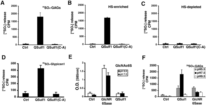
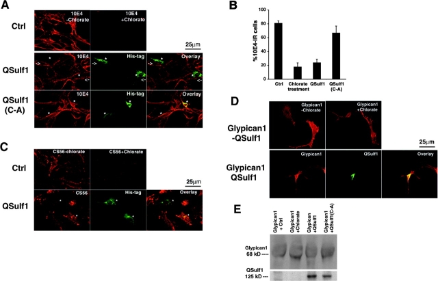
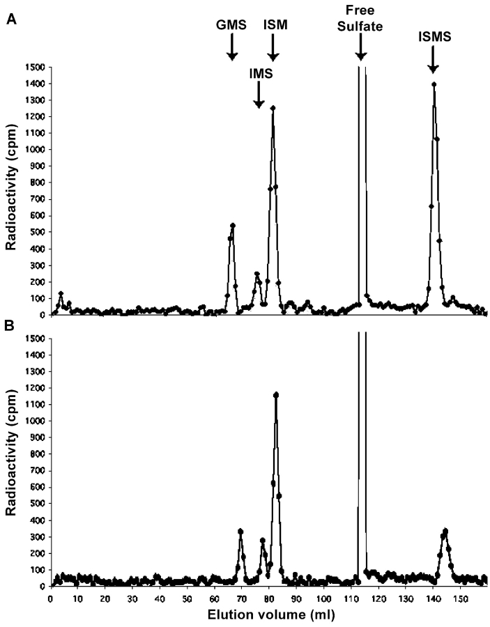
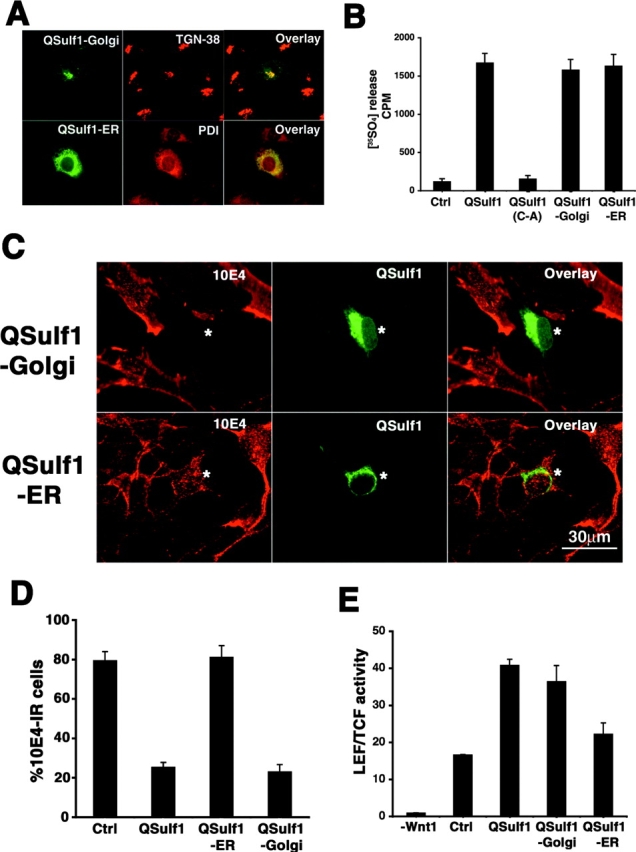
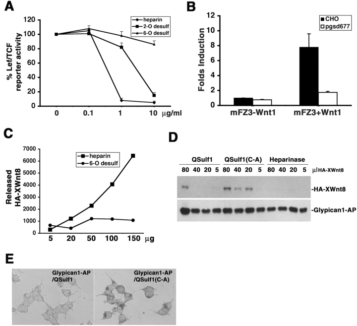
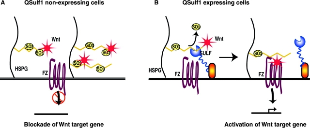
The 6-O sulfation states of cell surface heparan sulfate proteoglycans (HSPGs) are dynamically regulated to control the growth and specification of embryonic progenitor lineages. However, mechanisms for regulation of HSPG sulfation have been unknown. Here, we report on the biochemical and Wnt signaling activities of QSulf1, a novel cell surface sulfatase. Biochemical studies establish that QSulf1 is a heparan sulfate (HS) 6-O endosulfatase with preference, in particular, toward trisulfated IdoA2S-GlcNS6S disaccharide units within HS chains. In cells, QSulf1 can function cell autonomously to remodel the sulfation of cell surface HS and promote Wnt signaling when localized either on the cell surface or in the Golgi apparatus. QSulf1 6-O desulfation reduces XWnt binding to heparin and HS chains of Glypican1, whereas heparin binds with high affinity to XWnt8 and inhibits Wnt signaling. CHO cells mutant for HS biosynthesis are defective in Wnt-dependent Frizzled receptor activation, establishing that HS is required for Frizzled receptor function. Together, these findings suggest a two-state "catch or present" model for QSulf1 regulation of Wnt signaling in which QSulf1 removes 6-O sulfates from HS chains to promote the formation of low affinity HS-Wnt complexes that can functionally interact with Frizzled receptors to initiate Wnt signal transduction.


Figures

Figure 1.
Figure 1.
QSulf1 is an HS sulfatase with substrate specificities distinct from lysosomal GlcNR6Sase. (A) QSulf1 is enzymatically active in [35S]SO4 release from metabolically labeled [35S]GAGs. [35S]GAGs were reacted overnight (16 h) with QSulf1 in cell lysate. Myc bead–purified QSulf1 is similarly active. Control lysates were prepared from 293 cells transfected with either pAG empty vector (Ctrl) or a catalytic mutant (QSulf1[C-A]–MycHis). [35S]SO4 release was assayed by spin column fractionation of reaction products and quantitation by scintillation counting. QSulf1 released ∼5% of total radioactivity from the [35S]GAGs, whereas control extracts had no activity. (B) QSulf1 desulfates HS-enriched GAGs. CS was depleted from [35S]GAG preparations by enzymatic digestion with chondroitinase ABC, which eliminated ∼22% of the labeled GAGs. QSulf1 released ∼7% of the [35S]SO4 from HS-enriched GAGs, equivalent to the loss from total [35S]GAG substrates. (C) QSulf1 does not desulfate CS-enriched GAGs. HS was depleted from [35S]GAG preparations by enzymatic digestion with heparinases I and II. QSulf1 is inactive in [35S]SO4 release from CS-enriched GAG substrate. (D) Glypican1 is a substrate for QSulf1. [35S]Glypican1–Myc was prepared by metabolically labeling Glypican1–Myc-transfected cells with [35S]SO4, followed by Myc bead purification, and subsequently used as the substrate in the enzymatic assay. QSulf1 released [35S]SO4 from [35S]Glypican1, whereas control extracts from 293 cells transfected with pAG empty vector or catalytic mutant QSulf1(C-A)–MycHis plasmids had no [35S]SO4 release activity. (E) QSulf1 is inactive in 6-O sulfate release on monosaccharide GlcNAc6S substrates. GlcNAc6S was reacted with control, QSulf1, or lysosomal GlcNR6Sase at pH 5.5 and 7.5, in the presence of PbCl2. QSulf1 did not desulfate GlcNAc6S, whereas lysosomal GlcNR6Sase was highly active at both pHs. (F) QSulf1 showed pH optima at pH 7.5. Metabolically labeled [35S]GAGs were reacted overnight (16 h) with Ctrl, QSulf1, or lysosomal GlcNR6Sase at pH 5.5, 7.5, and 9.5. QSulf1 was most active in [35S]SO4 release at pH 7.5, whereas GlcNR6Sase showed low or little activity toward [35S]GAGs under these pHs.
Figure 2.
Figure 2.
QSulf1 desulfates cell surface HSPGs in living cells without affecting the stability of HSPG core proteins. (A) QSulf1 and chlorate desulfate cell surface HS, as assayed by IR to 10E4 antibody. Control 3T3 cells were transfected with empty vector, cultured with or without 25 mM chlorate to block sulfation, and then live cell stained with 10E4 antibody, and antibody reactivity was assayed by fluorescence microscopy. A majority of untransfected cells (Ctrl) express 10E4 IR on the cell surface, and chlorate treatment removes 10E4 IR. Cells transfected with QSulf1–MycHis or catalytic mutant QSulf1(C-A)–MycHis were live stained for extracellular 10E4 IR and then permeabilized to assay QSulf1 or QSulf1(C-A) with a His antibody. QSulf1-expressing cells (QSulf1) lose cell surface 10E4 IR, whereas QSulf1(C-A)-expressing cells remain immunoreactive, as shown in the overlay. Note that QSulf1 expression does not alter 10E4 IR on adjacent cells. An asterisk marks transfected cells and an arrow marks cells adjacent to QSulf1-expressing cells. (B) Quantitative analysis of the effects of QSulf1 expression and chlorate treatment on 10E4 IR. Cells stained with 10E4 were counted, and the percentage of 10E4 IR cells was calculated as the percent of total cells that were 10E4 stained in the control assay (Ctrl), or as the percent of transfected cells that expressed either QSulf1 or QSulf1(C-A). (C) QSulf1 expression does not alter the sulfation of cell surface CS. Extracellular CS was visualized with CS56 antibody in untransfected control (Ctrl) cultures treated with or without chlorate or in QSulf1-transfected cultures. An asterisk marks the cells transfected with QSulf1. Assays were conducted in duplicate in three independent experiments, counting >100 cells in each assay. (D) The protein core of Glypican1 remains on the cell surface of QSulf1-expressing cells and chlorate-treated cells. 3T3 cells cotransfected with Glypican1–Myc and untagged QSulf1 were live cell stained with Myc antibody to detect cell surface Glypican1–Myc. Cells were then permeabilized and immunostained for QSulf1 expression with QSulf1 antibody. Control cells were cotransfected with Glypican1–Myc and pAG empty vector plasmids, with or without chlorate treatment, followed by live cell staining to assay extracellular Glypican1–Myc. Similar Glypican1 staining was detected in control and QSulf1-transfected cells. (E) QSulf1 expression and chlorate treatment do not alter the stability or gel mobility of Glypican1. Western blot assays of cell extracts from 293 cells cotransfected with Glypican1–Myc, with pAG empty vector (Ctrl), QSulf1, or QSulf1(C-A) plasmids. Western blots were probed with anti-Myc and anti-QSulf1 antibodies.
Figure 3.
Figure 3.
QSulf1 selectively removes 6-O sulfates from HS in vivo. [35S]HS prepared from metabolically labeled stable 293T cell lines that expressed wild-type QSulf1 and enzymatically inactive QSulf1(C-A) mutant was prepared for disaccharide analysis. The resulting disaccharide fractions were resolved by HPLC anion exchange chromatography and analyzed as described previously. The arrows correspond to the elution positions of disaccharide components. (A) The disaccharide components of 293T cells that expressed inactive QSulf1(C-A) mutant protein. (B) The disaccharide components of QSulf1-expressing stable 293T cell lines. GMS, GlcA-GlcNS6S; IMS, IdoA-GlcNS6S; ISM, IdoA2S-GlcNS; ISMS, IdoA2S-GlcNS6S.
Figure 4.
Figure 4.
Golgi-targeted QSulf1 desulfates cell surface HSPGs and enhances Wnt1 signaling. (A) QSulf1 targeted to the Golgi apparatus or ER colocalizes with Golgi or ER markers. 3T3 cells transfected with QSulf1-Golgi or QSulf1-ER were permeabilized and double stained with QSulf1 antibody and antibody against TGN 38 or ER resident protein PDI. QSulf1-Golgi and QSulf1-ER were localized in the Golgi apparatus and ER, respectively, as shown in overlay. (B) QSulf1 targeted to the Golgi apparatus or ER is enzymatically active on cellular HS substrate. QSulf1-Golgi and QSulf1-ER expressed in 293T cells by transient transfection were incubated with [35S]GAGs overnight (16 h) and released similar amounts of radioactivity as QSulf1. (C) Golgi-targeted, but not ER-targeted, QSulf1 decreases cell surface 10E4 IR. 3T3 cells transfected with QSulf1-Golgi or QSulf1-ER were live cell stained with 10E4 antibody to assay cell surface 10E4 IR and then permeabilized and double stained with QSulf1 antibody. Expression of QSulf1-Golgi resulted in loss of 10E4 IR, whereas the expression of QSulf1-ER had no effect on 10E4 IR, as shown in overlay. An asterisk marks QSulf1-transfected cells. (D) QSulf1-Golgi expression decreases the percentage of cells with extracellular 10E4 IR. Cells stained with 10E4 were counted, and the percentage of 10E4 IR cells was calculated as the percent of total cells that were 10E4 stained in the control assay (Ctrl), or as the percent of transfected cells that expressed either QSulf1, QSulf1-Golgi, or QSulf1-ER. (E) QSulf1-Golgi enhances Wnt1 signaling activity. Lef/TCF luciferase reporter activity in C2C12 cells transfected with empty vector as control (Ctrl), QSulf1, QSulf1-Golgi, or QSulf1-ER. Luciferase activity was normalized to activities of extracts from control cells not induced by Wnt1.
Figure 5.
Figure 5.
QSulf1 regulates the interaction between HS and Wnt ligand to promote Wnt signaling. (A) 6-O– desulfated heparin does not inhibit Wnt signaling. C2C12 cells transfected with Tcf/LEF luciferase reporter to monitor Wnt signaling activity were stimulated by Wnt1-secreting cells in the presence of heparin or chemically 2-O–desulfated or 6-O–desulfated heparin. Luciferase activity was normalized to activities from cells cultured without heparin. 6-O–desulfated heparin had no effect on Wnt signaling, whereas heparin or 2-O– desulfated heparin completely inhibited Wnt signaling activity at a concentration of 10 μg/ml. (B) HS is required for Frizzled 3 receptor activation by Wnt1. Wild-type CHO cells or HS-deficient pgsd677 mutant CHO cells were transfected with a Frizzled 3 expression vector to initiate Wnt signaling. Expression of Frizzled 3 receptor alone did not activate Wnt signaling in wild-type CHO cells or mutant pgsd677 cells. Mutant pgsd677 cells are defective in Wnt1 signaling. (C) QSulf1 activity reduces the binding affinity between heparin and XWnt8. Heparin pretreated with QSulf1 or inactive QSulf1(C-A) mutant was incubated with HA–XWnt8 that was bound to heparin–agarose beads. The released HA–XWnt8 in the supernatant was measured by Western blot. Heparin treated with inactive QSulf1(C-A) mutant released HA–XWnt8 in a concentration-dependent manner, whereas QSulf1-desulfated heparin released much less HA–XWnt8. QSulf1 treatment reduced the competitive activity of heparin for HA–XWnt8 release consistently in four experiments, although the quantitative extent of research varied. (D) QSulf1 decreased XWnt8 binding to Glypican1. Glypican1–AP was treated by QSulf1, inactive QSulf1(C-A) mutant, or heparinase and then incubated with HA–XWnt8 to allow the binding. Glypican1-AP–HA-XWnt8 complex was separated by immunoprecipitation with AP antibodies and then analyzed by Western blot. QSulf1(C-A)-treated Glypican1 bound more HA–XWnt8 than QSulf1-treated Glypican1, and the binding was abolished by heparinase treatment. (E) QSulf1 decreased the binding of Glypican1 to HA–XWnt8-expressing cells. Glypican1–AP pretreated with either QSulf1 or QSulf1(C-A) was incubated with XWnt8-expressing cells. The bound Glypican1–AP on the cell surface was visualized by AP substrate staining.
Figure 6.
Figure 6.
A two-state catch or presentation model of QSulf1 regulation of Wnt signaling. (A) In QSulf1-nonexpressing embryonic cells, HS chains on cell surface HSPGs are in a 6-O–sulfated state, which binds with high affinities to catch Wnt ligands, preventing functional interactions of bound Wnts with their Frizzled receptors. (B) In QSulf1-expressing cells, selective 6-O desulfation activity of QSulf1 removes 6-O sulfates from HS chains on cell surface HSPGs to convert HS to a low affinity binding state for Wnts. 6-O–desulfated HS then can present Wnt ligands to Frizzled receptor and can form functionally active Wnt–HS–Frizzled receptor complexes for initiation of Wnt signal transduction.

References

    1. Avnur, Z., and B. Geiger. 1984. Immunocytochemical localization of native chondroitin-sulfate in tissues and cultured cells using specific monoclonal antibody. Cell. 38:811–822. - PubMed
    1. Baeg, G.H., X. Lin, N. Khare, S. Baumgartner, and N. Perrimon. 2001. Heparan sulfate proteoglycans are critical for the organization of the extracellular distribution of Wingless. Development. 128:87–94. - PubMed
    1. Bai, X., K.J. Bame, H. Habuchi, K. Kimata, and J.D. Esko. 1997. Turnover of heparan sulfate depends on 2-O sulfation of uronic acids. J. Biol. Chem. 272:23172–23179. - PubMed
    1. Bai, X., G. Wei, A. Sinha, and J.D. Esko. 1999. Chinese hamster ovary cell mutants defective in glycosaminoglycan assembly and glucuronosyltransferase I. J. Biol. Chem. 274:13017–13024. - PubMed
    1. Bernfield, M., M. Gotte, P.W. Park, O. Reizes, M.L. Fitzgerald, J. Lincecum, and M. Zako. 1999. Functions of cell surface heparan sulfate proteoglycans. Annu. Rev. Biochem. 68:729–777. - PubMed

Publication types

MeSH terms