. 2003 Aug;23(15):5165-73.
doi: 10.1128/MCB.23.15.5165-5173.2003.

CDK9 is constitutively expressed throughout the cell cycle, and its steady-state expression is independent of SKP2

Affiliations

CDK9 is constitutively expressed throughout the cell cycle, and its steady-state expression is independent of SKP2

Judit Garriga et al. Mol Cell Biol. 2003 Aug.

Abstract

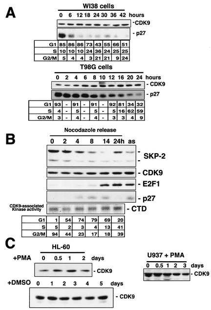
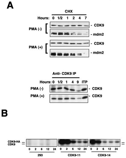
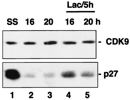
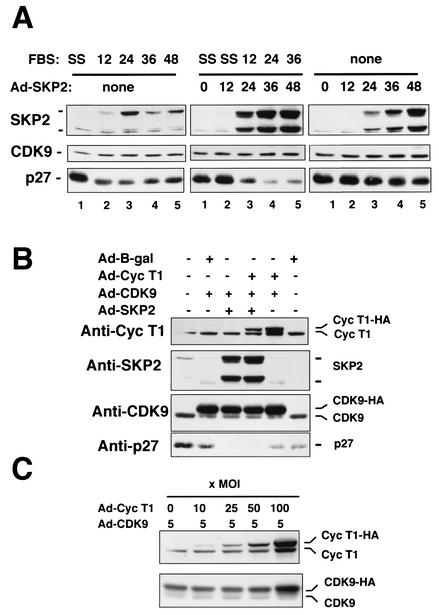
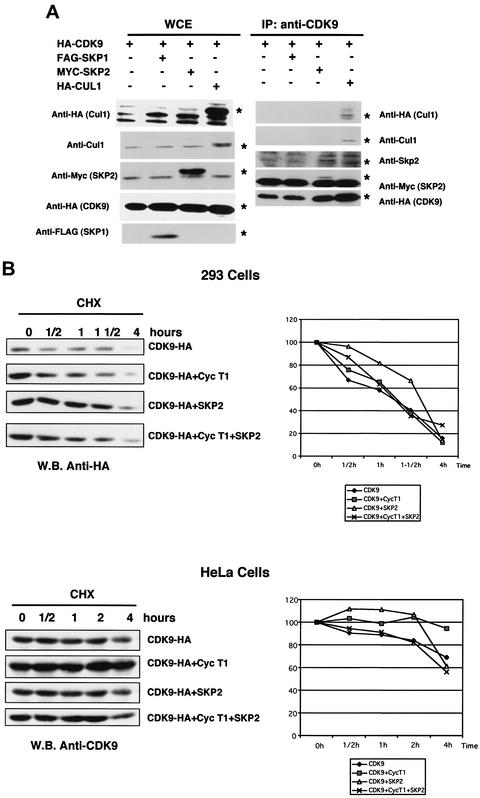
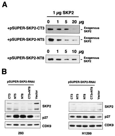
CDK9 is a CDC2-related kinase and the catalytic subunit of the positive-transcription elongation factor b and the Tat-activating kinase. It has recently been reported that CDK9 is a short-lived protein whose levels are regulated during the cell cycle by the SCF(SKP2) ubiquitin ligase complex (R. E. Kiernan et al., Mol. Cell. Biol. 21:7956-7970, 2001). The results presented here are in contrast to those observations. CDK9 protein levels remained unchanged in human cells entering and progressing through the cell cycle from G(0), despite dramatic changes in SKP2 expression. CDK9 levels also remained unchanged in cells exiting from mitosis and progressing through the next cell cycle. Similarly, the levels of CDK9 protein did not change as cells exited the cell cycle and differentiated along various lineages. In keeping with these observations, the kinase activity associated with CDK9 was found to not be regulated during the cell cycle. We have also found that endogenous CDK9 is a very stable protein with a half-life (t(1/2)) of 4 to 7 h, depending on the cell type. In contrast, when CDK9 is overexpressed, it is not stabilized and is rapidly degraded, with a t(1/2) of less than 1 h, depending on the level of expression. Treatment of cells with proteasome inhibitors blocked the degradation of short-lived proteins, such as p27, but did not affect the expression of endogenous CDK9. Ectopic overexpression of SKP2 led to reduction of p27 protein levels but had no effect on the expression of endogenous CDK9. Finally, downregulation of endogenous SKP2 gene expression by interfering RNA had no effect on CDK9 protein levels, whereas p27 protein levels increased dramatically. Therefore, the SCF(SKP2) ubiquitin ligase does not regulate CDK9 expression in a cell cycle-dependent manner.


Figures

FIG. 1.
FIG. 1.
CDK9 protein expression and kinase activity are not cell cycle regulated. (A) CDK9 protein levels in cells entering and progressing through the cell cycle from G0. WI38 primary human fibroblasts (upper panels) and T98G human glioblastoma cells (lower panels) were serum starved and restimulated as described in Materials and Methods, and cells were collected at the indicated time points. CDK9 and p27 protein levels were analyzed by Western blotting with anti-CDK9 and anti-p27 antibodies, respectively. The percentage of cells at each phase of the cell cycle was determined by flow cytometric analysis and is indicated in the tabular portions of the panels. (B) CDK9 protein expression and kinase activity in cells progressing through the cell cycle after mitotic arrest. HeLa cells were incubated in the presence of nocodazole for 16 h. Mitotic cells resumed the cell cycle after being placed in fresh, nocodazole-free medium. The cells were collected at the indicated time points. CDK9, SKP2, E2F1, and p27 protein levels were analyzed by Western blotting by using their respective antibodies. Kinase assays were performed for each time point after protein extracts were immunoprecipitated with anti-CDK9 antibodies. GST-RNAPII-CTD was used as the exogenous substrate (see Materials and Methods). as, asynchronous. (C) CDK9 protein expression does not change during myeloid cell differentiation. Exponentially growing HL60 and U937 cells were induced to differentiate to macrophages in the presence of PMA (3 ng/ml). HL-60 cells were also induced to differentiate to granulocytes in the presence of 1.3% DMSO. Cells were collected at the indicated time points, and the expression levels of CDK9 protein were assessed by Western blot analysis.
FIG. 2.
FIG. 2.
Endogenous CDK9 is not a short-lived protein. (A) CDK9 protein t1/2 was analyzed in exponentially growing HL60 cells (−PMA) or HL60 cells differentiating to macrophages (+PMA) by pulse-chase and CHX treatments. HL60 cells were treated with CHX (40 μg /ml) to block protein synthesis for the indicated time periods. CDK9 and mdm2 protein levels were analyzed by Western blotting by using anti-CDK9 and anti-mdm2 antibodies (upper panels). HL60 cells were labeled with [35S]Met and analyzed by pulse-chase. CDK9 was immunoprecipitated by using anti-CDK9 antibodies (lower panels). ITP, in vitro-translated protein. (B) The t1/2s of endogenous and ectopically expressed CDK9 proteins were analyzed by pulse-chase labeling (see above and Materials and Methods) in 293 cells and in two 293-derived cell lines (CDK9-11 and CDK9-14) that constitutively express CDK9-HA (ectopically expressed CDK9 carrying an HA tag).
FIG. 3.
FIG. 3.
Lactacystin does not affect the expression of endogenous CDK9. T98G cells were serum starved and restimulated as described in Materials and Methods. The cells were collected 16 and 20 h after restimulation. One set of samples was treated with lactacystin for 5 h before the cells were collected (11 and 15 h after restimulation). The levels of CDK9 and p27 proteins in extracts of cells treated with lactacystin or vehicle were determined by Western blot analysis using anti-CDK9 and anti-p27 antibodies, respectively. Lac/5h, treatment with lactacystin for 5 h; SS, serum starved.
FIG. 4.
FIG. 4.
Ectopic expression of SKP2 does not affect the expression of CDK9. (A) T98G cells were serum starved and restimulated (left panels); serum starved, infected with recombinant Ad expressing SKP2 (Ad-SKP2), and restimulated (middle panels); or serum starved and infected with Ad-SKP2 (right panels). Cells were collected at the indicated time points after restimulation or infection, and the expression levels of SKP2, CDK9, and p27 proteins were analyzed by Western blotting. SS, serum starved. (B) Exponentially growing HeLa cells were infected with the indicated recombinant Ad. The cells were collected 48 h after infection, and the expression levels of SKP2, CDK9, and p27 proteins were analyzed by Western blotting. (C) Effect of the ectopic expression of cyclin T1 on CDK9 stability. HeLa cells were infected with combinations of recombinant Ad expressing CDK9-HA (Ad-CDK9; constant MOI) and recombinant Ad expressing cyclin T1-HA (Ad-cyclin T1; increasing MOI). Forty-eight h postinfection, the cells were collected and the expression levels of cyclin T1 and CDK9 proteins were analyzed by Western blotting in the cell lysates.
FIG. 5.
FIG. 5.
CDK9 interacts with SKP2 and Cul-1 in transfected 293 cells. (A) Cells from the 293 cell line were transfected with pCMV-CDK9-HA (HA-CDK9), pCMV-Flag-SKP1 (FAG-SKP1), pCMV-myc-SKP2 (MYC-SKP2), and pCMV-HA-Cul1 (HA-Cul1) in the combinations indicated. Five micrograms of each plasmid was transfected together with 0.5 μg of a luciferase reporter plasmid by the calcium phosphate method. The cells were collected 48 h following transfection, and whole-cell extracts were immunoprecipitated with anti-CDK9 antibodies. Whole-cell extracts and immunoprecipitates were resolved by SDS-PAGE followed by Western blot analysis. The left panels show the expression levels of the transfected proteins, determined by using specific antibodies. The right panels show coimmunoprecipitation of Cul-1 and SKP2 with CDK9. SKP1 could not be detected in this experiment (see text for details). Asterisks indicate the migration of specific bands. WCE, whole-cell extracts; IP, immunoprecipitates. (B) Ectopic expression of SKP2 and cyclin T1 alone or in combination does not affect CDK9 t1/2. CDK9-, SKP2-, and cyclin T1-expressing plasmids were transfected into 293 cells alone or in combination, as indicated in the upper panels. In parallel experiments, HeLa cells were transduced with Ads expressing CDK9, SKP2, and cyclin T1 alone or in combination (lower panels). Twenty-four hours posttransfection and posttransduction, the cells were divided onto five identical plates. Twenty-four hours later, the cells were incubated with CHX (40 μg/ml) for the indicated periods of time. The expression levels of CDK9 at each time point were analyzed by Western blotting. In each case, Western blots from a representative experiment as well as quantification of the results of three independent experiments are shown. Expression levels are represented as percentages of the expression values at time zero. W.B., Western blot.
FIG. 6.
FIG. 6.
Downregulation of cellular SKP2 gene expression by RNAi has no effect on CDK9 protein levels. The mammalian expression vector pSUPER was used to synthesize SKP2-RNAi-like transcripts. (A) The effectiveness of each of the pSUPER-RNAi-SKP2 constructs was determined in transient-transfection experiments. H1299 cells were transiently cotransfected with 1 μg of pCDNA3-SKP2 and increasing amounts of the indicated pSUPER-RNAi-SKP2 constructs. Forty-eight hours after transfection, the cells were collected and whole protein extracts were resolved by SDS-10% PAGE followed by Western blot analysis using anti-SKP2 antibodies. (B) Cells from the 293 cell line and H1299 cells were cotransfected with 10 μg of different pSUPER-RNAi-SKP2 constructs and 1 μg of pSG5puro. After 1 week of selection with puromycin, pools of resistant cells were collected and whole protein extracts were resolved by SDS-12% PAGE followed by Western blot analysis using the indicated antibodies. The expression levels of cellular SKP2, p27, and CDK9 are shown.

References

    1. Ausubel, F. M., R. Brent, R. E. Kington, D. D. Moore, J. G. Seidman, J. A. Smith, and K. E. Struhl. 1988. Current protocols in molecular biology. Greene Publishing Associates and Wiley-Interscience, New York, N.Y.
    1. Barboric, M., R. M. Nissen, S. Kanazawa, N. Jabrane-Ferrat, and B. M. Peterlin. 2001. NF-κB binds P-TEFb to stimulate transcriptional elongation by RNA polymerase II. Mol. Cell 8:327-337. - PubMed
    1. Becker, T. C., R. J. Noel, W. S. Coats, A. M. Gomez-Foix, T. Alam, R. D. Gerard, and C. B. Newgard. 1994. Use of recombinant adenovirus for metabolic engineering of mammalian cells. Methods Cell Biol. 43:161-189. - PubMed
    1. Bhattacharya, S., J. Garriga, J. Calbó, T. Yong, D. S. Haines, and X. Graña. 2003. SKP2 associates with p130 and accelerates p130 ubiquitylation and degradation in human cells. Oncogene 22:2443-2451. - PubMed
    1. Brummelkamp, T., R. Bernards, and R. Agami. 2002. A system for stable expression of short interfering RNAs in mammalian cells. Science 296:550-553. - PubMed

Publication types

MeSH terms

LinkOut - more resources