. 2003 Aug;23(15):5198-207.
doi: 10.1128/MCB.23.15.5198-5207.2003.

Deletion of the SNARE vti1b in mice results in the loss of a single SNARE partner, syntaxin 8

Affiliations

Deletion of the SNARE vti1b in mice results in the loss of a single SNARE partner, syntaxin 8

Vadim Atlashkin et al. Mol Cell Biol. 2003 Aug.

Abstract

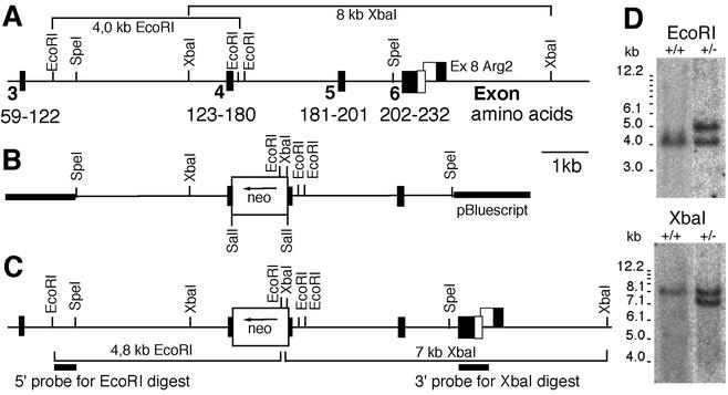
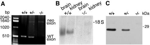
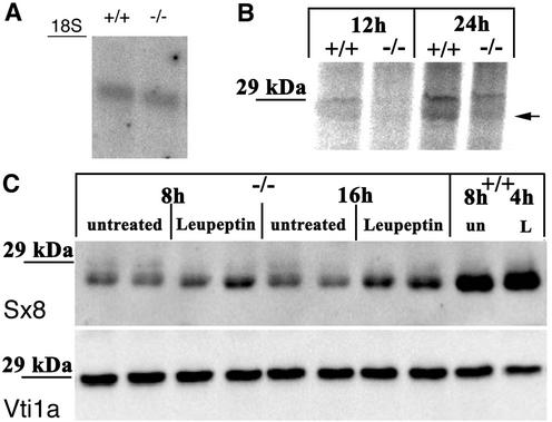
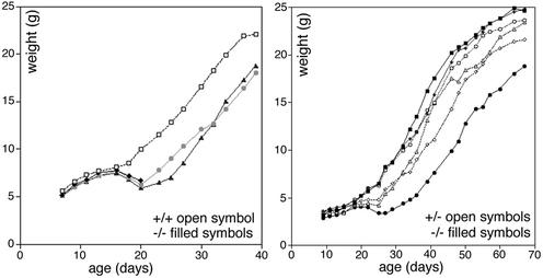
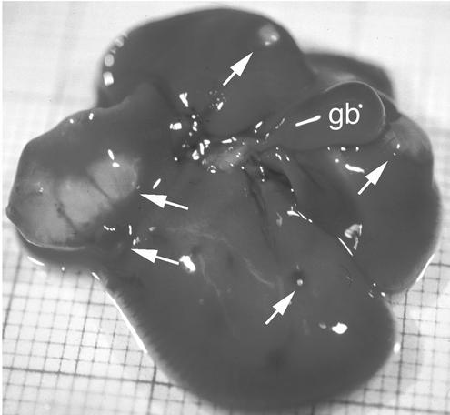
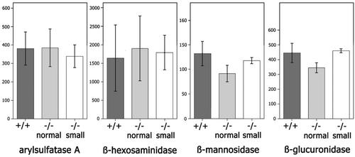
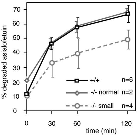
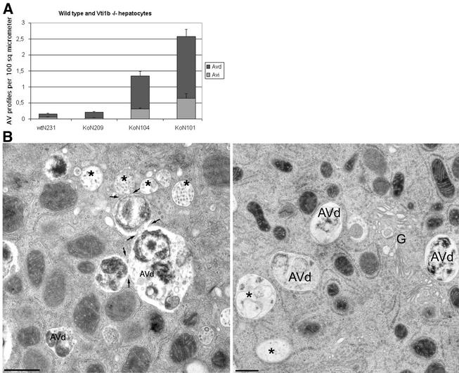
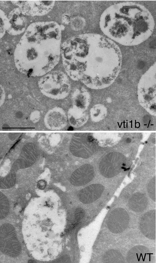
SNARE proteins participate in recognition and fusion of membranes. A SNARE complex consisting of vti1b, syntaxin 8, syntaxin 7, and endobrevin/VAMP-8 which is required for fusion of late endosomes in vitro has been identified recently. Here, we generated mice deficient in vti1b to study the function of this protein in vivo. vti1b-deficient mice had reduced amounts of syntaxin 8 due to degradation of the syntaxin 8 protein, while the amounts of syntaxin 7 and endobrevin did not change. These data indicate that vti1b is specifically required for the stability of a single SNARE partner. vti1b-deficient mice were viable and fertile. Most vti1b-deficient mice were indistinguishable from wild-type mice and did not display defects in transport to the lysosome. However, 20% of the vti1b-deficient mice were smaller. Lysosomal degradation of an endocytosed protein was slightly delayed in hepatocytes derived from these mice. Multivesicular bodies and autophagic vacuoles accumulated in hepatocytes of some smaller vti1b-deficient mice. This suggests that other SNAREs can compensate for the reduction in syntaxin 8 and for the loss of vti1b in most mice even though vti1b shows only 30% amino acid identity with its closest relative.


Figures

FIG. 1.
FIG. 1.
Targeted disruption of vti1b. (A) Genomic DNA for vti1b isolated from a phage library. According to information from the Mouse Genome Sequencing Consortium, exon 1 encodes 100 bp of 5′ untranslated region and amino acid residues 1 to 38 and exon 2 encodes amino acid residues 39 to 58. (B) Seven-kilobase targeting vector with exon 4 disrupted by insertion of the neomycin resistance cassette (neo). (C) vti1b locus after homologous recombination. Sections of DNA used as probes for Southern blot hybridizations are marked, and fragments detected after EcoRI and XbaI digestions are indicated for the targeted and wild-type (A) alleles. (D) Southern blots of ES cell DNA after EcoRI and XbaI digestions. An EcoRI fragment of 4.0 kb is detected in the wild-type (+/+) allele, and a 4.8-kb band is detected in the targeted allele. Insertion of the neo gene reduces an 8-kb wild-type XbaI fragment to 7 kb.
FIG. 2.
FIG. 2.
vti1b mRNA and protein were absent in animals homozygous for the targeted vti1b allele. (A) Genotyping of genomic DNA using PCR. A band of 500 bp including exon 4 of vti1b was amplified from the wild-type (WT) allele. After insertion of the neo gene, the fragment shifts to 1,600 bp. In addition, the neo gene was amplified using specific primers (not shown). (B) Northern blot for vti1b mRNA. RNA was isolated from the brains and kidneys of wild-type (+/+) and vti1b-deficient (−/−) animals, separated by agarose gel electrophoresis, blotted, and probed for vti1b. (C) Western blot for vti1b protein. Protein extracts from wild-type, heterozygous (+/−), and vti1b-deficient embryonic fibroblasts were separated by SDS-PAGE and stained with an antiserum directed against vti1b. The vti1b protein was not detected in vti1b−/− cells. Less vti1b was present in heterozygous than in wild-type cells.
FIG. 3.
FIG. 3.
Syntaxin 8 protein levels are reduced in vti1b-deficient mice. Homogenates were prepared from livers, brains, kidneys, and spleens of wild-type (+/+), heterozygous (+/−), and vti1b-deficient (−/−) animals; separated by SDS-PAGE; and stained with antisera directed against the indicated SNAREs. Liver homogenates from wild-type and vti1b-deficient animals were extracted with Triton X-114, and the detergent phase was separated by SDS-PAGE to allow the detection of endobrevin. No reduction in protein levels was observed for endobrevin, syntaxin 7, vti1a, and SNAP-29.
FIG. 4.
FIG. 4.
Syntaxin 8 mRNA was stable in vti1b-deficient mice, but the syntaxin 8 protein was degraded in lysosomes. (A) Similar levels of syntaxin 8 mRNA were detected in wild-type and vti1b-deficient mice. RNA was isolated from the kidneys of wild-type (+/+) and vti1b-deficient (−/−) animals, separated by agarose gel electrophoresis, blotted, and probed for syntaxin 8. (B) Syntaxin 8 protein was slowly synthesized in wild-type embryonic fibroblasts. Wild-type (+/+) and vti1b-deficient (−/−) embryonic fibroblasts were incubated with [35S]methionine for 12 or 24 h. Syntaxin 8 was immunoprecipitated, and the fractions were analyzed by SDS-PAGE and autoradiography. Even though syntaxin 8 protein could be precipitated, incorporation of radioactivity was detected only after 24 h (arrow), indicating that the protein is very stable. The upper band at 29 kDa was nonspecific. (C) Treatment with the lysosomal protease inhibitor leupeptin stabilized syntaxin 8 protein in vti1b-deficient fibroblasts. vti1b-deficient (−/−) and wild-type (+/+) embryonic fibroblasts were treated with 100 μM leupeptin as indicated. Extracts containing equal amounts of protein were analyzed by immunoblotting, using antiserum directed against syntaxin 8 or vti1a as a loading control. un, untreated; L, leupeptin.
FIG. 5.
FIG. 5.
vti1b-deficient mice showed a heterogeneous growth phenotype. Male mice from the same litter were compared. All of the mice gained weight at similar rates until day 16. Afterward, a few died (left, solid diamonds), and ∼20% of the vti1b-deficient mice lost weight and remained smaller than their littermates (left, shaded circles and solid triangles; right, solid circles). Most vti1b-deficient mice (right, solid squares and diamonds) grew at rates similar to those of their heterozygous (right, open symbols) or wild-type (left, open symbols) littermates.
FIG. 6.
FIG. 6.
Liver cysts in aged normal-size vti1b-deficient mice. Eight out of 23 vti1b-deficient mice between 15 and 21 months old had multiple liver cysts (arrows). These cysts were filled with a clear fluid, except for two small cysts containing a yellow liquid. gb, gall bladder.
FIG. 7.
FIG. 7.
Levels of lysosomal hydrolases were not affected by deletion of vti1b. The activities (in milliunits per gram of tissue) of the lysosomal hydrolases arylsulfatase A, β-hexosaminidase, β-mannosidase, and β-glucuronidase were determined in liver homogenates derived from wild-type (+/+; n = 5), normal-size (n = 3), and small (n = 2) vti1b-deficient (−/−) mice. The error bars indicate standard deviations.
FIG. 8.
FIG. 8.
Lysosomal degradation of endocytosed 125I-asialofetuin was slower in hepatocytes derived from small vti1b-deficient mice. No difference in degradation of 125I-asialofetuin was observed in hepatocytes from wild-type (+/+) and vti1b-deficient (−/−) mice of normal weight. Hepatocytes derived from wild-type or vti1b-deficient mice of normal or smaller size were cultured for 1 day. They were incubated with 125I-asialofetuin for 20 min at 37°C and chased for the indicated periods after removal of the radioactive medium. Degraded (soluble in TCA) and intact 125I-asialofetuins were separated by TCA precipitation of the medium and cells and quantified. About 8,000 to 18,000 cpm was endocytosed by the hepatocytes, depending on cell density but independent of the genotype. Three individual experiments were performed with double values. The error bars indicate standard deviations.
FIG. 9.
FIG. 9.
Hepatocytes of some smaller vti1b-deficient mice accumulated autophagic vacuoles and multivesicular bodies. (A) The numbers of early (Avi) and late (Avd) autophagic vacuole profiles per 100 μm2 of cell area on thin sections were determined for hepatocytes isolated from wild-type (wt) and three different smaller vti1b-deficient (Ko) mice. The error bars represent standard errors of the mean. (B) Fusion profiles of autophagic vacuoles (arrows), as well as multivesicular bodies (∗), were more prominent in hepatocytes from vti1b−/− mouse 101 (left) than in wild-type (right) hepatocytes. G, Golgi. Bar, 500 nm.
FIG.10.
FIG.10.
LAMP-1-positive structures had similar appearances in wild-type (WT) and vti1b-deficient hepatocytes. Ultrathin cryosections were obtained from hepatocytes of wild-type and smaller vti1b-deficient mice and were decorated with antibodies specific for LAMP-1 and 10-nm-diameter protein A-gold. LAMP-1 is found in lysosomes, late endosomes, and late autophagic vacuoles. vti1b−/− cells seemed to contain more LAMP-1-positive organelles, which were sometimes clustered. Bar, 1 μm.

Similar articles

Cited by

References

    1. Advani, R. J., H. R. Bae, J. B. Bock, D. S. Chao, Y. C. Doung, R. Prekeris, J. S. Yoo, and R. H. Scheller. 1998. Seven novel mammalian SNARE proteins localize to distinct membrane compartments. J. Biol. Chem. 273:10317-10324. - PubMed
    1. Advani, R. J., B. Yang, R. Prekeris, K. C. Lee, J. Klumperman, and R. H. Scheller. 1999. VAMP-7 mediates vesicular transport from endosomes to lysosomes. J. Cell Biol. 146:765-775. - PMC - PubMed
    1. Antonin, W., D. Fasshauer, S. Becker, R. Jahn, and T. Schneider. 2002. Crystal structure of the endosomal SNARE complex reveals common structural principles of all SNAREs. Nat. Struct. Biol. 9:107-111. - PubMed
    1. Antonin, W., C. Holroyd, D. Fasshauer, S. Pabst, G. Fischer von Mollard, and R. Jahn. 2000. A SNARE complex mediating fusion of late endosomes defines conserved properties of SNARE structure and function. EMBO J. 19:6453-6464. - PMC - PubMed
    1. Antonin, W., C. Holroyd, R. Tikkanen, S. Höning, and R. Jahn. 2000. The R-SNARE endobrevin/VAMP-8 mediates homotypic fusion of early and late endosomes. Mol. Biol. Cell 11:3289-3298. - PMC - PubMed

Publication types

MeSH terms

LinkOut - more resources