. 2003 Aug;23(15):5245-55.
doi: 10.1128/MCB.23.15.5245-5255.2003.

The actin-binding domain of Slac2-a/melanophilin is required for melanosome distribution in melanocytes

Affiliations

The actin-binding domain of Slac2-a/melanophilin is required for melanosome distribution in melanocytes

Taruho S Kuroda et al. Mol Cell Biol. 2003 Aug.

Abstract

Melanosomes containing melanin pigments are transported from the cell body of melanocytes to the tips of their dendrites by a combination of microtubule- and actin-dependent machinery. Three proteins, Rab27A, myosin Va, and Slac2-a/melanophilin (a linker protein between Rab27A and myosin Va), are known to be essential for proper actin-based melanosome transport in melanocytes. Although Slac2-a directly interacts with Rab27A and myosin Va via its N-terminal region (amino acids 1 to 146) and the middle region (amino acids 241 to 405), respectively, the functional importance of the putative actin-binding domain of the Slac2-a C terminus (amino acids 401 to 590) in melanosome transport has never been elucidated. In this study we showed that formation of a tripartite protein complex between Rab27A, Slac2-a, and myosin Va alone is insufficient for peripheral distribution of melanosomes in melanocytes and that the C-terminal actin-binding domain of Slac2-a is also required for proper melanosome transport. When a Slac2-a deletion mutant (DeltaABD) or point mutant (KA) that lacks actin-binding ability was expressed in melanocytes, the Slac2-a mutants induced melanosome accumulation in the perinuclear region, possibly by a dominant negative effect, the same as the Rab27A-binding-defective mutant of Slac2-a or the myosin Va-binding-defective mutant. Our findings indicate that Slac2-a organizes actin-based melanosome transport in cooperation with Rab27A, myosin Va, and actin.


Figures
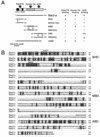
FIG. 1.
FIG. 1.
Domain structures of mouse Slac2-a/melanophilin. (A) Schematic representation of three functional domains of mouse Slac2-a and the deletion or point mutants of Slac2-a used in this study. The SHD is composed of two potential α-helical regions (SHD1 and SHD2; black boxes) separated by two zinc finger motifs (indicated by Zn2+), and it is necessary and sufficient for specific Rab27A/B recognition (7). The MBD and ABD are indicated by the shaded and hatched boxes, respectively (13). The Rab27A-, myosin Va-, and actin-binding activities of each mutant (−, ±, +, or ++) are indicated. (B) Sequence alignment of mouse Slac2-a and Slac2-c. Residues in the sequences that are conserved and similar are shown against black and shaded backgrounds, respectively. The SHD, MBD, and ABD are boxed. The solid lines indicate two SHDs. The pound signs indicate the positions of amino acid residues that are conserved between Slac2-a and Slac2-c, which was mutated in this study (see also panel A). The conserved Cys residues corresponding to two zinc finger motifs are indicated by asterisks. Amino acid numbers are indicated at the right of each line.
FIG. 2.
FIG. 2.
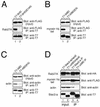
Identification of critical residues responsible for Rab27A, myosin Va, and actin binding of Slac2-a by Ala-based site-directed mutagenesis. (A to C) Loss of Rab27A-binding activity of the SHD(E14A) mutant (A), loss of myosin Va-binding activity of the MBD(EA) mutant (B), and reduced actin-binding activity of the ABD(KA) mutant (C). T7-tagged Slac2-a mutant and FLAG-tagged proteins (Rab27A or myosin Va tail) were coexpressed in COS-7 cells. Coimmunoprecipitated FLAG-tagged proteins and actin were first detected by anti-FLAG tag antibody and antiactin antibody, respectively (middle panels), and the immunoprecipitated (IP) T7-Slac2-a proteins were then visualized with anti-T7 tag antibody (lower panels). The upper panels show total expressed proteins (1/80 volume of the reaction mixtures; input) used for immunoprecipitation. (D) Deletion of the C-terminal actin-binding site (ΔABD) specifically impaired actin-binding activity but had no effect on Rab27A- or myosin Va-binding activity. pEF-T7-Slac2-a-ΔABD, pEF-HA-Rab27A, and pEF-FLAG-MC-myosin Va-tail were cotransfected into COS-7 cells. Coimmunoprecipitated HA-Rab27A, FLAG-myosin Va, and actin were detected by anti-HA tag, anti-FLAG tag, and antiactin antibodies, respectively (lanes 3 and 4 in the upper three panels). Lanes 1 and 2 represent the total proteins expressed (1/80 volume of the reaction mixtures; input) in lanes 3 and 4, respectively, used for immunoprecipitation. Note that the ΔABD mutant specifically impairs actin-binding activity (lane 4 in the third panel).
FIG. 3.
FIG. 3.
A black mouse-derived melanocyte cell line, melan-a, exhibits normal melanosome distribution at the cell periphery irrespective of expression of GFP or GFP-Slac2-a. An immortalized black melanocyte cell line, melan-a, was transfected with vectors encoding GFP alone (A to D) or GFP-tagged full-length Slac2-a (E to H). After cells were fixed and permeabilized, they were stained with anti-Rab27A and anti-myosin Va antibodies and visualized with Alexa Fluor secondary antibody conjugates. GFP was localized throughout the cell (A), whereas GFP-Slac2-a was localized at the periphery of the cell (E). Expression of both proteins affected neither the normal distribution of melanosomes at the periphery of the cells, as revealed by bright-field images (D and H), nor localization of Rab27A (B and F) and myosin Va (C and G) on the melanosomes. The inset in panel C is a merged image of Rab27A and myosin Va, where both proteins were colocalized (yellow signals) on melanosomes (see melanosome distribution in the inset in panel D). The inset in panel G is a merged image of GFP-Slac2-a (pseudocolored in light blue), Rab27A (green), and myosin Va (red), where three proteins were colocalized (white signals) on melanosomes (see melanosome distribution in the inset in panel H). Bars, 10 μm.
FIG. 4.
FIG. 4.
Effect of Rab27A-binding-defective mutants of Slac2-a on melanosome transport. Melan-a cells were transfected with vectors encoding GFP-SHD (A to D), GFP-SHD(E14A) (E to H), or GFP-Slac2-a(E14A) (I to L). After cells were fixed and permeabilized, they were stained with anti-Rab27A and anti-myosin Va antibodies followed by Alexa Fluor secondary antibody conjugates. Fluorescence of GFP-Slac2-a mutants (A, E, and I), Rab27A (B, F, and J), and myosin Va (C, G, and K) was analyzed by confocal microscopy. Bright-field images (D, H, and L) show the melanosome distribution in the cells, and the cells are outlined in yellow (D and L). The insets in panels C, G, and K are merged images of Rab27A and myosin Va, and bright-field images corresponding to each inset are shown in panels D, H, and L, respectively. Note that expression of GFP-SHD induced melanosome aggregation in the perinucleus (D) and segregation of Rab27A from melanosomes and myosin Va (inset in panel C), whereas GFP-SHD(E14A) had no effect on either melanosome distribution (H) or colocalization of Rab27A with myosin Va on melanosomes (yellow signals in the inset in panel G and melanosome distribution in the inset in panel H). Expression of GFP-Slac2-a(E14A) also caused melanosome accumulation in the perinucleus (L) and reduced myosin Va signals (K). Bars, 10 μm.
FIG. 5.
FIG. 5.
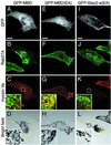
Effect of myosin Va-binding-defective mutants of Slac2-a on melanosome transport. Melan-a cells were transfected with vectors encoding GFP-MBD (A to D), GFP-MBD(EA) (E to H), or GFP-Slac2-a(EA) (I to L). After cells were fixed and permeabilized, they were stained with anti-Rab27A and anti-myosin Va antibodies followed by Alexa Fluor secondary antibody conjugates. Fluorescence of GFP-Slac2-a mutants (A, E, and I), Rab27A (B, F, and J), and myosin Va (C, G, and K) was analyzed by confocal microscopy. Bright-field images (D, H, and L) show the melanosome distribution in cells, and the cells were outlined in yellow (D and L). The insets in panels C, G, and K are merged images of Rab27A and myosin Va, and bright-field images corresponding to each inset are shown in panels D, H, and L, respectively. Note that expression of GFP-MBD induced melanosome aggregation in the perinucleus (D) and segregation of myosin Va from Rab27A and melanosomes (inset in panel C), whereas GFP-MBD(EA) affected neither melanosome distribution (H) nor colocalization of Rab27A with myosin Va on melanosomes (yellow signals in the inset in panel G and melanosome distribution in the inset in panel H). Expression of GFP-Slac2-a(EA) also caused melanosome accumulation in the perinucleus (L) and reduced myosin Va signals (K). Bars, 10 μm.
FIG. 6.
FIG. 6.
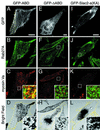
Effect of actin-binding-defective mutants of Slac2-a (KA and ΔABD) on melanosome transport. Melan-a cells were transfected with vectors encoding GFP-ABD (A to D), GFP-ΔABD (E to H), or GFP-Slac2-a(KA) (I to L). After cells were fixed and permeabilized, they were stained with anti-Rab27A and anti-myosin Va antibodies followed by Alexa Fluor secondary antibody conjugates. Fluorescence of GFP-Slac2-a mutants (A, E, and I), Rab27A (B, F, and J), and myosin Va (C, G, and K) was analyzed by confocal microscopy. Bright-field images (D, H, and L) show the melanosome distribution in cells, and the cells were outlined in yellow (D, H, and L). The insets in panels C, G, and K are merged images of Rab27A and myosin Va, and bright-field images corresponding to each inset are shown in panels D, H, and L, respectively. Note that expression of GFP-ABD did not alter distributions of Rab27A and myosin Va on melanosomes (yellow signals in the insets in panel C and melanosome distribution in the inset in panel D) but did result in exclusion of melanosomes from peripheral actin bundles without large clumps of melanosomes around the nucleus (A and D, arrowheads). Expression of GFP-ΔABD and GFP-Slac2-a(KA) caused melanosome accumulation in the perinucleus (H and L) and reduced myosin Va signals (G and K) even though these mutants still retained the ability to bind to both Rab27A and myosin Va (Fig. 2D and data not shown). These findings indicate that formation of a tripartite protein complex consisting of Rab27A, Slac2-a, and myosin Va is not sufficient for melanosome transport and that the actin-binding ability of ABD is also necessary for normal melanosome distribution at the cell periphery. Bars, 10 μm.
FIG. 7.
FIG. 7.
Summary of melanosome distribution assay results. Melan-a cells were transfected with a vector encoding the indicated Slac2-a protein tagged with GFP. Images of transfected melan-a cells were captured at random by using GFP fluorescence as a marker, and we judged whether melanosomes had aggregated in the perinucleus by examining the corresponding bright-field images. The results are expressed as the percentages of cells exhibiting perinuclear melanosome aggregation and are means ± standard deviations from three independent experiments (n > 150).
FIG. 8.
FIG. 8.
Schematic model illustrating possible roles of Slac2-a-actin interaction in actin-based melanosome transport. Slac2-a binding to actin could be involved in the following three steps of melanosome transport: melanosome transfer from microtubules to actin filaments (A), melanosome transport along actin filaments (B), and melanosome capture by actin filaments near the plasma membrane of the cell (C). Note that these three steps are not mutually exclusive. In this study we demonstrated that association between Slac2-a and actin is required for the transfer step (A). See Discussion for details.

References

    1. Bahadoran, P., E. Aberdam, F. Mantoux, R. Buscà, K. Bille, N. Yalman, G. de Saint-Basile, R. Casaroli-Marano, J. P. Ortonne, and R. Ballotti. 2001. Rab27a: a key to melanosome transport in human melanocytes. J. Cell Biol. 152:843-850. - PMC - PubMed
    1. Bahadoran, P., R. Buscà, C. Chiaverini, W. Westbroek, J. Lambert, K. Bille, G. Valony, M. Fukuda, J. M. Naeyaert, J. P. Ortonne, and R. Ballotti. 2003. Characterisation of the molecular defects in RAB27A, caused by RAB27A missense mutations found in patients with Griscelli syndrome. J. Biol. Chem. 278:11386-11392. - PubMed
    1. Bennett, D. C., P. J. Cooper, and I. R. Hart. 1987. A line of non-tumorigenic mouse melanocytes, syngeneic with the B16 melanoma and requiring a tumour promoter for growth. Int. J. Cancer 39:414-418. - PubMed
    1. Bock, J. B., H. T. Matern, A. A. Peden, and R. H. Scheller. 2001. A genomic perspective on membrane compartment organization. Nature 409:839-841. - PubMed
    1. Chen, D., J. Guo, T. Miki, M. Tachibana, and W. A. Gahl. 1997. Molecular cloning and characterization of rab27a and rab27b, novel human rab proteins shared by melanocytes and platelets. Biochem. Mol. Med. 60:27-37. - PubMed

Publication types

MeSH terms

LinkOut - more resources