. 2003 Aug;23(15):5256-68.
doi: 10.1128/MCB.23.15.5256-5268.2003.

Rb and N-ras function together to control differentiation in the mouse

Affiliations

Rb and N-ras function together to control differentiation in the mouse

Chiaki Takahashi et al. Mol Cell Biol. 2003 Aug.

Abstract

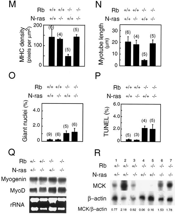
The product of the retinoblastoma tumor suppressor gene (Rb) can control cell proliferation and promote differentiation. Murine embryos nullizygous for Rb die midgestation with defects in cell cycle regulation, control of apoptosis, and terminal differentiation of several tissues, including skeletal muscle, nervous system, and lens. Previous cell culture-based experiments have suggested that the retinoblastoma protein (pRb) and Ras operate in a common pathway to control cellular differentiation. Here we have tested the hypothesis that the proto-oncogene N-ras participates in Rb-dependent regulation of differentiation by generating and characterizing murine embryos deficient in both N-ras and Rb. We show that deletion of N-ras rescues a unique subset of the developmental defects associated with nullizygosity of Rb, resulting in a significant extension of life span. Rb(-/-); N-ras(-/-) skeletal muscle has normal fiber density, myotube length and thickness, in contrast to Rb-deficient embryos. Additionally, Rb(-/-); N-ras(-/-) muscle shows a restoration in the expression of the late muscle-specific gene MCK, and this correlates with a significant potentiation of MyoD transcriptional activity in Rb(-/-); N-ras(-/-), compared to Rb(-/-) myoblasts in culture. The improved differentiation of skeletal muscle in Rb(-/-); N-ras(-/-) embryos occurs despite evidence of deregulated proliferation and apoptosis, as seen in Rb-deficient animals. Our findings suggest that the control of differentiation and proliferation by Rb are genetically separable.


Figures

FIG. 1.
FIG. 1.
Effects of N-ras loss on the appearance of Rb-deficient embryos. Embryos from the same litter with the indicated genotype were recovered at E13.5 (A to C) and E15.5 (D to F) and immediately photographed in saline. Bars, 150 μm.
FIG. 2.
FIG. 2.
Effect of N-ras loss on skeletal muscle development in E14.5 Rb-deficient embryos. (A to H) Longitudinal sections through the fibers of thoracic somite-associated skeletal muscle from sagittal sections of E14.5 embryos derived from the same litter with the indicated genotype were stained with H&E (A to D) or immunostained by using an antibody to MHC (E to H). Large nuclei (arrow) in myotubes are indicated in panel D. Magnification, ×40. (I to L) Apoptosis (TUNEL) observed in myoblasts of thoracic skeletal muscle of E14.5 embryos of the indicated genotype. Magnification, ×60. (M) The density of MHC in myotubes stained with antibody to MHC was quantified by using the NIH image 1.61 program. The strength of the signal was read as pixel number per square micrometer. Ten myotubes per embryo were analyzed, and the average density of the MHC signal ± standard error was calculated. In parentheses is the number of embryos used for the analysis. (N) The length of myotubes immunostained with an antibody to MHC was quantified. Longitudinal sections of thoracic skeletal muscle from E14.5 embryos were analyzed by microscopic observation. Twenty myotubes per embryo were measured, and average length ± standard error is presented. In parentheses is the number of embryos used for the analysis. (O) One hundred myotubes in the thoracic skeletal muscle were analyzed for the presence of giant nuclei following staining with H&E. The average percentage ± standard error is presented. In parentheses is the number of embryos used for the analysis. (P) The level of apoptosis was quantified by counting the frequency of TUNEL-positive cells per 300 nuclei analyzed in the thoracic muscle. The average percentage ± standard error is presented. In parentheses is the number of embryos used for the analysis. (Q) The levels of MyoD and myogenin transcripts in total RNA derived from carcasses of live E14.5 embryo littermates of the indicated genotype were determined by Northern blot analysis. (R) Expression of MCK determined by RNase protection assay with RNAs derived from carcasses of live E14.5 embryos of the indicated genotype from two sets of matched littermates, with lanes 1, 3, 5, and 7 showing data from one litter and lanes 2, 4, and 6 showing data from the other litter. The ratio of MCK to β-actin expression is shown at the bottom of the panel.
FIG. 2.
FIG. 2.
Effect of N-ras loss on skeletal muscle development in E14.5 Rb-deficient embryos. (A to H) Longitudinal sections through the fibers of thoracic somite-associated skeletal muscle from sagittal sections of E14.5 embryos derived from the same litter with the indicated genotype were stained with H&E (A to D) or immunostained by using an antibody to MHC (E to H). Large nuclei (arrow) in myotubes are indicated in panel D. Magnification, ×40. (I to L) Apoptosis (TUNEL) observed in myoblasts of thoracic skeletal muscle of E14.5 embryos of the indicated genotype. Magnification, ×60. (M) The density of MHC in myotubes stained with antibody to MHC was quantified by using the NIH image 1.61 program. The strength of the signal was read as pixel number per square micrometer. Ten myotubes per embryo were analyzed, and the average density of the MHC signal ± standard error was calculated. In parentheses is the number of embryos used for the analysis. (N) The length of myotubes immunostained with an antibody to MHC was quantified. Longitudinal sections of thoracic skeletal muscle from E14.5 embryos were analyzed by microscopic observation. Twenty myotubes per embryo were measured, and average length ± standard error is presented. In parentheses is the number of embryos used for the analysis. (O) One hundred myotubes in the thoracic skeletal muscle were analyzed for the presence of giant nuclei following staining with H&E. The average percentage ± standard error is presented. In parentheses is the number of embryos used for the analysis. (P) The level of apoptosis was quantified by counting the frequency of TUNEL-positive cells per 300 nuclei analyzed in the thoracic muscle. The average percentage ± standard error is presented. In parentheses is the number of embryos used for the analysis. (Q) The levels of MyoD and myogenin transcripts in total RNA derived from carcasses of live E14.5 embryo littermates of the indicated genotype were determined by Northern blot analysis. (R) Expression of MCK determined by RNase protection assay with RNAs derived from carcasses of live E14.5 embryos of the indicated genotype from two sets of matched littermates, with lanes 1, 3, 5, and 7 showing data from one litter and lanes 2, 4, and 6 showing data from the other litter. The ratio of MCK to β-actin expression is shown at the bottom of the panel.
FIG. 3.
FIG. 3.
Effect of N-ras loss on MyoD transcriptional activity in Rb-deficient myoblasts. (A) MEFs of the indicated genotypes were transfected with an MCK promoter-reporter construct (MCK-luc; 0.25 μg), pCSA-MyoD (1.25 μg), and pCMV-β-gal (0.25 μg). Twenty-four hours later the cells were allowed to differentiate for 48 h. Luciferase and β-galactosidase were determined, and normalized fold activations were calculated relative to the corrected luciferase activity in the absence of MyoD. Results are the means ± standard errors for three independent experiments performed in triplicate. The numbers at the top are for the particular MEFs used. Groups 1 and 5; 2 and 6; and 3, 4, 7, and 8 are each derived from different litters. (B) MEFs (1, 4, 5, and 7; designations are as described for panel A) were transfected as described for panel A except that a MEF2-luc reporter was used. Fold activations were calculated relative to the corrected luciferase activity in the absence of MyoD. Results are the means ± standard errors for three independent experiments performed in triplicate. (C) MEFs (3 and 7; designations are as described for panel A) were transfected as described for panel A. As indicated, included in the transfections were plasmids encoding pRb (pBPJTR2-Rb; 0.25 μg), N-ras (pBabe-N-ras; 0.5 μg), or H-rasV12 (pCXN2-H-rasV12; 0.5 μg). Fold activations were calculated relative to the corrected luciferase activity in the absence of MyoD. Results are the means ± standard errors for three independent experiments performed in triplicate. (D) MEFs (3 and 7; designations are as described for panel A) were transfected as described for panel B. As indicated, included in the transfections were plasmids encoding N-ras (pBabe-N-ras; 0.5 μg) or H-rasV12 (pCXN2-H-rasV12; 0.5 μg). Fold activations were calculated relative to the corrected luciferase activity in the absence of MyoD. Results are the means ± standard errors for three independent experiments performed in triplicate. (E to J) MEFs (3, 5, and 7; designations are described for panel A) were infected with a MyoD-encoding retrovirus. The quantity of retrovirus-containing medium was adjusted such that approximately 1% of the cells formed myotubes in wild-type MEFs. Following differentiation, immunofluorescence was performed for MyoD (with fluorescein isothiocyanate) and MHC (with rhodamine). Nuclei were stained with DAPI. (K) Quantification of the results shown in panels E to G. The percentage of MyoD-positive cells that also showed strong staining for MHC was determined by analyzing equivalent numbers of total cells (∼1,000) for each genotype in 10 fields (magnification, ×10). The average percentage ± standard error is presented. All cells showing strong MHC staining were also MyoD positive.
FIG. 3.
FIG. 3.
Effect of N-ras loss on MyoD transcriptional activity in Rb-deficient myoblasts. (A) MEFs of the indicated genotypes were transfected with an MCK promoter-reporter construct (MCK-luc; 0.25 μg), pCSA-MyoD (1.25 μg), and pCMV-β-gal (0.25 μg). Twenty-four hours later the cells were allowed to differentiate for 48 h. Luciferase and β-galactosidase were determined, and normalized fold activations were calculated relative to the corrected luciferase activity in the absence of MyoD. Results are the means ± standard errors for three independent experiments performed in triplicate. The numbers at the top are for the particular MEFs used. Groups 1 and 5; 2 and 6; and 3, 4, 7, and 8 are each derived from different litters. (B) MEFs (1, 4, 5, and 7; designations are as described for panel A) were transfected as described for panel A except that a MEF2-luc reporter was used. Fold activations were calculated relative to the corrected luciferase activity in the absence of MyoD. Results are the means ± standard errors for three independent experiments performed in triplicate. (C) MEFs (3 and 7; designations are as described for panel A) were transfected as described for panel A. As indicated, included in the transfections were plasmids encoding pRb (pBPJTR2-Rb; 0.25 μg), N-ras (pBabe-N-ras; 0.5 μg), or H-rasV12 (pCXN2-H-rasV12; 0.5 μg). Fold activations were calculated relative to the corrected luciferase activity in the absence of MyoD. Results are the means ± standard errors for three independent experiments performed in triplicate. (D) MEFs (3 and 7; designations are as described for panel A) were transfected as described for panel B. As indicated, included in the transfections were plasmids encoding N-ras (pBabe-N-ras; 0.5 μg) or H-rasV12 (pCXN2-H-rasV12; 0.5 μg). Fold activations were calculated relative to the corrected luciferase activity in the absence of MyoD. Results are the means ± standard errors for three independent experiments performed in triplicate. (E to J) MEFs (3, 5, and 7; designations are described for panel A) were infected with a MyoD-encoding retrovirus. The quantity of retrovirus-containing medium was adjusted such that approximately 1% of the cells formed myotubes in wild-type MEFs. Following differentiation, immunofluorescence was performed for MyoD (with fluorescein isothiocyanate) and MHC (with rhodamine). Nuclei were stained with DAPI. (K) Quantification of the results shown in panels E to G. The percentage of MyoD-positive cells that also showed strong staining for MHC was determined by analyzing equivalent numbers of total cells (∼1,000) for each genotype in 10 fields (magnification, ×10). The average percentage ± standard error is presented. All cells showing strong MHC staining were also MyoD positive.
FIG. 4.
FIG. 4.
Effect of N-ras loss on skeletal muscle development in E17.5 Rb-deficient embryos. (A to D) Thoracic (A and B) and cervical (C and D) skeletal muscle, immunostained with an antibody to MHC and counterstained with methyl green, derived from sagittal sections of E17.5 embryos of the indicated genotype from the same litter. Panels A and B show longitudinal sections of the muscle, while panels C and D show transverse sections through the muscle fibers. Magnification, ×20. (E to J) Immunostained and counterstained sections of intercostal muscle between the fourth and fifth ribs (E and F) and the diaphragm (G to J) derived from sagittal sections of embryos of the indicated genotype. Represented are the transverse sections of each muscle group. Magnifications, ×20 (E and F), ×10 (G and H), and ×40 (I and J). (K and L) Longitudinal sections through the fibers of thoracic muscle from sagittal sections of E17.5 embryos derived from the same litter with the indicated genotype were stained with H&E. Note the presence of abnormal large nuclei in Rb−/−; N-ras−/− muscle (arrow). Magnification, ×40.
FIG. 5.
FIG. 5.
Effects of N-ras loss on ectopic S-phase entry and apoptosis. (A to D) Transverse sections of the intermediate zone of the hindbrain from E13.5 embryos of the indicated genotype were stained for cells in S phase (with BrdU). Ventricular (v) and intermediate (i) zones are indicated. (E to H) Mid-sagittal sections of the cortical region around the fourth ventricle from E13.5 embryos of the indicated genotype were stained for apoptotic cells (with TUNEL). (I) The level of ectopic S phase was quantified by counting the frequency of BrdU-positive cells per unit area in tissue sections of the intermediate zone of the hindbrain (CNS, ectopic), dorsal root ganglia (PNS), and fiber compartment of lens. The total cell number was determined by counting cells counterstained with methyl green. The frequency for Rb−/− samples was set to 1.0, and the relative ratio of BrdU-positive cells is shown. Values are means ± standard errors for two to four embryos. (J) The level of apoptosis was quantified by counting the frequency of TUNEL-positive cells per unit area of tissue of the cortical region around the fourth ventricle of CNS, dorsal root ganglia of PNS, and fiber compartment of lens. The total cell number was determined by counting cells counterstained with methyl green. The frequency for Rb−/− samples was set to 1.0, and the relative ratio of TUNEL-positive cells is shown. Values are means ± standard errors for three to six embryos.
FIG. 6.
FIG. 6.
Effect of N-ras loss on lung and heart development in E17.5 Rb-deficient embryos. (A and B) H&E-stained sections from lung tissue from E17.5 embryos of the indicated genotypes. Terminal bronchioles (b) and primitive alveoli (a) are indicated. Magnification, ×10. (C and D) H&E-stained left ventricular wall of heart from E17.5 embryos of the indicated genotypes. The bar indicates the thinnest part of the heart wall. Magnification, ×10.

Similar articles

Cited by

References

    1. Bos, J. L. 1989. ras oncogenes in human cancer: a review. Cancer Res. 49:4682-4689. - PubMed
    1. Ceol, C. J., and H. R. Horvitz. 2001. dpl-1 DP and efl-1 E2F act with lin-35 Rb to antagonize Ras signaling in C. elegans vulval development. Mol. Cell 7:461-473. - PubMed
    1. Chen, P.-L., D. J. Riley, Y. Chen, and W.-H. Lee. 1996. Retinoblastoma protein positively regulates terminal adipocyte differentiation through direct interaction with C/EBPs. Genes Dev. 10:2794-2804. - PubMed
    1. Chen, P.-L., D. J. Riley, S. Chen-Kiang, and W.-H. Lee. 1996. Retinoblastoma protein directly interacts with and activates the transcription factor NF-IL6. Proc. Natl. Acad. Sci. USA 93:465-469. - PMC - PubMed
    1. Clarke, A. R., E. R. Maandag, M. van Roon, N. M. T. van der Lugt, M. van der Valk, M. L. Hooper, A. Berns, and H. te Riele. 1992. Requirement for a functional Rb-1 gene in murine development. Nature 359:328-330. - PubMed

Publication types

MeSH terms

LinkOut - more resources