. 2003 Aug;23(15):5320-30.
doi: 10.1128/MCB.23.15.5320-5330.2003.

Use of RNA interference and complementation to study the function of the Drosophila and human 26S proteasome subunit S13

Affiliations

Use of RNA interference and complementation to study the function of the Drosophila and human 26S proteasome subunit S13

Josefin Lundgren et al. Mol Cell Biol. 2003 Aug.

Abstract

The S13 subunit (also called Pad1, Rpn11, and MPR1) is a component of the 19S complex, a regulatory complex essential for the ubiquitin-dependent proteolytic activity of the 26S proteasome. To address the functional role of S13, we combined double-stranded RNA interference (RNAi) against the Drosophila proteasome subunit DmS13 with expression of wild-type and mutant forms of the homologous human gene, HS13. These studies show that DmS13 is essential for 26S function. Loss of the S13 subunit in metazoan cells leads to increased levels of ubiquitin conjugates, cell cycle defects, DNA overreplication, and apoptosis. In vivo assays using short-lived proteasome substrates confirmed that the 26S ubiquitin-dependent degradation pathway is compromised in S13-depleted cells. In complementation experiments using Drosophila cell lines expressing HS13, wild-type HS13 was found to fully rescue the knockdown phenotype after DmS13 RNAi treatment, while an HS13 containing mutations (H113A-H115A) in the proposed isopeptidase active site was unable to rescue. A mutation within the conserved MPN/JAMM domain (C120A) abolished the ability of HS13 to rescue the Drosophila cells from apoptosis or DNA overreplication. However, the C120A mutant was found to partially restore normal levels of ubiquitin conjugates. The S13 subunit may possess multiple functions, including a deubiquitinylating activity and distinct activities essential for cell cycle progression that require the conserved C120 residue.


Figures
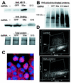
FIG. 1.
FIG. 1.
(A) Western blots showing RNAi knockdown of DmS13 and DmS5a in Drosophila S2 cells (top and middle, respectively). Control cells were untreated (−) or exposed to GFP dsRNA. RNAi against DmS5a leads to increased levels of DmS13. To confirm that equivalent amounts of total protein were present in each lane, the Western blots were Coomassie stained after Western detection (bottom). (B) Western blot of S2 cells exposed to dsRNA against DmS13 or DmS5a and probed with ubiquitin chain antibodies. The lane marked with a minus contains lysate from untreated cells. The cells were treated with dsRNA against GFP as a negative control and with dsRNA to the Prosβ2 20S proteasome subunit as a positive control. (C) Immunofluorescence image of S2 cells exposed to dsRNA against DmS13. The cells were fixed, permeabilized, and then stained for α-tubulin (red) and DNA (blue; DAPI). (D) Cell cycle arrest in Drosophila S2 cells treated with dsRNA against the proteasome subunit S13. The cells were labeled with BrdU to measure newly synthesized DNA (y axis) and stained with 7-AAD (x axis) for total cellular DNA.
FIG. 2.
FIG. 2.
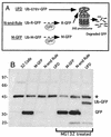
Use of short-lived GFP for measuring 26S proteasome activity in living cells. In vivo 26S activity assays were performed in stable S2 cell lines constitutively expressing ubiquitin-GFP fusion proteins. (A) Diagram of the two destabilized GFP constructs and the stable M-GFP (6). The UFD construct, UbG76VGFP, contains a ubiquitin sequence N-terminal of the GFP that is not cleaved by UPP but is further ubiquitinylated and degraded by the 26S proteasome. The N-end rule substrate, Ub-R-GFP, is first cleaved by UPPs to generate a destabilizing Arg residue at the N terminus and then degraded by the proteasome. In the control Ub-GFP fusion, UPPs generate an N-terminal Met residue yielding a long-lived GFP. (B) Steady-state expression of Ub-GFP in stable Drosophila cell lines and effect of proteasome inhibition. Stable cell lines were treated for 3 h with 50 μM proteasome inhibitor MG132 and then collected along with untreated samples. The stabilities of the GFP constructs were analyzed by Western blotting with anti-GFP antibodies. Both short-lived GFP substrates are stabilized by proteasome inhibitor treatment. A nonspecific band detected by the anti-GFP antibodies is marked (∗).
FIG. 3.
FIG. 3.
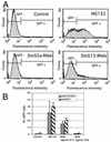
Effects of DmS13 RNAi and DmS5a RNAi on proteasome-dependent GFP degradation. Drosophila S2 cell lines constitutively expressing short-lived proteasome substrates were treated with dsRNA against the DmS13 subunit or DmS5a. Four days posttreatment, flow cytometric analysis was performed to detect cells positive for either UbG76VGFP (dark shading) or R-GFP (light shading). (A) Flow cytrometric profiles of S2 cells that were either mock treated (control) or exposed to 50 μM MG132 (MG132) or to RNAi against DmS5a or DmS13, as indicated. The region defined as GFP− corresponds to the fluorescence intensity obtained for control cells lacking GFP. (B) Quantification of GFP stabilization after RNAi against either DmS13 or DmS5a or after treatment with the proteasome inhibitor MG132. The values were obtained from the treatments shown in panel A performed in triplicate. The error bars indicate standard deviations.
FIG. 4.
FIG. 4.
Effects of DmS13 RNAi on proteasome activity and subunit expression. (A) RNAi-treated and control extracts were prepared in the absence (−) or presence (+) of 4 mM ATP. In ATP− samples, endogenous ATP was depleted by apyrase treatment. Equivalent amounts of total protein were separated by native gel electrophoresis, and in-gel peptidase assays were carried out by overlaying with the fluorogenic proteasome substrate LLVY-MCA, followed by exposure to UV light. Elevated 20S activity becomes apparent after DmS13 or DmS5a RNAi treatment. The total amount of 20S proteasome in untreated cells was estimated by the fluorescence in the 20S band of the apyrase-treated control (left lane). (B) Extracts from control cells with and without ATP (two left lanes) and cells exposed to DmS5a RNAi (middle lane) or DmS13 RNAi (right lane) were separated by native gel electrophoresis, transferred to membranes, and probed with anti-α-proteasome antibodies. (C) Extracts from control (−) and DmS13 RNAi-treated cells were separated by SDS-PAGE and immunoblotted with anti-S5a antibodies. Loss of S13 leads to the up-regulation of S5a and to the appearance of a slower-migrating band (arrow). (D) Two-dimensional PAGE of DmS13 RNAi-treated extracts immunoblotted with anti-S5a antibodies (right). Extracts from control or epoxomicin-treated cells are shown in the left and middle panels, respectively.
FIG. 5.
FIG. 5.
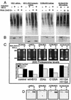
HS13 is a functional homolog of Drosophila S13. Four distinct stable S2 cell lines containing inducible forms of the wild-type HS13, HS13Δ224, HS13C120A, or HS13 H113A-H115A were subjected to RNAi to eliminate the Drosophila S13 subunit. Immunoblots were carried out using anti-ubiquitin chain antibodies or anti-S13 antibodies. (A) Top section of the Western blot probed with anti-ubiquitin antibodies to compare ubiquitin conjugate pool levels. The HS13C120A mutant can partially restore ubiquitin conjugate pool levels. +, present; −, absent. (B) Western blot probed with anti-S13 antibodies showing levels of endogenous DmS13 and of induced HS13 in RNAi+c experiments. To confirm that equivalent amounts of total protein were loaded, the blot shown in panel A was stained for total protein after Western blot development (not shown). (C) Equivalent amounts of total protein were separated by native gel electrophoresis, and in-gel peptidase assays were carried out by overlaying with the fluorogenic-proteasome substrate LLVY-MCA, followed by exposure to UV light. The rescue of elevated 20S activity after RNAi treatment by the expression of HS13 wild type or mutants was estimated by the 20S fluorescence intensities, and a digitized image was quantified using Image Gauge analysis software (bottom). (D) From in-gel peptidase assays, the 26S bands were cut out and then separated by SDS-PAGE, followed by immunoblotting with anti-S13 antibodies. The expected migration position for HS13Δ224 is marked (∗).
FIG. 6.
FIG. 6.
Wild-type (wt) HS13 restores cell cycle progression in Drosophila S13-depleted cells. Stable Drosophila S2 cell lines containing inducible forms of the HS13 gene were induced with copper sulfate and also treated with dsRNA to remove the Drosophila host subunit. The cells were labeled with BrdU to measure newly synthesized DNA (y axis) and stained with 7-AAD (x axis) for total cellular DNA. The specific cell cycle regions are marked in the first control sample (OR, overreplicating cells). The sub-G0 fraction was confirmed to represent apoptotic cells by comparison with apoptotic S2 cells (Apo) induced by staurosporine. Top row, untreated cells; middle row, cells exposed to DmS13 RNAi only; bottom row, cells exposed to DmS13 RNAi and expressing HS13, HS13Δ224, HS13C120A, or HS13 H113A-H115A, as indicated.
FIG. 7.
FIG. 7.
Mutant forms of HS13 do not rescue DmS13-depleted cells from apoptosis or DNA overreplication. Stable Drosophila S2 cells containing inducible wild-type or mutant forms of HS13 were treated with dsRNA to remove endogenous DmS13 and exposed to copper sulfate to induce HS13 expression. Apoptotic-cell subpopulations and cells undergoing DNA overreplication were quantified by flow cytometric analysis using constant gating parameters. The graph shows averages (plus standard deviations) from experiments carried out in triplicate.

Similar articles

Cited by

References

    1. Arioka, M., M. Kouhashi, K. Yoda, A. Takatsuki, M. Yamasaki, and K. Kitamoto. 1998. Multidrug resistance phenotype conferred by overexpressing bfr2+/pad1+/sks1+ or pap1+ genes and mediated by bfr1+ gene product, a structural and functional homologue of P-glycoprotein in Schizosaccharomyces pombe. Biosci. Biotechnol. Biochem. 62:390-392. - PubMed
    1. Bosher, J. M., and M. Labouesse. 2000. RNA interference: genetic wand and genetic watchdog. Nat. Cell Biol. 2:E31-E36. - PubMed
    1. Braun, B. C., M. Glickman, R. Kraft, B. Dahlmann, P. M. Kloetzel, D. Finley, and M. Schmidt. 1999. The base of the proteasome regulatory particle exhibits chaperone-like activity. Nat. Cell Biol. 1:221-226. - PubMed
    1. Clemens, J. C., C. A. Worby, N. Simonson-Leff, M. Muda, T. Maehama, B. A. Hemmings, and J. E. Dixon. 2000. Use of double-stranded RNA interference in Drosophila cell lines to dissect signal transduction pathways. Proc. Natl. Acad. Sci. USA 97:6499-6503. - PMC - PubMed
    1. Cope, G. A., G. S. Suh, L. Aravind, S. E. Schwarz, S. L. Zipursky, E. V. Koonin, and R. J. Deshaies. 2002. Role of predicted metalloprotease motif of Jab1/Csn5 in cleavage of Nedd8 from Cul1. Science 298:608-611. - PubMed

Publication types

MeSH terms

LinkOut - more resources