. 2003 Aug;23(15):5331-45.
doi: 10.1128/MCB.23.15.5331-5345.2003.

RNF5, a RING finger protein that regulates cell motility by targeting paxillin ubiquitination and altered localization

Affiliations

RNF5, a RING finger protein that regulates cell motility by targeting paxillin ubiquitination and altered localization

Christine Didier et al. Mol Cell Biol. 2003 Aug.

Abstract

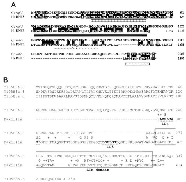
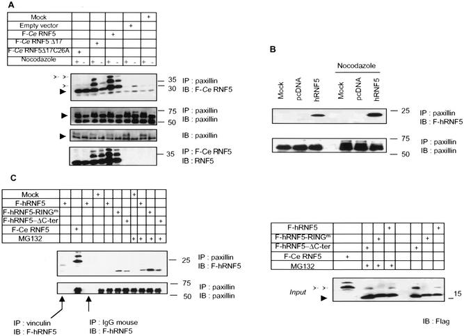
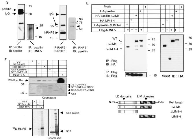
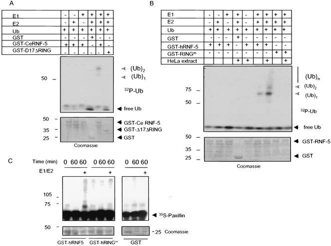
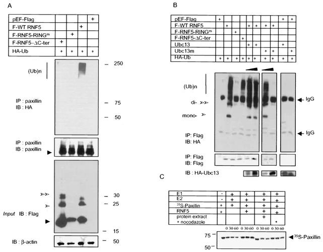
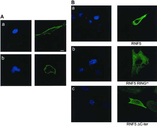
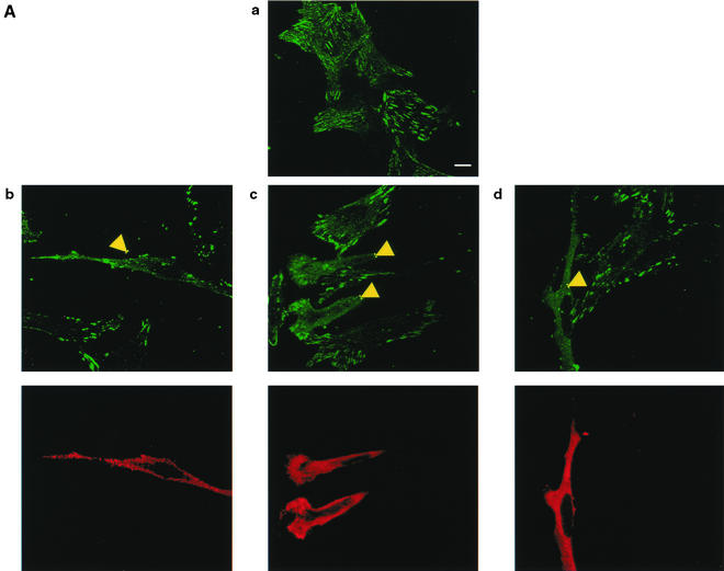
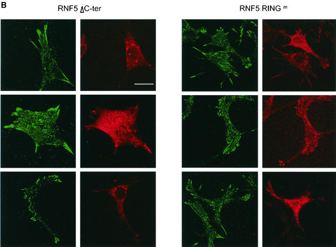
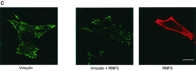
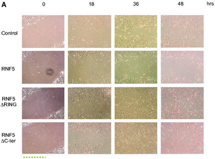
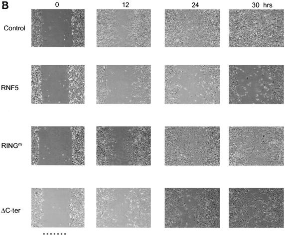
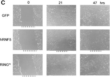
RNF5 is a RING finger protein found to be important in the growth and development of Caenorhabditis elegans. The search for RNF5-associated proteins via a yeast two-hybrid screen identified a LIM-containing protein in C. elegans which shows homology with human paxillin. Here we demonstrate that the human homologue of RNF5 associates with the amino-terminal domain of paxillin, resulting in its ubiquitination. RNF5 requires intact RING and C-terminal domains to mediate paxillin ubiquitination. Whereas RNF5 mediates efficient ubiquitination of paxillin in vivo, protein extracts were required for in vitro ubiquitination, suggesting that additional modifications and/or an associated E3 ligase assist RNF5 targeting of paxillin ubiquitination. Mutant Ubc13 efficiently inhibits RNF5 ubiquitination, suggesting that RNF5 generates polychain ubiquitin of the K63 topology. Expression of RNF5 increases the cytoplasmic distribution of paxillin while decreasing its localization within focal adhesions, where it is primarily seen under normal growth. Concomitantly, RNF5 expression results in inhibition of cell motility. Via targeting of paxillin ubiquitination, which alters its localization, RNF5 emerges as a novel regulator of cell motility.


Figures

FIG. 1.
FIG. 1.
Alignment of C. elegans and human RNF5. (A) The amino acid sequences of human (Hs) and C. elegans (Ce) forms of RNF5 were aligned. Important domains in RNF5 are underlined (RING, central, and carboxyl-terminal domains). (B) Alignment of the C. elegans LIM-containing protein Y105E8a.6 and human paxillin. Homology between the two proteins was found in the LIM domain (highest in LIM1 and LIM4 of paxillin; boxed area) and also in the region between the LD4 and LD5 domains of paxillin.
FIG. 2.
FIG. 2.
RNF5 association with paxillin. (A) C. elegans RNF5 associates with paxillin. Flag-tagged C. elegans RNF5 (F-Ce RNF5), a spliced variant lacking 17 amino acids, and the RING mutant C26A forms were transfected into 293T cells, and 24 h later the cells were subjected to treatment with nocodazole (100 ng/ml) or mock treatment, as indicated in the figure. Proteins were prepared 18 h later and subjected to immunoprecipitation (IP) with antibodies to endogenous paxillin. Immunoprecipitated material was analyzed via Western blots (IB) with the antibodies indicated in the figure. Arrowheads point to the corresponding protein. Empty arrows point to mono- and diubiquitin forms of RNF5. (B) Human RNF5 (hRNF5) binds to paxillin. Flag-tagged human RNF5 (F-hRNF5) was transfected into 293T cells that were mock or nocodazole treated. Proteins (1 mg) were subjected to immunoprecipitation with antibodies to paxillin, following Western blot analysis with the antibodies indicated in the figure. (C) Mutant forms of RNF5 exhibit stronger binding to paxillin than wild-type RNF5. Flag-tagged forms of RNF5 (human wild-type, RING mutant, and C-terminally deleted [ΔC-ter] forms and C. elegans RNF5) were transfected into 293T cells; 24 h later, proteins were prepared and subjected to immunoprecipitation with antibodies to paxillin, followed by immunoblot analysis with the antibodies indicated in the figure. The relative intensity of the ECL reaction was reduced compared with that in panel B to emphasize the difference in association of paxillin with the different forms of RNF5. Control reactions with vinculin antibodies and control IgG for immunoprecipitation followed by immunoblotting with Flag-RNF5 are shown. Expression of RNF5 forms (input) is shown on the right panel. (D) In vivo association of RNF5 and paxillin. Endogenous human RNF5 was immunoprecipitated with monoclonal antibodies raised against RNF5, followed by immunoblot analysis with the antibodies indicated in the figure. NS, nonspecific. (E) RNF5 associates with paxillin via the amino-terminal domain. Expression vectors containing the LIM or amino-terminal domain were coexpressed with RNF5 in 293T cells. Immunoprecipitations with the indicated antibodies identified an association with paxillin lacking the LIM domain (ΔLIM1-4) and containing only the amino-terminal domain. (F) In vitro association between paxillin and RNF5. GST-RNF5 (human wild-type and RING mutant forms and C. elegans wild-type and Δ17ΔRING forms) fusion proteins were incubated with 35S-labeled paxillin, and reciprocally, GST-paxillin was incubated with 35S-labeled RNF5. The amount of bound material shown reflects direct association that was followed by extensive high-salt washes.
FIG. 2.
FIG. 2.
RNF5 association with paxillin. (A) C. elegans RNF5 associates with paxillin. Flag-tagged C. elegans RNF5 (F-Ce RNF5), a spliced variant lacking 17 amino acids, and the RING mutant C26A forms were transfected into 293T cells, and 24 h later the cells were subjected to treatment with nocodazole (100 ng/ml) or mock treatment, as indicated in the figure. Proteins were prepared 18 h later and subjected to immunoprecipitation (IP) with antibodies to endogenous paxillin. Immunoprecipitated material was analyzed via Western blots (IB) with the antibodies indicated in the figure. Arrowheads point to the corresponding protein. Empty arrows point to mono- and diubiquitin forms of RNF5. (B) Human RNF5 (hRNF5) binds to paxillin. Flag-tagged human RNF5 (F-hRNF5) was transfected into 293T cells that were mock or nocodazole treated. Proteins (1 mg) were subjected to immunoprecipitation with antibodies to paxillin, following Western blot analysis with the antibodies indicated in the figure. (C) Mutant forms of RNF5 exhibit stronger binding to paxillin than wild-type RNF5. Flag-tagged forms of RNF5 (human wild-type, RING mutant, and C-terminally deleted [ΔC-ter] forms and C. elegans RNF5) were transfected into 293T cells; 24 h later, proteins were prepared and subjected to immunoprecipitation with antibodies to paxillin, followed by immunoblot analysis with the antibodies indicated in the figure. The relative intensity of the ECL reaction was reduced compared with that in panel B to emphasize the difference in association of paxillin with the different forms of RNF5. Control reactions with vinculin antibodies and control IgG for immunoprecipitation followed by immunoblotting with Flag-RNF5 are shown. Expression of RNF5 forms (input) is shown on the right panel. (D) In vivo association of RNF5 and paxillin. Endogenous human RNF5 was immunoprecipitated with monoclonal antibodies raised against RNF5, followed by immunoblot analysis with the antibodies indicated in the figure. NS, nonspecific. (E) RNF5 associates with paxillin via the amino-terminal domain. Expression vectors containing the LIM or amino-terminal domain were coexpressed with RNF5 in 293T cells. Immunoprecipitations with the indicated antibodies identified an association with paxillin lacking the LIM domain (ΔLIM1-4) and containing only the amino-terminal domain. (F) In vitro association between paxillin and RNF5. GST-RNF5 (human wild-type and RING mutant forms and C. elegans wild-type and Δ17ΔRING forms) fusion proteins were incubated with 35S-labeled paxillin, and reciprocally, GST-paxillin was incubated with 35S-labeled RNF5. The amount of bound material shown reflects direct association that was followed by extensive high-salt washes.
FIG. 3.
FIG. 3.
RNF5 E3 ligase activities and ubiquitination of paxillin in vitro. (A) E3 ligase activities of C. elegans RNF5 in vitro. In vitro ubiquitination assays were carried out with bacterially expressed and purified GST fused to full-length or Δ17C26(A) (17 amino acids within the RING domain mutated) C. elegans RNF5. The purified forms of E1, E2 (UbcH5c), and bacterially produced 32P-labeled ubiquitin (Ub) were added in the presence of ATP. GST-RNF5 bound to glutathione beads was washed before separation on SDS-PAGE and analysis via autoradiography. The positions of mono- and diubiquitin are indicated. The lower panel depicts Coomassie blue staining, reflecting the quantity of proteins used for the reaction. (B) E3 ligase activities of human RNF5 in vitro. The in vitro ubiquitination reaction was carried out as indicated above except that the human forms of wild-type and RING mutant RNF5 were used. As indicated in the figure, GST-hRNF5 was also incubated with HeLa cell extracts that were immunodepleted of RNF5 following extensive washes before adding E1, E2, 32P-ubiquitin, and ubiquitination buffer. Arrows point to the positions of the mono-, di-, and polyubiquitin forms. The lower panel depicts Coomassie blue staining, reflecting the quantity of proteins used for the reaction. (C) RNF5 mediates ubiquitination of paxillin in vitro. In vitro ubiquitination assays were performed with bacterially expressed and purified forms of GST-hRNF5 (wild type or RING mutant) and 35S-labeled in vitro-translated paxillin in the presence of E1 and E2 for the period indicated. Following the reaction, GST-hRNF5 was spun, and supernatant-containing [35S]paxillin was subjected to separation on SDS-PAGE. Shown is the autoradiograph of ubiquitinated paxillin. The lower panel depicts input of GST-hRNF5 (left panel) or GST (right panel).
FIG. 4.
FIG. 4.
RNF5 ubiquitinates paxillin in vivo without affecting its half-life. (A) RNF5 promotes paxillin ubiquitination in vivo. Wild-type (WT) and mutant forms of RNF5 (RING mutant and C-terminally deleted) were cotransfected with HA-tagged ubiquitin into 293T cells. Proteins were prepared 24 h after transfection and subjected to immunoprecipitation, followed by immunoblot analysis with the antibodies indicated in the figure. Sizes and polyubiquitin chains are marked on the side panels. Western blot analysis of RNF5 input is presented on the bottom panel. β-Actin blot was used to assure equal loading. (B) RNF5 E3 ligase in vivo is Ubc13 dependent. Wild-type and mutant forms of human RNF5 were cotransfected with HA-ubiquitin and wild-type or mutant forms of Ubc13 into 293T cells. Proteins prepared under denaturing conditions were subjected to immunoprecipitation with antibodies to Flag-tagged RNF5, followed by immunoblot with the antibodies indicated in the figure. The lower panel depicts expression of Ubc13. (C) RNF5 does not affect paxillin half-life in vitro. The half-life of paxillin was monitored in vitro with in vitro-translated 35S-labeled paxillin. Following its translation, paxillin was immunoprecipitated and incubated with E1, E2, and RNF5 as indicated in the figure. RNF5 was also incubated with protein extracts of mock-treated (+) and nocodazole-treated (∗) cells to enable posttranslational modifications that increase the efficiency of association with and ubiquitination of paxillin.
FIG. 5.
FIG. 5.
Cellular localization of endogenous and exogenously expressed RNF5. (A) Cellular localization of endogenous RNF5. NIH 3T3 (a) or HeLa (b) cells were subjected to immunocytochemistry-based confocal microscopic analysis with monoclonal antibodies raised against RNF5. 4′,6′-Diamidino-2-phenylindole (DAPI) staining is shown in the left panels. Scale bar. 10 μm. (B) Cellular localization of exogenously expressed RNF5. NIH 3T3 cells were transfected with Flag-tagged wild-type (a), RING mutant (b), and C-terminally deleted (c) forms of RNF5; 30 h later the cells were subjected to confocal microscopy-based immunocytochemistry analysis with antibodies to Flag. The left panel depicts DAPI staining. The picture represents multiple fields from independent experiments.
FIG. 6.
FIG. 6.
RNF5 expression affects cellular distribution of paxillin. (A) RNF5 alters paxillin localization. Localization of endogenous paxillin was monitored in RNF5 transfected cells. Panel a depicts staining of endogenous paxillin. Note the difference in paxillin localization in cells that express RNF5 (panels b to d) with those that do not (compare to the corresponding cells that express RNF5 [yellow arrows]). The figure represents multiple fields and data observed in five independent experiments. Yellow arrows point to cells which expressed RNF5 and exhibited altered localization of paxillin. Scale bar, 10 μm. (B) Mutant forms of RNF5 do not affect paxillin localization. NIH 3T3 cells were transfected with mutant Flag-RNF5 forms (RING mutant or C-terminally deleted forms), as indicated in the figure. Antibodies to Flag (RNF5) were recognized by red fluorescence, whereas antibodies to paxillin were recognized by green fluorescence. (C) RNF5 and vinculin localization. Analysis of vinculin was carried out on cells that were transfected or not with RNF5. Red staining depicts RNF5, whereas green reveals vinculin expression.
FIG. 6.
FIG. 6.
RNF5 expression affects cellular distribution of paxillin. (A) RNF5 alters paxillin localization. Localization of endogenous paxillin was monitored in RNF5 transfected cells. Panel a depicts staining of endogenous paxillin. Note the difference in paxillin localization in cells that express RNF5 (panels b to d) with those that do not (compare to the corresponding cells that express RNF5 [yellow arrows]). The figure represents multiple fields and data observed in five independent experiments. Yellow arrows point to cells which expressed RNF5 and exhibited altered localization of paxillin. Scale bar, 10 μm. (B) Mutant forms of RNF5 do not affect paxillin localization. NIH 3T3 cells were transfected with mutant Flag-RNF5 forms (RING mutant or C-terminally deleted forms), as indicated in the figure. Antibodies to Flag (RNF5) were recognized by red fluorescence, whereas antibodies to paxillin were recognized by green fluorescence. (C) RNF5 and vinculin localization. Analysis of vinculin was carried out on cells that were transfected or not with RNF5. Red staining depicts RNF5, whereas green reveals vinculin expression.
FIG. 7.
FIG. 7.
RNF5 inhibits migration of NIH 3T3 cells. (A) Cells were transfected with the RNF5 constructs indicated in the figure, and 48 h later cells were subjected to the wound assay. With a pipette tip, scratches were made in the centers of equally confluent cells. Time lapse photographs were made at the indicated time points, revealing the cells' ability to fill the wound (gap). Pictures shown represent multiple analyses. (B) The experiment was carried out as indicated for panel A with the exception that the plates were first coated with fibronectin (10 μg/ml). (C). Effect of RNF5 on cell motility in paxillin-null cells. Paxillin-null cells were subjected to transfection with the constructs indicated in the figure, and time lapse analysis was carried out to monitor changes in the migration of the cells as indicated above.
FIG. 7.
FIG. 7.
RNF5 inhibits migration of NIH 3T3 cells. (A) Cells were transfected with the RNF5 constructs indicated in the figure, and 48 h later cells were subjected to the wound assay. With a pipette tip, scratches were made in the centers of equally confluent cells. Time lapse photographs were made at the indicated time points, revealing the cells' ability to fill the wound (gap). Pictures shown represent multiple analyses. (B) The experiment was carried out as indicated for panel A with the exception that the plates were first coated with fibronectin (10 μg/ml). (C). Effect of RNF5 on cell motility in paxillin-null cells. Paxillin-null cells were subjected to transfection with the constructs indicated in the figure, and time lapse analysis was carried out to monitor changes in the migration of the cells as indicated above.

References

    1. Borden, K. L. B. 2000. RING domains: master builders of molecular scaffolds? J. Biol. Chem. 295:1103-1112. - PubMed
    1. Brown, M. C., M. S. Curtis, and C. E. Turner. 1998. Paxillin LD motifs may define a new family of protein recognition domains. Nat. Struct. Biol. 5:677-678. - PubMed
    1. Brown, M. C., J. A. Perrotta, and C. E. Turner. 1998. Serine and threonine phosphorylation of the paxillin LIM domains regulates paxillin focal adhesion localization and cell adhesion to fibronectin. Mol. Biol. Cell 9:1803-1816. - PMC - PubMed
    1. Deshaies, R. J. 1999. SCF and Cullin/Ring H2-based ubiquitin ligases. Annu. Rev. Cell. Dev. Biol. 15:435-467. - PubMed
    1. Fuchs, S. Y., B. Xie, V. Adler, V. A. Fried, R. J. Davis, and Z. Ronai. 1997. c-Jun NH2-terminal kinases target the ubiquitination of their associated transcription factors. J. Biol. Chem. 272:32163-32168. - PubMed

Publication types

MeSH terms