. 2003 Aug;23(15):5354-65.
doi: 10.1128/MCB.23.15.5354-5365.2003.

Acetylation-dependent chromatin reorganization by BRDT, a testis-specific bromodomain-containing protein

Affiliations

Acetylation-dependent chromatin reorganization by BRDT, a testis-specific bromodomain-containing protein

Christophe Pivot-Pajot et al. Mol Cell Biol. 2003 Aug.

Abstract

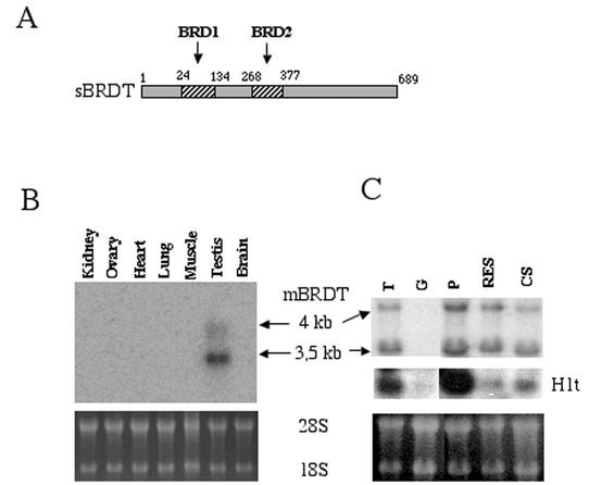
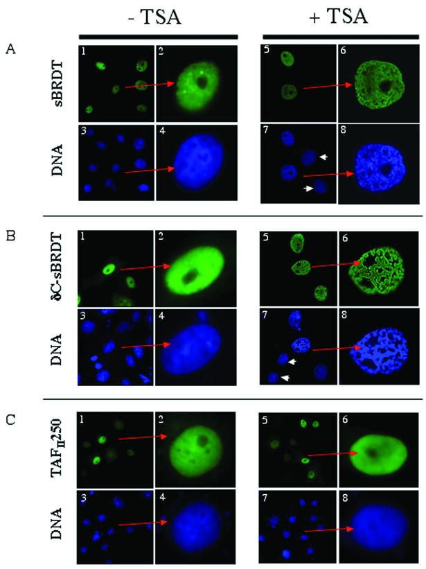
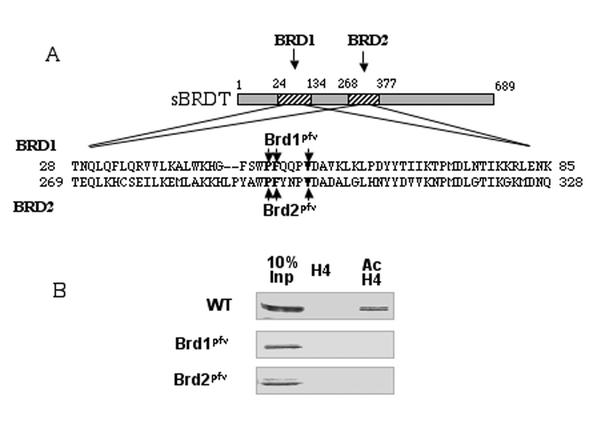
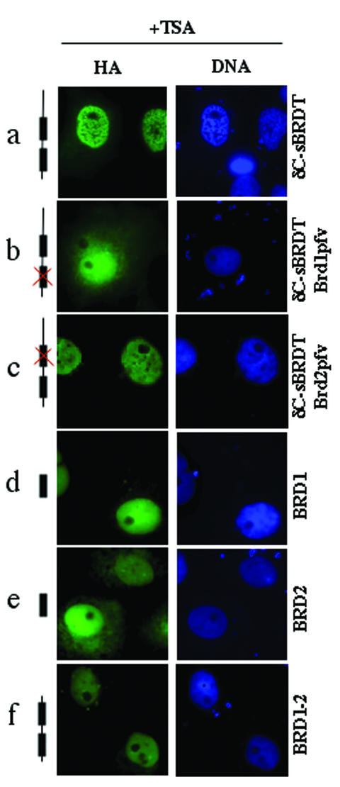
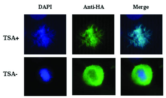
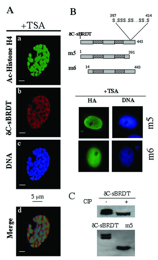
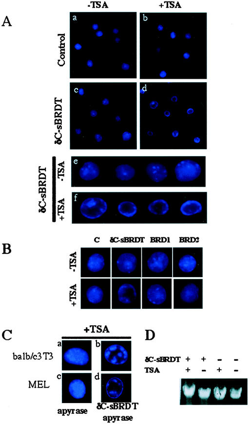
The association between histone acetylation and replacement observed during spermatogenesis prompted us to consider the testis as a source for potential factors capable of remodelling acetylated chromatin. A systematic search of data banks for open reading frames encoding testis-specific bromodomain-containing proteins focused our attention on BRDT, a testis-specific protein of unknown function containing two bromodomains. BRDT specifically binds hyperacetylated histone H4 tail depending on the integrity of both bromodomains. Moreover, in somatic cells, the ectopic expression of BRDT triggered a dramatic reorganization of the chromatin only after induction of histone hyperacetylation by trichostatin A (TSA). We then defined critical domains of BRDT involved in its activity. Both bromodomains of BRDT, as well as flanking regions, were found indispensable for its histone acetylation-dependent remodelling activity. Interestingly, we also observed that recombinant BRDT was capable of inducing reorganization of the chromatin of isolated nuclei in vitro only when the nuclei were from TSA-treated cells. This assay also allowed us to show that the action of BRDT was ATP independent, suggesting a structural role for the protein in the remodelling of acetylated chromatin. This is the first demonstration of a large-scale reorganization of acetylated chromatin induced by a specific factor.


Figures

FIG. 1.
FIG. 1.
Murine BRDT is specifically expressed during spermatogenesis. (A) The cDNA corresponding to the most abundant BRDT mRNA was cloned and sequenced. The schematic representation shows the domain organization of the cDNA-encoded protein (sBRDT) and positions of the two bromodomains (BRD1 [amino acids 24 to 134] and BRD2 [amino acids 268 and 377]) are indicated. (B and C) BRDT transcripts were detected by Northern blots of several mouse tissues (B) and of mouse germinal cell populations (C), including 6-day-old mouse testis containing spermatogonia and Sertoli cells (G), pachytene spermatocytes (P), round and elongating spermatids (RES), and condensed spermatids (CS). RNA from adult mouse testis (T) was also used. The blot was reprobed with a H1t probe. Ethidium bromide staining of 18S and 28S RNA in the gels prior to transfer is shown at the bottom.
FIG. 2.
FIG. 2.
TSA-dependent chromatin reorganization after ectopic expression of sBRDT. Cos7 cells were transfected by vectors expressing the indicated proteins, and cells were either treated with TSA (100 ng/ml) (+ TSA) or not treated with TSA (− TSA). A higher magnification of a transfected cell is shown in the right panels (indicated by a red arrow) to better visualize the chromatin reorganization. Arrowheads show TSA-treated nontransfected cells.
FIG. 3.
FIG. 3.
The two bromodomains of sBRDT are involved in the specific recognition of the acetylated histone H4 tail. (A) Schematic representation of sBRDT proteins bearing mutations in the first bromodomain (Brd1pfv) or in the second bromodomain (Brd2pfv) were expressed in Cos7 cells. In the Brd1pfv mutant, P50, F51 and V55 were replaced by A. In the Brd2pfv mutant, P293, F294, and V298 were replaced by A (the mutations are shown in boldface type). (B) Wild-type (WT) or mutant δC-sBRDT proteins bearing mutations in the first bromodomain (Brd1pfv) or in the second bromodomain (Brd2pfv) were expressed in Cos7 cells. Twenty-four hours posttransfection, an extract was prepared and subjected to a peptide pull-down assay using beads bound to either nonmodified or hyperacetylated (Ac) histone H4 N-terminal tails. Proteins retained on the beads were then analyzed by Western blotting using an anti-HA antibody. The Inp lane contains 10% of the input material.
FIG. 4.
FIG. 4.
The integrity of the two bromodomains is necessary but not sufficient to induce TSA-dependent reorganization of the chromatin. Cos7 cells were transfected with vectors expressing the indicated proteins. Twenty-four hours posttransfection, protein expression and chromatin reorganization were visualized in control cells (not shown) or cells treated with TSA.
FIG. 5.
FIG. 5.
BRDT remains associated with mitotic chromosomes in TSA-treated cells. Cos7 cells expressing δC-sBRDT were left untreated (TSA−) or treated for 6 h with TSA (TSA+), and the localization of the protein was recorded in mitotic cells. The TSA+ panels show a tight association of BRDT with the mitotic chromosomes, whereas in the absence of TSA, BRDT appears to be excluded from the condensing chromosomes.
FIG. 6.
FIG. 6.
Chromatin remodelling by BRDT depends on bromodomains and flanking regions. (A) Confocal analysis of the chromatin in BRDT-expressing and TSA-treated cells shows that δC-sBRDT (shown in red in panel b) is distributed around the condensed hyperacetylated chromatin regions (detected by an anti-acetylated H4 antibody [shown in green in panel a] and also visible after DAPI staining [panel c]). The images of panels a, b, and c were merged (panel d). (B) Deletions of the 52 amino acids in the C-terminal serine-rich region of δC-sBRDT (m5 mutant) or the 13 N-terminal amino acids of δC-sBRDT (m6 mutant) both inhibit the δC-sBRDT's remodelling activity of acetylated chromatin in overexpressing TSA-treated Cos7 cells. (C) Western blots show that δC-sBRDT appears as a double band and as a single band after CIP treatment, indicating a phosphorylation of the protein. The serine-rich region of δC-sBRDT removed in the m5 mutant appears to be the site of phosphorylation, since in contrast to δC-sBRDT, this mutant migrates as a single band (this band was not sensitive to CIP treatment [not shown]).
FIG.7.
FIG.7.
Recombinant δC-sBRDT induced in vitro remodelling of hyperacetylated chromatin. (A) A total of 105 purified nuclei from untreated (−TSA) or TSA-treated (+TSA) MEL cells were incubated alone (control [panels a and b]) or with 5 μg of recombinant δC-sBRDT (panels c and d) for 1 h, DAPI stained, and examined under a microscope. Panels e and f show higher-magnification views of the structures of several nuclei present in panels c and d, respectively. (B) δC-sBRDT induces an in vitro remodelling of hyperacetylated MEL nuclei, which is absent from control (C) nuclei, and not detected in hyperacetylated nuclei (+TSA) treated with the same amount of recombinant proteins containing isolated bromodomains (BRD1 or BRD2). (C) In vitro remodelling of nuclei from BALB/c 3T3 or MEL cells containing hyperacetylated chromatin in the absence of ATP. Nuclei were incubated either with apyrase alone or with apyrase and recombinant δC-sBRDT. (D) δC-sBRDT treatment is not associated with the degradation of DNA. Nuclei from MEL cells were either treated with TSA (+) or not treated with TSA (−) and then incubated with (+) or without (−) δC-sBRDT as described above. DNA was then purified and analyzed on an agarose gel.

References

    1. Agalioti, T., G. Chen, and D. Thanos. 2002. Deciphering the transcriptional histone acetylation code for a human gene. Cell 111:381-392. - PubMed
    1. Bannister, A. J., P. Zegerman, J. F. Partridge, E. A. Miska, J. O. Thomas, R. C. Allshire, and T. Kouzarides. 2001. Selective recognition of methylated lysine 9 on histone H3 by the HP1 chromo domain. Nature 410:120-124. - PubMed
    1. Bellvé, A. R. 1993. Purification, culture, and fractionation of spermatogenic cells. Methods Enzymol. 225:84-113. - PubMed
    1. Christensen, M. E., and G. H. Dixon. 1982. Hyperacetylation of histone H4 correlates with the terminal, transcriptionally inactive stages of spermatogenesis in rainbow trout. Dev. Biol. 93:404-415. - PubMed
    1. Christensen, M. E., J. B. Rattner, and G. H. Dixon. 1984. Hyperacetylation of histone H4 promotes chromatin decondensation prior to histone replacement by protamines during spermatogenesis in rainbow trout. Nucleic Acids Res. 12:4575-4592. - PMC - PubMed

Publication types

MeSH terms