. 2003 Aug;23(15):5388-400.
doi: 10.1128/MCB.23.15.5388-5400.2003.

Cdc34 self-association is facilitated by ubiquitin thiolester formation and is required for its catalytic activity

Affiliations

Cdc34 self-association is facilitated by ubiquitin thiolester formation and is required for its catalytic activity

Xaralabos Varelas et al. Mol Cell Biol. 2003 Aug.

Abstract

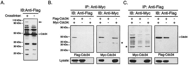
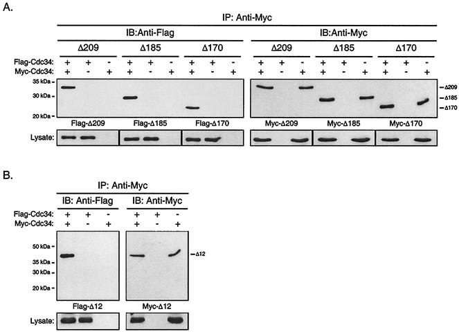
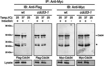
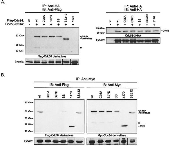
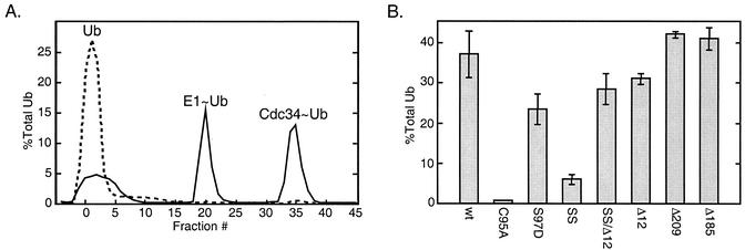
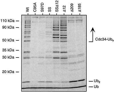
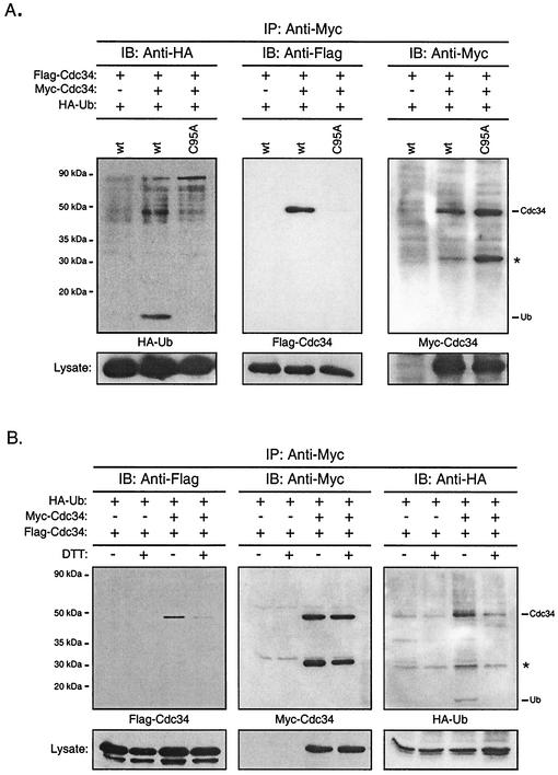
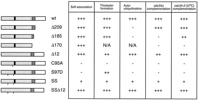
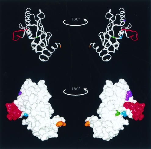
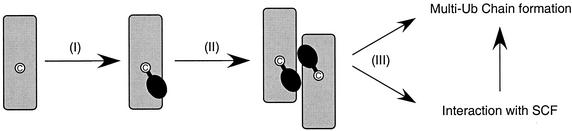
Using a coimmunoprecipitation strategy, we showed that the Cdc34 ubiquitin (Ub)-conjugating enzyme from Saccharomyces cerevisiae self-associates in cell lysates, thereby indicating an in vivo interaction. The ability of Cdc34 to interact with itself is not dependent on its association with the ubiquitin ligase Skp1-Cdc53/Cul1-Hrt1-F-box complex. Rather, this interaction depends upon the integrity of the Cdc34-Ub thiolester. Furthermore, several principal determinants within the Cdc34 catalytic domain, including the active-site cysteine, amino acid residues S73 and S97, and its catalytic domain insertion, also play a role in self-association. Mutational studies have shown that these determinants are functionally important in vivo and operate at the levels of both Cdc34-Ub thiolester formation and Cdc34-mediated multi-Ub chain assembly. These determinants are spatially situated in a region that is close to the active site, corresponding closely to the previously identified E2-Ub interface. These observations indicate that the formation of the Cdc34-Ub thiolester is important for Cdc34 self-association and that the interaction of Cdc34-Ub thiolesters is in turn a prerequisite for both multi-Ub chain assembly and Cdc34's essential function(s). A conclusion from these findings is that the placement of ubiquitin on the Cdc34 surface is a structurally important feature of Cdc34's function.


Figures

FIG. 1.
FIG. 1.
Cdc34 self-associates in vivo. (A) Cross-linking. Cell lysates from YPH499 yeast cells expressing Flag-Cdc34 were treated with or without the chemical cross-linker DSS. Cell lysates were subsequently analyzed by immunoblotting (IB) with an anti-Flag antibody. The position of Flag-Cdc34 is indicated, as is the position of a unique cross-linked product containing Flag-Cdc34 (arrow). (B and C) Coimmunoprecipitation. Total cell extracts of YPH499 cells expressing Flag-Cdc34, Myc-Cdc34, or both Flag-Cdc34 and Myc-Cdc34 were prepared. (B) Myc-Cdc34 was immunoprecipitated (IP) with an anti-Myc antibody, followed by immunoblotting with an anti-Myc antibody (right panel) to detect the amount of Myc-Cdc34 that had immunoprecipitated and with an anti-Flag antibody (left panel) to detect the amount of Flag-Cdc34 that had coimmunoprecipitated. (C) The reciprocal coimmunoprecipitation experiment to that for panel B was performed. Flag-Cdc34 was immunoprecipitated with an anti-Flag antibody, followed by immunoblotting with an anti-Flag antibody (right panel) and with an anti-Myc antibody (left panel). The position of Cdc34 is indicated, as is the position of a proteolytic product of Cdc34 (*). Protein expression levels within the lysates used for immunoprecipitations are shown at the bottoms of panels B and C.
FIG. 2.
FIG. 2.
The Cdc34 carboxy-terminal extension and catalytic domain insertion are dispensable with respect to Cdc34 self-association. The same coimmunoprecipitation strategy as employed for Cdc34 (Fig. 1B) was employed for the Δ209 (residues 1 to 209), Δ185 (residues 1 to 185), and Δ170 (residues 1 to 170) carboxy-terminal truncation derivatives (A) as well as for the Δ12 (residues 103 to 114 deleted) catalytic domain deletion derivative (B). Flag- and Myc-tagged versions of these truncation derivatives were expressed in YPH499 cells. Cell lysates were extracted and subjected to immunoprecipitation (IP) with an anti-Myc antibody followed by immunoblotting (IB) with an anti-Myc antibody (right panels) and an anti-Flag antibody (left panels). Protein expression levels within the lysates used for immunoprecipitations are shown at the bottoms of the respective panels.
FIG. 3.
FIG. 3.
Cdc34 self-association is not dependent upon a functional SCF complex. Flag-Cdc34 and Myc-Cdc34 were expressed in 15DaubΔ (wild type [wt]) or DSY1105 (cdc53-1) cells at either the permissive (25°C) or nonpermissive (37°C) temperature. Cell extracts were prepared, and Myc-Cdc34 was immunoprecipitated (IP) with an anti-Myc antibody, followed by immunoblotting (IB) with either an anti-Myc antibody (right panel) or an anti-Flag antibody (left panel). The asterisk indicates a proteolytic product of Cdc34. Protein expression levels within the lysates used for immunoprecipitations are shown at the bottoms of the respective panels.
FIG. 4.
FIG. 4.
(A) Catalytic domain derivatives of Cdc34 interact with Cdc53 of the SCF complex. Total cell extracts were prepared from YPH499 cells coexpressing Cdc53-3xHA and Flag-Cdc34 or one of the Flag-tagged Cdc34 derivatives: C95A, S97D, S73K/S97D (SS), S73K/S97D/Δ12 (SSΔ12), or Δ170. Cdc53-3xHA was immunoprecipitated (IP) with an anti-HA antibody and was detected by immunoblotting (IB) with an anti-HA antibody (right panel). The amount of Flag-Cdc34 or Flag-Cdc34 derivative that coimmunoprecipitated was detected by immunoblotting with an anti-Flag antibody (left panel). The positions of Cdc53, Cdc34, and the Cdc34 derivatives are indicated. The asterisk indicates a proteolytic product of Cdc34. Protein expression levels within the lysates used for immunoprecipitations are shown at the bottoms of the respective panels. (B) Residues within the Cdc34 catalytic domain critical for self-association. The same coimmunoprecipitation strategy as employed for Cdc34 (Fig. 1B) was used for the C95A, S97D, S73K/S97D (SS), and S73K/S97D/Δ12 (SSΔ12) catalytic domain derivatives. Flag- and Myc-tagged versions of these derivatives were expressed in YPH499 cells. Cell lysates were extracted and subjected to immunoprecipitation with anti-Myc antibody followed by immunoblotting with anti-Myc antibody (right panel) and anti-Flag antibody (left panel). The asterisk indicates a proteolytic product of Cdc34. Protein expression levels within the lysates used for immunoprecipitations are shown at the bottoms of the respective panels. wt, wild type.
FIG. 5.
FIG. 5.
Ub thiolester formation. In vitro Ub thiolester formation was compared for a number of Cdc34 derivatives. Reaction mixtures containing Cdc34 or one of its derivatives (100 nM), Uba1 (10 nM), 35S-Ub (200 nM), and an ATP cocktail were incubated for 5 min at 30°C. Reactions were stopped by the addition of 50 mM EDTA, and the mixtures were then immediately loaded onto an anion-exchange column to separate reaction products. The Δ209 and Δ185 derivatives were separated by using a Superdex 75 HR10/30 size exclusion column (see Materials and Methods). (A) Example of the elution profile generated for a reaction mixture containing Cdc34 (solid line). Peaks containing 35S-Ub correspond to free Ub, Ub incorporated into Uba1 (E1∼Ub), and Cdc34 (Cdc34∼Ub) thiolester. In a separate reaction mixture, 10 mM DTT was added to disrupt thiolester (dashed line). (B) Incorporation of 35S-Ub into thiolester was determined for each Cdc34 derivative as a percentage of the total 35S-Ub added to each reaction mixture. The mean and standard deviation observed for three separate reactions for each derivative is shown. SS, S73K/S97D derivative; SSΔ12, S73K/S97D/Δ12 derivative; Δ12, catalytic domain insert deletion (residues 103 to 114 deleted); Δ209 and Δ185, carboxy-terminal truncation derivatives (residues 1 to 209 and 1 to 185, respectively).
FIG. 6.
FIG. 6.
Autoubiquitination. Cdc34 autoubiquitination was assayed for Cdc34 and its various derivatives by using an in vitro ubiquitination reaction. Reaction mixtures contained Cdc34 or one of its derivatives (100 nM), Uba1 (10 nM), 35S-Ub (200 nM), and an ATP cocktail and were incubated for 8 h at 30°C, representing an end point assay for autoubiquitination. DTT (100 mM) was added to stop the reactions, and the reaction products were analyzed by SDS-PAGE followed by autoradiography. The positions of free Ub (Ub), di-Ub (Ub2), and multi-Ub chains covalently linked to Cdc34 (Cdc34-Ubn) are indicated. SS, S73K/S97D derivative; SSΔ12, S73K/S97D/Δ12 derivative; Δ12, catalytic domain insert deletion (residues 103 to 114 deleted); Δ209 and Δ185, carboxy-terminal truncation derivatives (residues 1 to 209 and 1 to 185, respectively). A downward shift in molecular mass in the Ub chains on the Δ12 derivatives is observed, consistent with the catalytic domain insert deletion. Bands not indicated are degradation products of Cdc34-Ub.
FIG. 7.
FIG. 7.
Cdc34 self-association is facilitated by Ub thiolester formation. (A) Presence of Ub in Cdc34 coimmunoprecipitates. Myc- and Flag-tagged Cdc34 were coexpressed together with HA-tagged Ub in YPH499 cells. Myc-Cdc34 was immunoprecipitated (IP) from cell extracts by using an anti-Myc antibody. The Myc-Cdc34, Flag-Cdc34, and HA-Ub contained in the immunoprecipitate were separated by SDS-PAGE and detected by immunoblotting (IB) with anti-Myc (left panel), anti-Flag (middle panel), and anti-HA (left panel) antibodies. The replacement of Cdc34 by Cdc34 C95A served as the negative control. The asterisk indicates a proteolytic product of Cdc34. Protein expression levels within the lysates used for immunoprecipitations are shown at the bottoms of the respective panels. wt, wild type. (B) Dependence of Cdc34 self-association on thiolester formation. The procedure was the same as for panel A except that extracts were treated with or without DTT followed by dialysis to remove DTT prior to immunoprecipitation.
FIG. 8.
FIG. 8.
Functional comparison of the Cdc34 derivatives. The primary structures of various Cdc34 derivatives are shown, with the catalytic domain shown in light gray and the 12-amino-acid insert that is present within the catalytic domain shown in black (amino acids 103 to 114). The carboxy-terminal extension is shown in white, and residues of this domain previously shown to be necessary and sufficient for Cdc34 function are shown in dark gray (amino acids 170 to 209) (21). A line at the appropriate position along the block diagram indicates the position of the point substitution(s) present in each derivative. The various derivatives are scored based on their abilities to self-associate, form Cdc34∼Ub thiolester, build multi-Ub chains in an autoubiquitination reaction, and complement either a cdc34 disruption strain (cdc34Δ) or a cdc34(Ts) strain (cdc34-2) (3, 19, 26). Each derivative was scored relative to wild-type (wt) Cdc34 (+++) such that partial function was scored as ++ or + and the absence of function was scored as −. N/A, not applicable.
FIG. 9.
FIG. 9.
Determinants of Cdc34 self-association. The crystal structure of the S. cerevisiae Ubc7 catalytic domain (6) is used here as a template for highlighting key residues within Cdc34 and is represented as both a ribbon diagram (top) and a surface model (bottom). The amino terminus is found at the top of the structures and the carboxy terminus is found at the bottom. The residue that corresponds to residue 170, where the carboxy-terminal extension of Cdc34 would start, is highlighted (orange). Amino acid residues that play a role in Cdc34 self-association are also highlighted and include the active-site residue C95 (yellow), S73 (purple), S97 (light blue), and the catalytic domain insert (amino acids 103 to 114) (red).
FIG. 10.
FIG. 10.
Hypothetical scheme for Cdc34 self-association and multi-Ub chain formation. Cdc34 is represented in gray, the catalytic cysteine residue is shown in white, and Ub is shown in black. (I) Ub is transferred to Cdc34 from an activated Uba1∼Ub thiolester to form Cdc34∼Ub thiolester. (II) Cdc34∼Ub thiolester formation mediates the self-association of two or more Cdc34s. (III) Self-associated Cdc34∼Ub thiolester is charged and positioned in such a way as to interact with a target either alone or through an Ub ligase, such as the SCF complex, and will proceed to build multi-Ub chains.

Similar articles

Cited by

References

    1. Arnason, T., and M. J. Ellison. 1994. Stress resistance in Saccharomyces cerevisiae is strongly correlated with assembly of a novel type of multiubiquitin chain. Mol. Cell. Biol. 14:7876-7883. - PMC - PubMed
    1. Ausubel, F., R. Brent, R. E. Kingston, D. D. Moore, J. G. Seidman, J. A. Smith, and K. Struhl. 1995. Short protocols in molecular biology, 3rd ed. John Wiley & Sons, Inc., New York, N.Y.
    1. Banerjee, A., R. J. Deshaies, and V. Chau. 1995. Characterization of a dominant negative mutant of the cell cycle ubiquitin-conjugating enzyme Cdc34. J. Biol. Chem. 270:26209-26215. - PubMed
    1. Banerjee, A., L. Gregori, Y. Xu, and V. Chau. 1993. The bacterially expressed yeast CDC34 gene product can undergo autoubiquitination to form a multiubiquitin chain-linked protein. J. Biol. Chem. 268:5668-5675. - PubMed
    1. Chen, P., P. Johnson, T. Sommer, S. Jentsch, and M. Hochstrasser. 1993. Multiple ubiquitin-conjugating enzymes participate in the in vivo degradation of the yeast MAT alpha 2 repressor. Cell 74:357-369. - PubMed

Publication types

MeSH terms