. 2003 Jul;112(2):152-9.
doi: 10.1172/JCI17740.

Cathepsin B inactivation attenuates hepatic injury and fibrosis during cholestasis

Affiliations

Cathepsin B inactivation attenuates hepatic injury and fibrosis during cholestasis

Ali Canbay et al. J Clin Invest. 2003 Jul.

Abstract

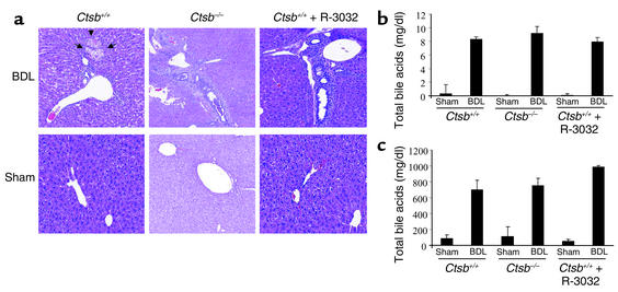
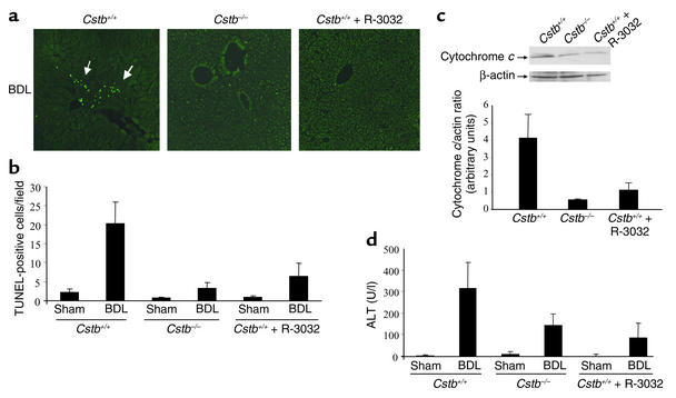
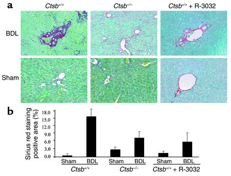
Although a lysosomal, cathepsin B-dependent (Ctsb-dependent) pathway of apoptosis has been described, the contribution of this pathway to tissue damage remains unclear. Our aim was to ascertain if Ctsb inactivation attenuates liver injury, inflammation, and fibrogenesis after bile duct ligation (BDL). In 3-day BDL mice, hepatocyte apoptosis, mitochondrial cytochrome c release, and serum alanine aminotransferase (ALT) values were reduced in Ctsb-/- versus Ctsb+/+ animals. Likewise, R-3032 (a Ctsb inhibitor) also reduced these parameters in BDL WT mice. Both genetic and pharmacologic inhibition of Ctsb in the BDL mouse reduced (a). hepatic inflammation, as assessed by transcripts for CXC chemokines and neutrophil infiltration, and (b). fibrogenesis, as assessed by transcripts for stellate cell activation and sirius red staining for hepatic collagen deposition. These differences could not be ascribed to alterations in cholestasis. These findings support a prominent role for the lysosomal pathway of apoptosis in tissue injury and link apoptosis to inflammation and fibrogenesis. Ctsb inhibition may be therapeutic in liver diseases.


Figures

Figure 1
Figure 1
The magnitude of cholestasis is similar in Ctsb+/+, Ctsb–/–, and R-3032–treated Ctsb+/+ mice. Ctsb+/+, Ctsb–/–, and R-3032–treated Ctsb+/+ mice underwent a sham operation (control) or ligation of their common bile duct. Three days after the surgical procedure, the mice were anesthetized, and liver tissue and serum were obtained. (a) Fixed liver specimens from all mice were stained by conventional hematoxylin and eosin. Bile duct proliferation, portal edema, and mild portal infiltrates were present in all BDL mice. However, bile infarcts only occurred in Ctsb+/+ BDL mice. (b and c) Serum total bilirubin and total bile acids in BDL mice were comparable among the three groups.
Figure 2
Figure 2
Liver injury and cytochrome c release are reduced in Ctsb–/– and R-3032–treated Ctsb+/+ mice. Ctsb+/+ and Ctsb–/– mice were BDL for 3 days as described in Methods. (a) Fixed liver specimens were analyzed by TUNEL assay to identify apoptotic hepatocytes (arrows). (b) The number of TUNEL-positive cells was significantly higher in Ctsb+/+ BDL mice than in sham-operated control or Ctsb–/– and R-3032–treated Ctsb+/+ BDL mice (P < 0.0001, n = 4 for each experimental group). (c) Cytosolic and mitochondrial fractions were prepared as described in Methods. Aliquots of 50 μg of cytosolic protein were subjected to SDS-PAGE and immunoblotted for cytochrome c. Release of cytochrome c in the cytosol was higher in Ctsb+/+ BDL livers compared with Ctsb–/– and R-3032–treated Ctsb+/+ BDL mice. A representative immunoblot from one animal from each group is shown, together with densitometric analysis of multiple immunoblots (n = 3 animals for each group). (d) Serum ALT values are significantly greater in Ctsb+/+ than in Ctsb–/– (P < 0.005, n = 4 for each experimental group) and R-3032–treated Ctsb+/+ mice 3 days after BDL (P < 0.0001, n = 4 for each experimental group). U/l, units per liter.
Figure 3
Figure 3
Neutrophilic infiltration and chemokine expression are increased in Ctsb+/+ compared with Ctsb–/– and R-3032–treated Ctsb+/+ BDL mice. Fixed liver specimens from mice were stained for MPO, a neutrophil marker. Neutrophil infiltration was present only in Ctsb+/+ BDL mice (a) (original magnification ×20). Three days after the surgical procedure, liver tissue was procured and total hepatic RNA was isolated as described in Methods. (b and c) MIP-2 and KC expression was quantitated by real-time PCR. The expression was normalized as a ratio using GAPDH mRNA as a housekeeping gene. A value of 1 for this ratio was arbitrarily assigned to the data obtained from sham-operated Ctsb+/+ mice. KC mRNA expression in Ctsb+/+ BDL was higher than in Ctsb–/– (P < 0.0001) and R-3032–treated Ctsb+/+ BDL mice (P < 0.0001, n = 4 for each group). The expression of MIP-2 mRNA was greater in Ctsb+/+ than in Ctsb–/– (P < 0.0001) and R-3032–treated Ctsb+/+ BDL mice (P < 0.0001, n = 4 for each group).
Figure 4
Figure 4
Markers for HSC activation are increased in Ctsb+/+ compared with Ctsb–/– and R-3032–treated Ctsb+/+ BDL mice. Three days after the surgical procedure, liver tissue was procured and total hepatic RNA was isolated as described in Methods. α-SMA, TGF-β1, COL1A1, and TIMP were quantitated by real-time PCR. The expression was normalized as a ratio using 18S and GAPDH mRNA as housekeeping genes. A value of 1 for this ratio was arbitrarily assigned to the data obtained from sham-operated Ctsb+/+ mice. (a) α-SMA mRNA expression in Ctsb+/+ BDL was higher than in Ctsb–/– (P < 0.0001) and R-3032–treated Ctsb+/+ BDL mice (P < 0.0003, n = 4 for each group). (b) The expression of TGF-β1 mRNA was greater in Ctsb+/+ than in Ctsb–/– (P < 0.0001) and R-3032–treated Ctsb+/+ BDL mice (P < 0.0001, n = 4 for each group). (c) The expression of COL1A1 mRNA was significantly elevated in Ctsb+/+ BDL mice and markedly attenuated in Ctsb–/– (P < 0.0001) and R-3032–treated Ctsb+/+ BDL mice (P < 0.0001, n = 4 for each group). (d) The expression of TIMP mRNA was not significantly different in Ctsb+/+, Ctsb–/–, and R-3032–treated Ctsb+/+ BDL mice (P = NS, n = 4 for each group).
Figure 5
Figure 5
Hepatic fibrosis is increased in Ctsb+/+ BDL compared with Ctsb–/– BDL mice. (a) Two weeks after the surgical procedure, liver tissue was obtained from BDL and sham-operated Ctsb+/+, Ctsb–/–, and R-3032–treated Ctsb+/+ BDL mice, and collagen fibers were stained with sirius red as described in Methods. (b) The surface area stained with sirius red was quantitated using digital image analysis. Sirius red staining was quantitatively greater in Ctsb+/+ BDL than in Ctsb–/– and R-3032–treated Ctsb+/+ BDL mice (P < 0.001, n = 4 for each group). Only minimal sirius red staining was observed in sham-operated mice from the three groups of animals. (Original magnification ×20.)

References

    1. Kim WR, Brown RS, Jr, Terrault NA, El-Serag H. Burden of liver disease in the United States: summary of a workshop. Hepatology. 2002;36:227–242. - PubMed
    1. Sandler RS, et al. The burden of selected digestive diseases in the United States. Gastroenterology. 2002;122:1500–1511. - PubMed
    1. Galle PR, et al. Involvement of the CD95 (APO-1/Fas) receptor and ligand in liver damage. J. Exp. Med. 1995;182:1223–1230. - PMC - PubMed
    1. Luster AD. Chemokines: chemotactic cytokines that mediate inflammation. N. Engl. J. Med. 1998;338:436–445. - PubMed
    1. Miwa K, et al. Caspase 1-independent IL-1beta release and inflammation induced by the apoptosis inducer Fas ligand. Nat. Med. 1998;4:1287–1292. - PubMed

Publication types

MeSH terms