. 2003 Jul;112(2):160-9.
doi: 10.1172/JCI17902.

HGF, SDF-1, and MMP-9 are involved in stress-induced human CD34+ stem cell recruitment to the liver

Affiliations

HGF, SDF-1, and MMP-9 are involved in stress-induced human CD34+ stem cell recruitment to the liver

Orit Kollet et al. J Clin Invest. 2003 Jul.

Abstract

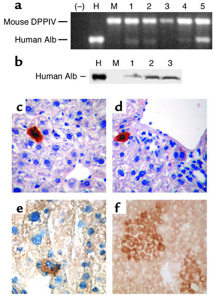
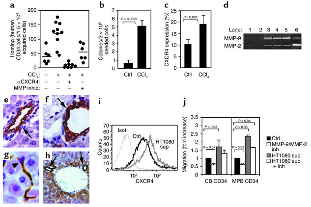
Hematopoietic stem cells rarely contribute to hepatic regeneration, however, the mechanisms governing their homing to the liver, which is a crucial first step, are poorly understood. The chemokine stromal cell-derived factor-1 (SDF-1), which attracts human and murine progenitors, is expressed by liver bile duct epithelium. Neutralization of the SDF-1 receptor CXCR4 abolished homing and engraftment of the murine liver by human CD34+ hematopoietic progenitors, while local injection of human SDF-1 increased their homing. Engrafted human cells were localized in clusters surrounding the bile ducts, in close proximity to SDF-1-expressing epithelial cells, and differentiated into albumin-producing cells. Irradiation or inflammation increased SDF-1 levels and hepatic injury induced MMP-9 activity, leading to both increased CXCR4 expression and SDF-1-mediated recruitment of hematopoietic progenitors to the liver. Unexpectedly, HGF, which is increased following liver injury, promoted protrusion formation, CXCR4 upregulation, and SDF-1-mediated directional migration by human CD34+ progenitors, and synergized with stem cell factor. Thus, stress-induced signals, such as increased expression of SDF-1, MMP-9, and HGF, recruit human CD34+ progenitors with hematopoietic and/or hepatic-like potential to the liver of NOD/SCID mice. Our results suggest the potential of hematopoietic CD34+/CXCR4+cells to respond to stress signals from nonhematopoietic injured organs as an important mechanism for tissue targeting and repair.


Figures

Figure 1
Figure 1
SDF-1/CXCR4 interactions mediate homing and engraftment of irradiated NOD/SCID mouse liver by human CD34+ cells. (a) Homing of human CB or MPB enriched CD34+ cells to the murine BM, spleen (Spl), and liver is inhibited by neutralizing CXCR4. Data present inhibition as percentage of control. P ≤ 0.008, comparing anti-CXCR4–treated samples with their control counterparts. (b) A representative homing experiment shows human CD34+/CD38–/low homing cells (gated) in the liver of mice transplanted with nontreated cells (top), or CXCR4-neutralized cells (middle). A noninjected (Non-inj) mouse served as a negative control (bottom). Numbers indicate human homing cells/1.5 × 106 acquired cells. (c) Four-hour homing of CXCR4-neutralized or nontreated CD34+ cells to the liver of nonirradiated mice. Human SDF-1 was injected into the liver parenchyma as indicated. Cells were collected from the injected lobe to determine the homing of human CD34+ cells. (d) Human cell engraftment of the liver by CB nonstimulated CD34+ cells or CD34+ cells that migrated toward SDF-1, determined by Southern blot specific for human DNA. A representative blot is shown (*mouse transplanted with CD34+ cells migrating toward SDF-1). (e) Data as presented in d, summarizing three independent experiments (n = 37 mice). (f) SDF-1 levels in liver extracts of mice with no irradiation (ctrl), 24 hours and 48 hours after irradiation, determined by ELISA. Data summarize three experiments.
Figure 2
Figure 2
SDF-1 expression and engrafting cell accumulation within the liver. (ad) NOD/SCID mice transplanted with CB CD34+ cells. (a) Hematoxylin and eosin staining 5–6 weeks after transplantation shows a bile duct (large arrow) adjacent to a large portal vein (pv) with cells surrounding the duct (dashed arrow) that are not observed in normal mouse liver. (b) Identification of SDF-1 in the bile ducts of NOD/SCID mouse liver. All epithelial cells in the bile ducts are strongly positive for SDF-1 (large arrow); scattered bile ductule cells (arrowheads) are also SDF-1–positive. (c) Human CD45+ hematopoietic cells (small arrows) are present in large numbers surrounding the bile ducts (large arrows) and accumulate to very high density between the ducts and the adjacent portal veins. (d) Human CD45+ cells are also observed as single cells or small clusters in random distribution in the hepatic sinusoids. Original magnification for ad, ×400. (e and f) SDF-1 expression in normal adult human liver and in the liver of a patient with chronic hepatitis resulting from HCV infection was detected by immunohistochemistry. (e) Normal liver shows a single mature bile duct stained positive for SDF-1 (arrow) and absence of SDF-1 expression in endothelial cells lining a venous channel (arrowheads). (f) Liver from a patient with chronic liver disease from HCV infection shows extensive proliferation of bile ducts positive for SDF-1 expression (arrows) as well as expression of SDF-1 in bile ductule epithelium and/or canal of Hering or oval cells (arrowheads). Magnification for e and f, ×200.
Figure 3
Figure 3
Hepatic differentiation of human albumin–producing cells within the liver of transplanted NOD/SCID mice. (a) Detection of human albumin (Alb) mRNA by RT-PCR using human albumin–specific oligonucleotides and mouse dipeptidyl peptidase IV (DPPIV-specific) oligonucleotides as an internal control. (–), no RNA included; H, human liver RNA; M, RNA from a control, nontransplanted mouse liver; lanes 1, 2, 3, 4, and 5, RNA from the liver of mice transplanted with human CB CD34+ enriched cells. (b) Detection of human albumin by Western blot in the liver of NOD/SCID mice engrafted with human CD34+ CB cells using a mouse mAb specific for human albumin. Shown are a control human liver protein extract (H), liver protein extract from a control nontransplanted mouse (M), and extract from three mice transplanted with human CB CD34+ cells (lanes 1, 2, and 3). (c and d) Sections of formaldehyde-fixed, paraffin-embedded liver tissue analyzed for detection of human albumin using human albumin–specific mAb. (e and f) Sections of frozen liver stained for human albumin, as described in c and d, counterstained with hematoxylin (e) and not counterstained (f). Individual cells (c and d) and small to medium-sized clusters of cells (e and f) with the morphologic appearance of hepatocytes and expressing human albumin were present in the liver of NOD/SCID mice transplanted with human CB CD34+ cells, but not in the liver of control, nontransplanted NOD/SCID mice (data not shown). Original magnification: c, ×200; d, ×100; e and f, ×400.
Figure 4
Figure 4
Stress-induced MMP-2 and MMP-9 increase progenitor cell motility and recruitment to the liver. (a) Four-hour homing of human MPB CD34+ cells to the liver of nonirradiated mice injected 24 hours earlier with 15 μl of CCl4. Anti-CXCR4 pretreatment of transplanted cells or intraperitoneal injection of MMP-2/MMP-9 inhibitor (1.5 hours before transplantation). Bars indicate the mean number of homing cells per 1.5 × 10⁶ acquired cells (n = 3 experiments). (b and c) PBMCs of chimeric mice 1 day after injection of 10 μl CCl4 assayed for the level of human progenitors (b) and human CXCR4 expression (c), (n = 3 experiments). (d) Representative zymography shows MMPs activity in the mouse liver. Control mice (lanes 1 and 2), mice 1 day after injection of 15 μl CCl4 (lane 3), 2 days after 30 μl CCl4 (lane 4), 2 days after 15 μl CCl4 (lane 5), and conditioned media of the human cell line HT1080 (lane 6). (eh) SDF-1 immunostaining of untreated mouse liver (e) or mouse liver after injection of CCl4 (fh). Arrows indicate positively stained bile duct epithelium. Arrowhead indicates positively stained bile ductule or canal of Hering oval cells. Original magnifications: ×1,008, ×1,575, ×1,575, and ×500, respectively. (i) CB CD34+ cells were incubated for 5 hours before CXCR4 staining: isotype control (Isot), cells cultured with RPMI 1640 (Ctrl), cells cultured with HT1080 conditioned media (sup). Representative data of three experiments. (j) Transwell migration toward SDF-1 with CB and MPB CD34+ cells, preincubated with RPMI 1640 (Ctrl), MMP-2/MMP-9 inhibitor (inh), HT1080 conditioned media (sup), or HT1080 supernatant together with MMP-2/MMP-9 inhibitor. Data represent fold-increased migration compared with control cells.
Figure 5
Figure 5
HGF facilitates CD34+ cell motility, CXCR4 expression, and SDF-1–mediated directional migration. CB CD34+ cells were cultured for 40 hours in RPMI 1640 supplemented with 10% FCS alone (Ctrl) or in the presence of SCF (50 ng/ml), HGF (100 ng/ml), or SCF plus HGF. (a) CD34+ cultured cells indirectly immunolabeled with anti-CXCR4 Ab (green) and stained for polymerized actin (red). Merged images are also presented. Arrowhead indicates cell surface protrusion. Arrow indicates lamellipodia. (b) CXCR4 expression analyzed by flow cytometry. (c) Transwell migration toward a gradient of SDF-1 with CB CD34+ cultured cells. Data represent percentage of migration. (–) indicates spontaneous migration without SDF-1.

References

    1. Alison MR, et al. Hepatocytes from non-hepatic adult stem cells. Nature. 2000;406:257. - PubMed
    1. Theise ND, et al. Liver from bone marrow in humans. Hepatology. 2000;32:11–16. - PubMed
    1. Korbling M, et al. Hepatocytes and epithelial cells of donor origin in recipients of peripheral-blood stem cells. N. Engl. J. Med. 2002;346:738–746. - PubMed
    1. Petersen BE, et al. Bone marrow as a potential source of hepatic oval cells. Science. 1999;284:1168–1170. - PubMed
    1. Theise ND, et al. Derivation of hepatocytes from bone marrow cells in mice after radiation-induced myeloablation. Hepatology. 2000;31:235–240. - PubMed

Publication types

MeSH terms