. 2003 Jul;112(2):181-8.
doi: 10.1172/JCI17778.

Essential role of the cryptic epitope SLAYGLR within osteopontin in a murine model of rheumatoid arthritis

Affiliations

Essential role of the cryptic epitope SLAYGLR within osteopontin in a murine model of rheumatoid arthritis

Nobuchika Yamamoto et al. J Clin Invest. 2003 Jul.

Abstract

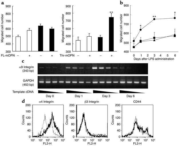
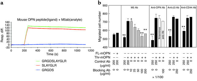
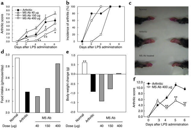
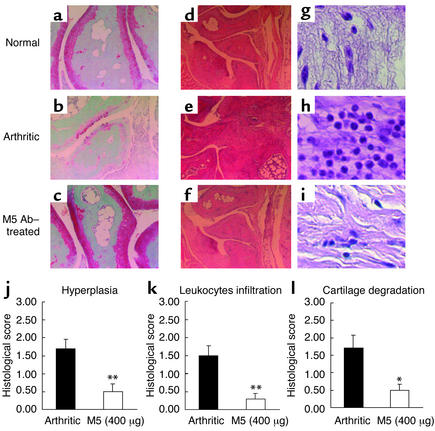
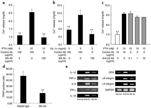
It has been shown that osteopontin (OPN) plays a pivotal role in the pathogenesis of rheumatoid arthritis (RA). However, the molecular mechanism of OPN action is yet to be elucidated. Splenic monocytes obtained from arthritic mice exhibited a significant capacity for cell migration toward thrombin-cleaved OPN but not toward full-length OPN. Migratory monocytes expressed alpha9 and alpha4 integrins. Since cleavage of OPN by thrombin exposes the cryptic epitope recognized by alpha9 and alpha4 integrins, we investigated the role of the cryptic epitope SLAYGLR in a murine RA model by using a specific antibody (M5) reacting to SLAYGLR sequence. The M5 antibody could abrogate monocyte migration toward the thrombin-cleaved form of OPN. Importantly, M5 antibody could inhibit the proliferation of synovium, bone erosion, and inflammatory cell infiltration in arthritic joints. Thus, we demonstrated that a cryptic epitope, the SLAYGLR sequence of murine OPN, is critically involved in the pathogenesis of a murine model of RA.


Figures

Figure 1
Figure 1
Monocyte migration in response to thrombin-cleaved OPN. (a) Splenic monocytes from normal (white bars) and arthritic (black bars) mice on day 6 of migration in response to full-length murine OPN (FL-mOPN) and thrombin-cleaved murine OPN (Thr-mOPN) at 10 μg/ml. (b) The time course of the migration response toward medium (square), full-length (triangle), and thrombin-cleaved OPN (circle) was evaluated. The results are expressed as mean numbers ± SEM of migrated cells. *P < 0.05 and **P < 0.01. (c) Expression of α9 integrin mRNA levels in spleen of arthritic mice on days 0, 1, 3, and 6 after LPS administration was analyzed using RT-PCR and GAPDH as a housekeeping gene. Template cDNA was diluted sequentially (left to right) and amplified by PCR. (d) Expressions of α4 and β3 integrins and CD44 on splenocytes obtained from normal (dotted line) and arthritic (solid line) mice were analyzed by FACS. Histograms represent the mean fluorescence intensity. Controls are from isotype-matched irrelevant antibodies.
Figure 2
Figure 2
M5 Ab inhibited migration of splenic monocytes from arthritic mice. (a) Specificity of M5 Ab against OPN peptides was examined by analysis using BIAcore analysis of M5 Ab. Vertical and horizontal axes indicate surface plasmon resonance intensity (units) and flow time of HBS buffer, respectively. M5 Ab was injected at 200 seconds over the surface of the chip and then washed with HBS buffer. The binding of M5 Ab to GRGDSLAYGLR peptide (green), SLAYGLR peptide (red), and GRGDS peptide (blue) is shown. (b) Inhibition by M5 Ab of cell migration toward thrombin-cleaved OPN. M5 Ab, antipolyclonal OPN Abs, anti-β3 integrin Ab, anti-CD44 Ab, and isotype-matched control antibodies were added at the indicated concentrations. Anti–N-terminal OPN was added at a 1:100 dilution to wells. The results are expressed as mean numbers ± SEM of migrated cells. *P < 0.05 and **P < 0.01. Resp. diff., response difference.
Figure 3
Figure 3
Prophylactic and therapeutic treatment with M5 Ab ameliorated symptoms of arthritis. M5 Ab was administered intravenously at doses of 40, 150, and 400 μg per mouse before the onset of clinical symptoms on days 0 and 3. The arthritic group was intravenously administered 400 μg of rabbit IgG. Arthritic score (a), incidence of arthritis (b), food intake (d), and body weight (e) were monitored. Representative gross appearances of the forepaw are shown (c). Arthritic mice were therapeutically treated with M5 Ab after the onset of symptoms on day 3, and arthritic scores were monitored (f). Each point represents the mean score ± SEM of five mice. *P < 0.05 and **P < 0.01 for comparison by Mann-Whitney U test with arthritic mice; ##P < 0.01 for comparison by Dunnet’s multiple-comparison test with arthritic mice.
Figure 4
Figure 4
Histology of arthritic joints after M5 Ab treatment. Mice receiving prophylactic treatment of 400 μg per mouse (intravenous injection) of M5 Ab (Figure 3) (n = 5) were analyzed. The sections of ankle joints in fore and hind paws were stained with either safranin O (ac) or hematoxylin and eosin (di), and representative histological images of hind paws are shown: normal (a and d), arthritic (b and e) and M5 Ab treated (c and f). The image of leukocytes from an arthritic joint was magnified (gi). Hyperplasia of synovium (j), leukocyte infiltration (k), and cartilage degeneration (l) were quantified. Histological scores are expressed as means ± SEM of four paws of five mice. *P < 0.05 and **P < 0.01 for comparison by Mann-Whitney U test with arthritic mice.
Figure 5
Figure 5
M5 Ab prevented osteoclast-mediated bone resorption and osteoclast formation in vitro. (a and b) PTH or IL-1α induced calcium releases. PTH and IL-1 induced calcium release from bone in murine neonatal calvaria culture in the presence of M5 Ab (a and b) or anti-β3 integrin Ab (c). Results are expressed as means ± SEM. **P < 0.01 for comparison by Dunnet’s multiple-comparison test with isotype control IgG plus PTH or IL-1α. (d) Mouse bone marrow cells were cultured for 7 days in the presence of M-CSF and RANKL in the presence of control antibody or M5 Ab (200 μg/ml), and TRAP-positive mature osteoclasts numbers were counted and expressed as mean numbers ± SEM of cells. (e) Arthritic mice prophylactically treated with M5 Ab on day 0 and 3 in Figure 3 were used. Expressions of proinflammatory cytokines, OPN, and integrin mRNA in joint extracts were analyzed using RT-PCR and GAPDH as a housekeeping gene. These are representative data from three separate experiments.

Comment in

Similar articles

Cited by

References

    1. van der Heijde DM. Joint erosions and patients with early rheumatoid arthritis. Br. J. Rheumatol. 1995;34 (Suppl. 2):74–78. - PubMed
    1. Bathon JM, et al. A comparison of etanercept and methotrexate in patients with early rheumatoid arthritis. N. Engl. J. Med. 2000;343:1586–1593. - PubMed
    1. Campion GV, Lebsack ME, Lookabaugh J, Gordon G, Catalano M. Dose-range and dose-frequency study of recombinant human interleukin-1 receptor antagonist in patients with rheumatoid arthritis. The IL-1Ra Arthritis Study Group. Arthritis Rheum. 1996;39:1092–1101. - PubMed
    1. Paget SA. Efficacy of anakinra in bone: comparison to other biologics. Adv. Ther. 2002;19:27–39. - PubMed
    1. Genovese MC, et al. Etanercept versus methotrexate in patients with early rheumatoid arthritis: Two-year radiographic and clinical outcomes. Arthritis Rheum. 2002;46:1443–1450. - PubMed

MeSH terms