. 2003 Sep 15;551(Pt 3):927-43.
doi: 10.1113/jphysiol.2003.046847. Epub 2003 Jul 15.

Distinct electrophysiological properties of glutamatergic, cholinergic and GABAergic rat septohippocampal neurons: novel implications for hippocampal rhythmicity

Affiliations

Distinct electrophysiological properties of glutamatergic, cholinergic and GABAergic rat septohippocampal neurons: novel implications for hippocampal rhythmicity

F Sotty et al. J Physiol. .

Abstract

The medial septum-diagonal band complex (MSDB) contains cholinergic and non-cholinergic neurons known to play key roles in learning and memory processing, and in the generation of hippocampal theta rhythm. Electrophysiologically, several classes of neurons have been described in the MSDB, but their chemical identity remains to be fully established. By combining electrophysiology with single-cell RT-PCR, we have identified four classes of neurons in the MSDB in vitro. The first class displayed slow-firing and little or no Ih, and expressed choline acetyl-transferase mRNA (ChAT). The second class was fast-firing, had a substantial Ih and expressed glutamic acid decarboxylase 67 mRNA (GAD67), sometimes co-localized with ChAT mRNAs. A third class exhibited fast- and burst-firing, had an important Ih and expressed GAD67 mRNA also occasionally co-localized with ChAT mRNAs. The ionic mechanism underlying the bursts involved a low-threshold spike and a prominent Ih current, conductances often associated with pacemaker activity. Interestingly, we identified a fourth class that expressed transcripts solely for one or two of the vesicular glutamate transporters (VGLUT1 and VGLUT2), but not ChAT or GAD. Some putative glutamatergic neurons displayed electrophysiological properties similar to ChAT-positive slow-firing neurons such as the occurrence of a very small Ih, but nearly half of glutamatergic neurons exhibited cluster firing with intrinsically generated voltage-dependent subthreshold membrane oscillations. Neurons belonging to each of the four described classes were found among septohippocampal neurons by retrograde labelling. We provide results suggesting that slow-firing cholinergic, fast-firing and burst-firing GABAergic, and cluster-firing glutamatergic neurons, may each uniquely contribute to hippocampal rhythmicity in vivo.


Figures

Figure 1
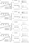
Figure 1. ChAT-positive neurons display electrophysiological properties of slow-firing neurons
A, agarose gel analysis of the RT-mPCR products obtained from a single MSDB cell. The only PCR-generated fragment was that of ChAT. B and C, current-clamp recordings in the same MSDB neuron of the membrane responses to injection of depolarizing current pulses applied from a membrane potential of −60 mV (B) or −80 mV (C). This neuron displays a slow and regular firing activity. D, response recorded in current-clamp mode to injection of a hyperpolarizing current pulse from −60 mV. Note the absence of depolarizing sag in response to membrane hyperpolarization, and of rebound firing when returning to −60 mV. E, currents recorded in voltage-clamp mode evoked by a series of hyperpolarizing voltage steps applied from a holding potential of −50 mV. Notice the small amplitude inward current in this cell, as shown by the difference between the amplitudes of the instantaneous current (open circle) and steady-state current (filled circle). F, instantaneous and steady-state I-V plots derived from the data in E. (B, C, D: vertical bars: 20 mV; horizontal bars: 200 ms.)
Figure 2
Figure 2. GAD67-positive neurons display electrophysiological properties of either burst-firing or fast-firing neurons
A1-F1 (left panel), example of a GAD-positive, fast-firing neuron. This particular neuron expressed only GAD67 mRNA (A1). This neuron displayed a fast and regular firing activity when depolarized from both −60 mV (B1) and −80 mV (C1). Injection of hyperpolarizing current pulses in this neuron induced a moderate depolarizing sag (D1). Rebound firing was weak without bursts (D1). Voltage-clamp recordings of currents evoked by a series of hyperpolarizing voltage steps applied from a holding potential of −50 mV show the presence of a moderate inward current in this cell (E1), indicated by a difference in amplitude between the instantaneous current (open circle) and steady-state current (filled circle). F1, instantaneous and steady-state I-V plots derived from the data in E1. A2-F2 (right panel), example of a GAD-positive, burst-firing neuron. Agarose gel analysis of the RT-mPCR products obtained from a single MSDB cell shows that this particular neuron expressed only GAD67 mRNA (A2). This neuron displayed a fast and regular firing activity when depolarized from −60 mV (B2), and burst firing when depolarized from −80 mV (C2). Burst firing was characterized by few action potentials riding on a slow depolarizing wave (inset C2). Injection of hyperpolarizing current pulses in this neuron induced a profound depolarizing sag (D2). Rebound firing appeared as a burst of action potentials (inset D2). E2, voltage-clamp recordings of currents elicited by a series of hyperpolarizing voltage steps applied from a holding potential of −50 mV. Note the presence of a rapidly activating and large inward current in this cell, as shown by the difference between the amplitude of the instantaneous current (open circle) and steady-state current (filled circle). F2, instantaneous and steady-state I-V plots derived from the data in E2. (B, C, D: vertical bars: 20 mV; horizontal bars: 200 ms.)
Figure 3
Figure 3. Most ChAT and GAD67- positive neurons display electrophysiological properties of either burst-firing or fast-firing neurons
A1-F1, example of a ChAT and GAD-positive, fast-firing neuron. This particular neuron expressed ChAT and GAD67 mRNAs (A1). In current-clamp mode, this neuron displayed a fast and regular firing activity when depolarized from both −60 mV (B1) and −80 mV (C1) Injection of a hyperpolarizing current pulse in this neuron induced a moderate depolarizing sag (D1). Rebound firing appeared as a single action potential (D1). E1, voltage-clamp recordings of currents evoked by a series of hyperpolarizing voltage steps applied from a holding potential of −50 mV show the presence of a moderate inward current in this cell, corresponding to the difference between the amplitude of the instantaneous current (open circle) and steady-state current (filled circle). F1, instantaneous and steady-state I-V plots derived from the data in E1. A2-F2, example of a ChAT and GAD-positive, burst-firing neuron. Agarose gel analysis of the RT-mPCR products obtained from a single MSDB cells shows that this particular neuron expressed both ChAT- and GAD67 mRNAs, as well as VGLUT1 and VGLUT2 mRNAs (A2). This neuron displayed a fast and regular firing activity when depolarized from −60 mV (B2), and burst firing when depolarized from −80 mV (C2). Injection of hyperpolarizing current pulses in this neuron induced a fast and large depolarizing sag (D2). Rebound firing appeared as a burst of action potentials (D2). Voltage-clamp recordings of currents evoked by a series of hyperpolarizing voltage steps applied from a holding potential of −50 mV showing the presence of a fast and large inward current in this cell (E2), as shown by the difference between the amplitude of the instantaneous current (open circle) and steady-state current (filled circle). F2, instantaneous and steady-state I-V plots derived from the data in E2. (B, C, D, vertical bars: 20 mV; horizontal bars: 200 ms.)
Figure 4
Figure 4. VGLUT-positive neurons display electrophysiological properties of cluster-firing neurons
A, agarose gel analysis of the RT-mPCR products obtained from a single MSDB cell showing the coexpression of both VGLUT1 and VGLUT2 mRNAs. B and C, current-clamp recordings in the same MSDB neuron of the membrane responses to injection of depolarizing current pulses applied from a membrane potential of −60 mV (B) or −80 mV (C). This neuron displayed a slow firing activity when depolarized from both membrane potentials. D, depolarizations up to −45 mV elicited cluster firing and subthreshold membrane oscillations between clusters. E, current-clamp recordings of the membrane response to injection of a hyperpolarizing current pulse from −60 mV. Note the absence of depolarizing sag in response to membrane hyperpolarization, and the absence of rebound firing when returning to −60 mV. F, voltage-clamp recordings of currents evoked by a series of hyperpolarizing voltage steps applied from a holding potential of −50 mV. Note the absence of inward current in this cell, as shown by the difference between the amplitude of the instantaneous current (open circle) and steady-state current (filled circle). G, instantaneous and steady-state I-V plots derived from the data in E. (B, C, D: vertical bars: 20 mV; horizontal bars: 200 ms.)
Figure 6
Figure 6. Intracluster, intercluster and subthreshold oscillations frequencies in VGLUT-positive, septohippocampal cluster-firing neurons are voltage dependent
A, FITC-labelled septohippocampal neuron is shown with a patch pipette. B, this neuron recorded in current-clamp mode showed cluster-firing in response to injection of depolarizing current pulses of increasing amplitude. An enlargement of subthreshold oscillations is shown on the right (a-c) for each trace depicted to the left. The frequency of the membrane oscillations increased with depolarized membrane potentials (−54 to −47 mV). In this cluster-firing neuron, the frequency of subthreshold oscillations was near 25 Hz at −47 mV (oscillations indicate by dots are shown enlarged in the right panel), and decreased to about 10 Hz at −54 mV. At −50 mV, the intracluster, intercluster and subthreshold oscillations frequencies were lower than at −47 mV. The enlargement in the right panel (b) shows the subthreshold oscillations emerging and preceding firing of action potentials (arrows). At −54 mV, the intraclusters and interclusters frequencies were very low but subthreshold oscillations were still present (right panel). C, histograms showing that the intracluster frequency, calculated from the first two clusters evoked by a 4 s depolarizing current pulse (open bars) increased with depolarization of the membrane potential (correlation coefficient: r2= 0.993), and that the frequency of subthreshold oscillations (filled bars) similarly increased with depolarization of the membrane potential (correlation coefficient r2= 0.991). D, histogram showing that the voltage dependence of the intercluster frequency, calculated from the first two to three clusters evoked by a 4 s depolarizing current pulse, increased slightly with membrane depolarization (correlation coefficient r2= 0.911). The junction potential was corrected.
Figure 5
Figure 5. Slow-firing, fast-firing, burst-firing and cluster-firing neurons project to the hippocampus
Septohippocampal neurons were labelled by prior injection of fluorescent latex microspheres at several sites within the hippocampus. A1-D1: FITC-labelled neurons were vizualized in MSDB slices by confocal imaging (left images), and the overlay of both fluorescence and transmission images showing the patch pipette (right images). Electrophysiological recordings in current-clamp mode are shown for slow-firing (A2-A4), fast-firing (B2-B4,) burst-firing (C2-C4) and cluster-firing (D2-D4) retrogradely labelled neurons. Neuronal response to injection of depolarizing current pulses applied from −60 mV (A2-D2) or −80 mV (A3-D3), as well as to injection of hyperpolarizing current pulses applied from −60 mV (A4-D4) are shown. (B, C, D: vertical bars: 20 mV; horizontal bars: 200 ms.)
Figure 8
Figure 8. Ih significantly contributes to the mean firing rate, the delay of occurrence of rebound firing, and the instantaneous frequency of rebound firing in fast-firing and burst-firing neurons
A, ZD 7288 (100 μM) significantly decreased the mean firing rate of fast-firing (t test, P < 0.01) and burst-firing (t test, P < 0.05) neurons, but not of slow-firing neurons. B, ZD 7288 increased the delay of occurrence of rebound firing in fast-firing and burst-firing neurons, but not in slow-firing neurons. In burst-firing neurons, this effect was significantly more profound than in both fast-firing (P < 0.05) and slow-firing (P < 0.001) neurons. In fast-firing neurons, this effect of ZD was significantly greater than in slow-firing neurons (P < 0.05). C, ZD 7288 decreased the instantaneous frequency of rebound firing in fast-firing and burst-firing neurons, but not in slow-firing neurons. In both fast-firing and burst-firing neurons, the effects of ZD 7288 were significantly different from the effect in slow firing neurons (P < 0.001). D, Ih current amplitude, determined for a hyperpolarizing voltage step from −50 to −120 mV, was plotted as a function of the amplitude of the depolarizing sag (rectification), measured for a hyperpolarizing current pulse inducing an initial hyperpolarization of the membrane to −95 mV, for each neuronal population. This plot shows a perfect linear correlation between these two parameters for each neuronal population (r2= 0.9726). E, the mean firing rate, determined for a depolarizing current pulse applied from −60 mV, was plotted as a function of the amplitude of the depolarizing sag for each neuronal population, showing a perfect linear correlation (r2= 0.9968) between these two parameters. F, the mean firing rate, determined for a depolarizing current pulse applied from −60 mV, was plotted as a function of the amplitude of the Ih current for each neuronal population, showing again a perfect linear correlation (r2= 0.9879) between these two parameters. These analyses showed that burst-firing exhibited the largest Ih current and depolarizing sag, correlated to the fastest firing rate; slow-firing neurons displayed the smallest Ih current and depolarizing sag, associated to the slowest firing rate; fast-firing neurons were intermediate between burst-firing and slow-firing neurons for all three parameters.
Figure 7
Figure 7. Ih current blockade by ZD 7288 profoundly affects electrophysiological properties of fast-firing and burst-firing neurons but not slow-firing and cluster-firing neurons
In a fast-firing cell, ZD 7288 (100 μM) totally blocked Ih current as shown by the instantaneous (open symbols) and steady-state (filled symbols) I-V plots (A) derived from voltage-clamp recordings of the membrane currents evoked by hyperpolarizing voltage steps applied from a holding potential of −50 mV before (squares) and during (circles) bath application of ZD 7288 (100 μM) (not shown). In the same neuron, ZD 7288 totally abolished the depolarizing sag induced by a hyperpolarizing current pulse (B, left panel) and decreased the mean firing rate in response to a depolarizing current pulse applied from −80 mV (B, right panel). In a burst-firing cell, ZD 7288 (100 μM) also induced a complete blockade of Ih current as shown by the instantaneous (open symbols) and steady-state (filled symbols) I-V plots (C), as well as a complete blockade of the depolarizing sag (D, left panel). In addition, ZD 7288 induced an increase of the delay at which rebound firing occurred, as well as a decrease of the instantaneous frequency calculated from the time interval between the first two spikes occurring in rebound firing (D, left panel). ZD 7288 finally decreased the mean firing rate of this burst-firing neuron when depolarized from −80 mV (D, right panel). In contrast, ZD 7288 (100 μM) induced a small blockade of the small Ih current in slow-firing (E) and cluster-firing (G) neurons, as shown by the instantaneous (open symbols) and steady-state (filled symbols) I-V plots derived from voltage-clamp recordings of the membrane currents evoked by hyperpolarizing voltage steps applied from a holding potential of −50 mV before (squares) and during (circles) bath application of ZD 7288 (100 μM) (not shown). In slow-firing neurons, ZD 7288 did not affect the membrane response to injection of hyperpolarizing current pulses (F, left panel), or their mean firing rate when depolarized from −80 mV (F, right panel). In the same way, ZD 7288 did not affect the membrane response to injection of hyperpolarizing current pulses in cluster-firing neurons (H, left panel), and more importantly, did not affect clustering of action potentials (H, right panel). (Vertical bars: 20 mV; horizontal bars: 200 ms.)

References

    1. Alreja M, Wu M, Liu W, Atkins JB, Leranth C, Shanabrough M. Muscarinic tone sustains impulse flow in the septohippocampal GABA but not cholinergic pathway: implications for learning and memory. J Neurosci. 2000;20:8103–8110. - PMC - PubMed
    1. Apartis E, Poindessous-Jazat FR, Lamour YA, Bassant MH. Loss of rhythmically bursting neurons in rat medial septum following selective lesion of septo-hippocampal cholinergic system. J Neurophysiol. 1998;79:1633–1642. - PubMed
    1. Bland BH, Oddie SD. Theta band oscillation and synchrony in the hippocampal formation and associated structures: the case for its role in sensorimotor integration. Behav Brain Res. 2001;127:119–136. - PubMed
    1. Bonansco C, Buno W. Cellular mechanisms underlying the rhythmic bursts induced by NMDA microiontophoresis at the apical dendrites of CA1 pyramidal neurons. Hippocampus. 2003;13:150–163. - PubMed
    1. Brashear HR, Zaborsky L, Heimer L. Distribution of GABAergic and cholinergic neurons in the rat diagonal band. Neuroscience. 1986;17:439–451. - PubMed

Publication types

MeSH terms

LinkOut - more resources