. 2003 Jul 21;89(2):374-84.
doi: 10.1038/sj.bjc.6601098.

Downregulation of urokinase plasminogen activator receptor expression inhibits Erk signalling with concomitant suppression of invasiveness due to loss of uPAR-beta1 integrin complex in colon cancer cells

Affiliations

Downregulation of urokinase plasminogen activator receptor expression inhibits Erk signalling with concomitant suppression of invasiveness due to loss of uPAR-beta1 integrin complex in colon cancer cells

N Ahmed et al. Br J Cancer. .

Abstract

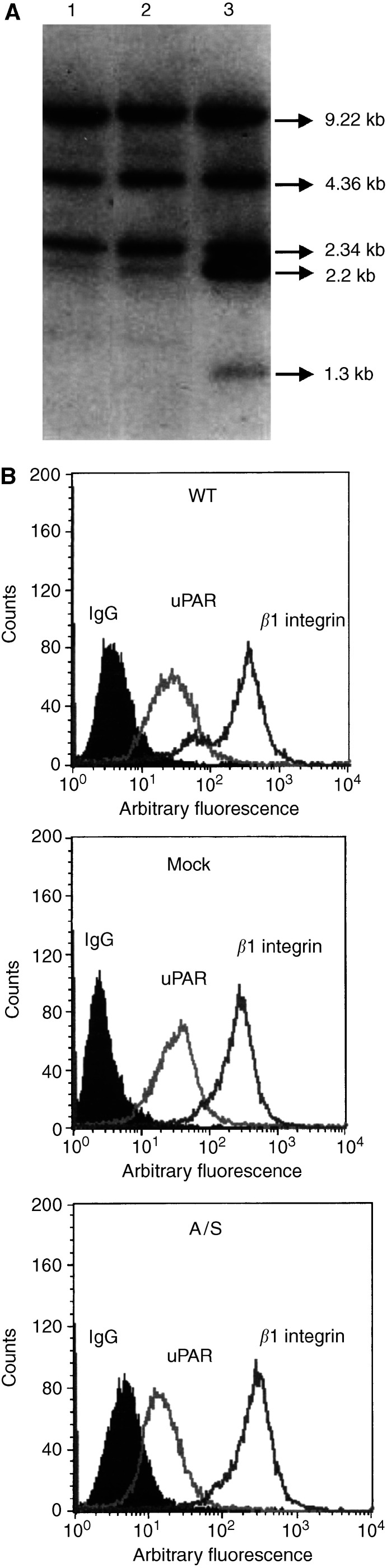
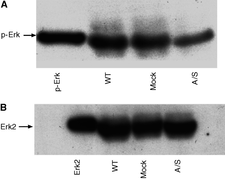
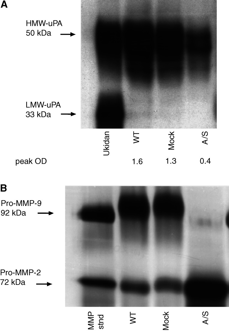
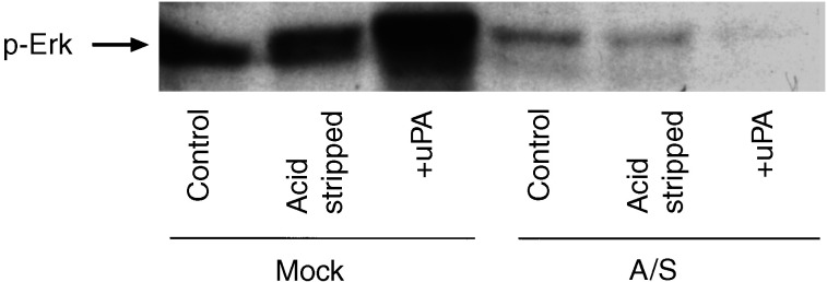
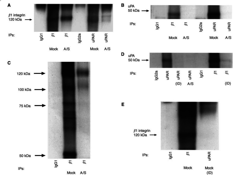
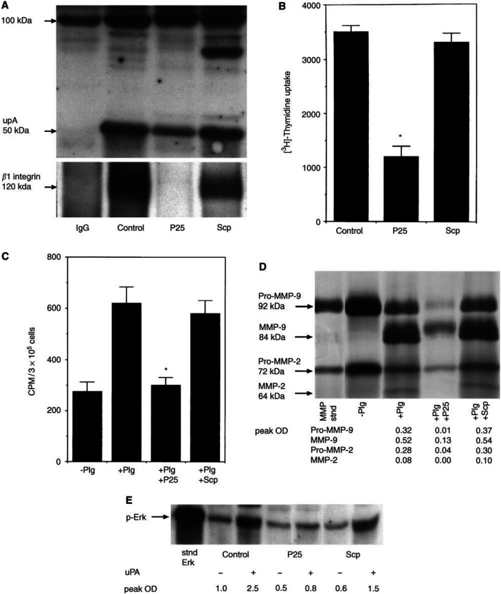
Cancer invasion is regulated by cell surface proteinases and adhesion molecules. Interaction between specific cell surface molecules such as urokinase plasminogen activator receptor (uPAR) and integrins is crucial for tumour invasion and metastasis. In this study, we examined whether uPAR and beta1 integrin form a functional complex to mediate signalling required for tumour invasion. We assessed the expression of uPAR/beta1 integrin complex, Erk signalling pathway, adhesion, uPA and matrix metalloproteinase (MMP) expression, migration/invasion and matrix degradation in a colon cancer cell line in which uPAR expression was modified. Antisense inhibition of the cell surface expression of uPAR by 50% in human colon carcinoma HCT116 cells (A/S) suppressed Erk-MAP kinase activity by two-fold. Urokinase plasminogen activator receptor antisense treatment of HCT116 cells was associated with a 1.3-fold inhibition of adhesion, approximately four-fold suppression of HMW-uPA secretion and inhibition of pro-MMP-9 secretion. At a functional level, uPAR antisense resulted in a four-fold decline in migration/invasion and abatement of plasmin-mediated matrix degradation. In empty vector-transfected cells (mock), uPA strongly elevated basal Erk activation. In contrast, in A/S cells, uPA induction of Erk activation was not observed. Urokinase plasminogen activator receptor associated with beta1 integrin in mock-transfected cells. Disruption of uPAR-beta1 integrin complex in mock-transfected cells with a specific peptide (P25) inhibited uPA-mediated Erk-MAP kinase pathway and inhibited migration/invasion and plasmin-dependent matrix degradation through suppression of pro-MMP-9/MMP-2 expression. This novel paradigm of uPAR-integrin signalling may afford opportunities for alternative therapeutic strategies for the treatment of cancer.


Figures

Figure 1
Figure 1
Characterisation of HCT116 cells. (A) Southern blot analyses of genomic DNAs isolated from transfected and untransfected HCT116 cells. Lane 1, WT HCT116 cells; lane 2, mock-transfected HCT116 cells; and lane 3 is 5′A/S HCT116 cells. (B) Flow-cytometric analyses of uPAR and β1 integrin in HCT116 cell lines. The median intensity of fluorescence (MIF, arbitary units, log scale) was measured. Results are representative of three independent experiments.
Figure 2
Figure 2
(A) Erk-MAP kinase activity in HCT116 cell lines. Cell lysates used for in vitro kinase assay were analysed by equal protein loading on 10% SDS–PAGE (under nonreducing conditions) followed by Western blotting using antibodies against phosphorylated Erk-MAP kinase (E10 antibody, New England Biolabs) and (B) total Erk (Santa Cruz, CA, USA).
Figure 3
Figure 3
(A) Expression of HMW-uPA in conditioned medium of WT, mock- and A/S-transfected HCT116 cell lines. Conditioned medium was analysed by equal protein loading on 10% SDS–PAGE (under nonreducing conditions) followed by Western blotting using monoclonal anti-uPA antibody. Ukidan was used as standard reference uPA (Serono, Australia). The expression of HMW-uPA was quantified by densitometry and is expressed as peak optical density (peak OD). Results are representative of three experiments. (B) Gelatin zymography showing the amounts of pro-MMP-2 and pro-MMP-9 secreted in conditioned medium (concentrated 90–100-fold) from HCT116 cell lines. The positions of purified pro-MMP-2 and pro-MMP-9 are shown on the left. Results are representative of three independent experiments.
Figure 4
Figure 4
Effect of uPA on the activation of Erk. Subconfluent cultures of mock and A/S HCT116 cells were serum starved for 24 h, acid stripped for 1 min and incubated with uPA (20 nM) for 30 min. The level of phospho Erk1/2 was determined by Western blot using equal protein loading. Results are representative of one experiment. The experiment was repeated three times.
Figure 5
Figure 5
Coimmunoprecipitation of β1 integrin and uPAR. Mock- and A/S-transfected HCT116 cells were lysed in Triton X-100 buffer (see Materials and Methods). Cell protein (500 μg) was mixed with anti-β1 antibody (PD52) or anti-uPAR antibody (3936) or isotype-matched mouse IgG and the resulting immunoprecipitates were analysed by Western blotting with (A) anti-β1-integrin antibody (PD52) or (B) anti-uPAR antibody (3936). (C) Cells were surface biotinylated and cell extracts were subjected to immunoprecipitation with anti-β1 antibody or with isotype-matched IgG antibodies and analysed by streptavidin–HRP binding to biotinylated proteins after SDS–PAGE and transfer to nitrocellulose membranes. (D and E) Mock-transfected cell lysates were immunodepleted (ID)of β1 after five rounds of sequential β1 and uPAR immunoprecipitation. Cell lysates were resolved by 10% SDS–PAGE followed by blotting with (D) anti-uPAR and (E) anti-β1 integrin antibody. The experiments were repeated at least three times.
Figure 6
Figure 6
uPAR/β1 integrin complex and the affect of P25 peptide. (A) P25 peptide but not scrambled peptide (Scp) disrupts uPAR/β1 integrin complex in mock-transfected HCT116 cells. Cells were treated for 16 h with P25 peptide (100 μM) and Scp (100 μM). Immunoprecipitates of uPAR were prepared and coimmunoprecipitated β1 integrin band was analysed. (B) Migration/invasion of HCT116 mock cells in the presence of P25 peptide and its scrambled analogue (100 μM each). (C) Plasminogen-dependent matrix degradation of HCT116 mock cells in the presence of P25 peptide and its scrambled analogue (100 μM each). Results for both Figures 6B and C are shown as mean + S.E.M. of three different experiments performed in triplicate (*P<0.001, compared to control cells in the presence of Plg). (D) Effects of P25 peptide and its scrambled analogue on the secretion and activation of pro-MMP-2/MMP-9. Conditioned medium in the presence and absence of Plg, P25 peptide and Scp (100 μM each) was prepared as described in the Materials and Methods section. The samples were analysed by equal protein loading by gelatin-zymography for the activation of pro-MMP-2/MMP-9. Quantification of MMP secretion in the tumour-conditioned medium was performed by densitometry and the results are expressed as peak OD. Results are representative of one experiment. The experiment was repeated three times. (E) Effect of P25 peptide and its scrambled analogue (100 μM each) on uPA-induced activation of Erk in mock-transfected HCT116 cells. Subconfluent cultures of mock-transfected HCT116 cells were serum starved for 24 h, acid stripped for 1 min and incubated with uPA (20 nM) for 30 min. The level of phospho Erk1/2 was determined by Western blot using equal protein loading. Quantification of phospho Erk1/2 expression was performed by densitometry and expressed as peak OD. Results are representative of one experiment. The experiment was repeated three times.

References

    1. Aguirre Ghiso JA, Alonso DF, Farias EF, Gomez DE, de Kier Joffe EB (1999a) Deregulation of the signaling pathways controlling urokinase-production. Its relationship with the invasive phenotype. Eur J Biochem 263: 295–304 - PubMed
    1. Aguirre Ghiso JA, Kovalski K, Ossowski L (1999b) Tumor dormancy induced by downregulation of urokinase receptor in human carcinoma involves integrin and MAPK signaling. J Cell Biol 147: 89–104 - PMC - PubMed
    1. Ahmed N, Niu J, Dorahy DJ, Gu X, Andrews S, Meldrum CJ, Scott RJ, Baker MS, Macreadie IG, Agrez MV (2002a) Direct integrin alphavbeta6-ERK binding: implications for tumour growth. Oncogene 21: 1370–1380 - PubMed
    1. Ahmed N, Pansino F, Baker M, Rice G, Quinn M (2002b) Association between [alpha]v[beta]6 integrin expression, elevated p42/44 kDa MAPK, and plasminogen-dependent matrix degradation in ovarian cancer. J Cell Biochem 84: 675–686 - PubMed
    1. Ahmed N, Pansino F, Clyde R, Murthi P, Quinn MA, Rice GE, Agrez MV, Mok S, Baker MS (2002c) Overexpression of alphavbeta6 integrin in serous epithelial ovarian cancer regulates extracellular matrix degradation via the plasminogen activation cascade. Carcinogenesis 23: 237–244 - PubMed

Publication types

MeSH terms