. 2003 Aug;185(15):4402-9.
doi: 10.1128/JB.185.15.4402-4409.2003.

YjdE (AdiC) is the arginine:agmatine antiporter essential for arginine-dependent acid resistance in Escherichia coli

Affiliations

YjdE (AdiC) is the arginine:agmatine antiporter essential for arginine-dependent acid resistance in Escherichia coli

Shimei Gong et al. J Bacteriol. 2003 Aug.

Abstract

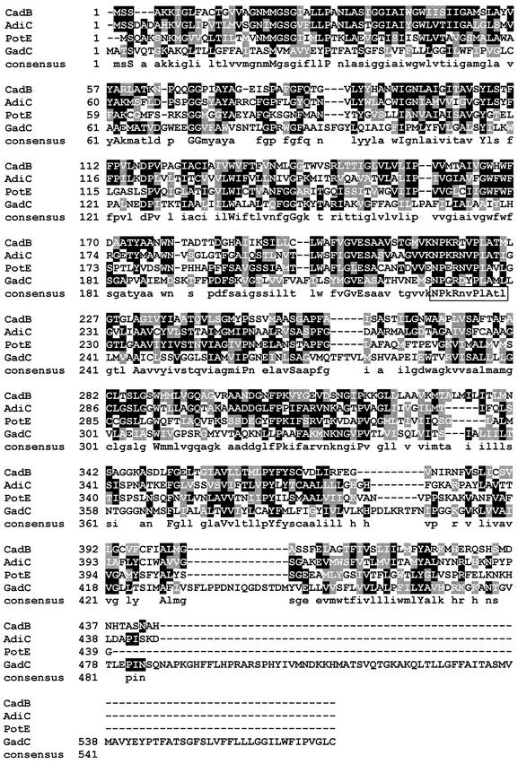
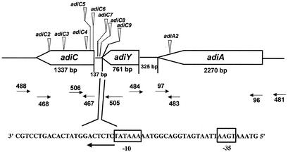
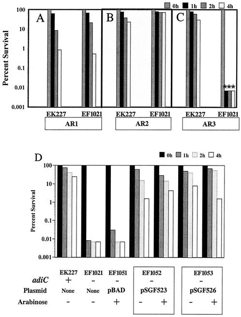
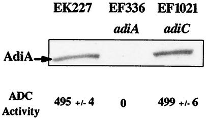
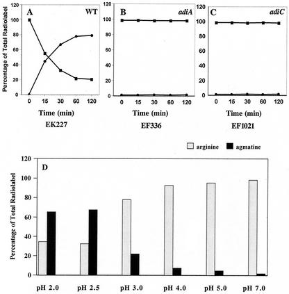
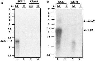
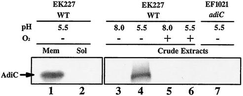
To survive in extremely acidic conditions, Escherichia coli has evolved three adaptive acid resistance strategies thought to maintain internal pH. While the mechanism behind acid resistance system 1 remains enigmatic, systems 2 and 3 are known to require external glutamate (system 2) and arginine (system 3) to function. These latter systems employ specific amino acid decarboxylases and putative antiporters that exchange the extracellular amino acid substrate for the intracellular by-product of decarboxylation. Although GadC is the predicted antiporter for system 2, the antiporter specific for arginine/agmatine exchange has not been identified. A computer-based homology search revealed that the yjdE (now called adiC) gene product shared an overall amino acid identity of 22% with GadC. A series of adiC mutants isolated by random mutagenesis and by targeted deletion were shown to be defective in arginine-dependent acid resistance. This defect was restored upon introduction of an adiC(+)-containing plasmid. An adiC mutant proved incapable of exchanging extracellular arginine for intracellular agmatine but maintained wild-type levels of arginine decarboxylase protein and activity. Western blot analysis indicated AdiC is an integral membrane protein. These data indicate that the arginine-to-agmatine conversion defect of adiC mutants was at the level of transport. The adi gene region was shown to be organized into two transcriptional units, adiAY and adiC, which are coordinately regulated but independently transcribed. The data also illustrate that the AdiA decarboxylase:AdiC antiporter system is designed to function only at acid levels sufficient to harm the cell.


Figures

FIG. 1.
FIG. 1.
Homology-based prediction of a putative arginine:agmatine antiporter. CLUSTAL W amino acid sequence alignments of AdiC (YjdE), the predicted arginine:agmatine antiporter; CadB, the lysine:cadeverine antiporter of the lysine decarboxylase system; PotE, the ornithine:putrescine antiporter of the ornithine decarboxylase system; and GadC, the putative glutamate:GABA antiporter of the glutamate decarboxylase system. Identical amino acids are shown in black boxes, while similar amino acids are shown in gray boxes. The consensus line shows identical (uppercase) and similar (lowercase) amino acids.
FIG. 2.
FIG. 2.
Genetic organization of the E. coli adi cluster and the locations of acid sensitive Tn10dTc insertions. Block arrows representing genes point in the direction of transcription. The small arrows indicate relative binding locations of the oligonucleotide primers used in this study. Vertical arrowheads represent approximate positions of acid-sensitive Tn10dTc insertions as determined via PCR analysis. Each designation represents a class of Tn10 insertions. The inset sequence at the bottom shows a predicted adiC promoter as determined by using the Neural Network Promoter Prediction site (http://www.fruitfly.org/seq_tools/nnppHelp.html). Putative −10 and −35 regions are marked.
FIG. 3.
FIG. 3.
An adiC mutant is specifically defective in arginine-dependent acid resistance. Acid resistance assays of EK227 (wild-type) and EF1021 (ΔadiC::Km). (A) AR 1. Cells were grown in LB-MES (pH 5.5) to stationary phase and challenged to EG medium (pH 2.5). (B) AR 2. Cells were grown in LB-glucose to stationary phase and challenged to EG medium (pH 2.0) in the presence of 0.7 mM glutamate. (C and D) AR 3. Cells were grown in BHIG to stationary phase (22 h) and challenged to EG medium (pH 2.5) with 1.5 mM arginine. Control cells grown in LB-MOPS (pH 8) (the control for AR 1) or cells challenged without glutamate or arginine (controls for AR 2 and 3, respectively) were completely inviable at 1 h post challenge (data not shown). (D) Cloned E. coli adiC+ complements the ΔadiC arginine-dependent acid resistance defect. EF1051 (ΔadiC::Km/pBAD24), EF1052 (ΔadiC::Km/pSGF523), and EF1053 (ΔadiC::Km/pSGF526) were used. Values represent average survival after 0, 1, 2, and 4 h. All assays were performed in triplicate. Representative results are shown. Asterisks indicate that survival was below detection limits.
FIG. 4.
FIG. 4.
An adiC mutation does not affect ADC levels or activity. Top, Western blot analysis of stationary-phase EK227 (wild-type strain), EF336 (adiA::mudJ), and EF1021(ΔadiC::Km). Whole-cell lysates prepared from BHIG-grown cells were separated on a 10% SDS-PAGE gel and blotted with anti-AdiA antibody. Bottom, ADC activity was measured at pH 5 in Triton X-100-solubilized cells. Units given are nanomolar of agmatine formed per hour per cell density unit (optical density at 600 nm).
FIG. 5.
FIG. 5.
Exchange of extracellular arginine for intracellular agmatine. Cells of strains EK227 (wild-type) (A), EF1021(ΔadiC::Km) (B), and EF336(adiA::MudJ) (C) were grown in BHIG for 22 h and adjusted to a cell density of 108/ml in EG medium at pH 2.5. The exchange of extracellular agmatine for arginine was measured by using 1 mM radiolabeled arginine. At various times, cell-free supernatants were collected from the cultures, adjusted to pH 7, spiked with unlabeled arginine and agmatine, and separated by paper chromatography. Spots corresponding to arginine and agmatine were cut into strips and counted for radioactivity. (D) Optimal pH for whole-cell exchange of arginine to agmatine by EK227.
FIG. 6.
FIG. 6.
Acid induction of adiC and adiA. Cells were grown anaerobically in ADC media to log phase (108 CFU/ml). Five micrograms of RNA was loaded per lane. Blots were probed either with adiC (A) or adiA (B). An RNA ladder (kilobases) is shown to the left of each panel. The 1.2-kb RNA band seen in panel B is unrelated to adiA, as it is seen in both the wild type and adiA mutants.
FIG. 7.
FIG. 7.
Membrane location of the AdiC antiporter. Cells (EK227) were grown to exponential phase in ADC media with or without oxygen at the pH values indicated. Cells were harvested by centrifugation, sonicated, and separated into membrane (lane 1) and soluble (lane 2) fractions. Crude extracts were analyzed in lanes 3 through 7.

References

    1. Auger, E. A., K. E. Redding, T. Plumb, L. C. Childs, S.-Y. Meng, and G. N. Bennett. 1989. Construction of lac fusions to the inducible arginine and lysine decarboxylase genes of Escherichia coli K-12. Mol. Microbiol. 3:609-620. - PubMed
    1. Bocker, E. A., and E. E. Snell. 1972. Amino acid decarboxylases, p. 217-253. In P. Boyer (ed.), The enzymes, vol. 6. Academic Press, New York, N.Y.
    1. Castanie-Cornet, M. P., and J. W. Foster. 2001. Escherichia coli acid resistance: cAMP receptor protein and a 20-bp cis-acting sequence control pH and stationary phase expression of the gadA and gadBC glutamate decarboxylase genes. Microbiology 147:709-715. - PubMed
    1. Castanie-Cornet, M. P., T. A. Penfound, D. Smith, J. F. Elliott, and J. W. Foster. 1999. Control of acid resistance in Escherichia coli. J. Bacteriol. 181:3525-3535. - PMC - PubMed
    1. Datsenko, K. A., and B. L. Wanner. 2000. One-step inactivation of chromosomal genes in Escherichia coli K-12 using PCR products. Proc. Natl. Acad. Sci. USA 97:6640-6645. - PMC - PubMed

Publication types

MeSH terms

LinkOut - more resources