. 2003 Aug;185(15):4418-23.
doi: 10.1128/JB.185.15.4418-4423.2003.

The glycosyltransferase domain of penicillin-binding protein 2a from Streptococcus pneumoniae catalyzes the polymerization of murein glycan chains

Affiliations

The glycosyltransferase domain of penicillin-binding protein 2a from Streptococcus pneumoniae catalyzes the polymerization of murein glycan chains

Anne Marie Di Guilmi et al. J Bacteriol. 2003 Aug.

Abstract

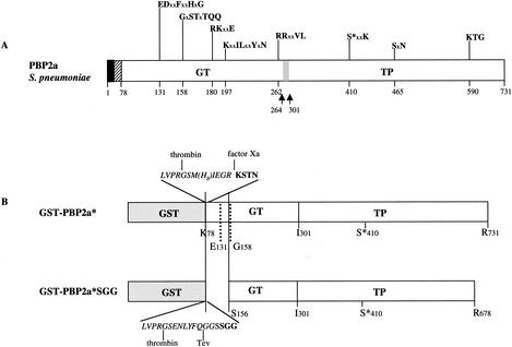
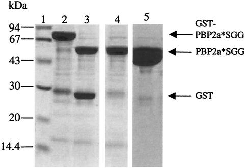
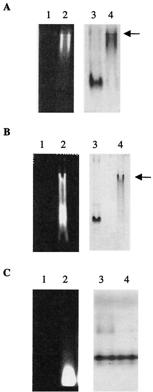
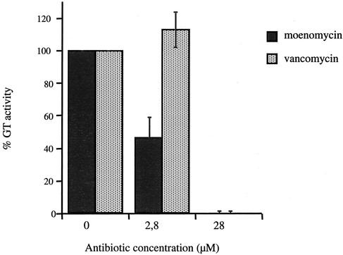
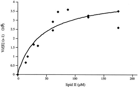
The bacterial peptidoglycan consists of glycan chains of repeating beta-1,4-linked N-acetylglucosaminyl-N-acetylmuramyl units cross-linked through short peptide chains. The polymerization of the glycans, or glycosyltransfer (GT), and transpeptidation (TP) are catalyzed by bifunctional penicillin-binding proteins (PBPs). The beta-lactam antibiotics inhibit the TP reaction, but their widespread use led to the development of drug resistance in pathogenic bacteria. In this context, the GT catalytic domain represents a potential target in the antibacterial fight. In this work, the in vitro polymerization of glycan chains by the extracellular region of recombinant Streptococcus pneumoniae PBP2a, namely, PBP2a* (the asterisk indicates the soluble form of the protein) is presented. Dansylated lipid II was used as the substrate, and the kinetic parameters K(m) and k(cat)/K(m) were measured at 40.6 micro M (+/- 15.5) and 1 x 10(-3) M(-1) s(-1), respectively. The GT reaction catalyzed by PBP2a* was inhibited by moenomycin and vancomycin. Furthermore, the sequence between Lys 78 and Ser 156 is required for enzymatic activity, whereas it is dispensable for lipid II binding. In addition, we confirmed that this region of the protein is also involved in membrane interaction, independently of the transmembrane anchor. The characterization of the catalytically active GT domain of S. pneumoniae PBP2a may contribute to the development of new inhibitors, which are urgently needed to renew the antibiotic arsenal.


Figures

FIG. 1.
FIG. 1.
Schematic diagrams of S. pneumoniae PBP2a. (A) Topology of the native PBP2a protein. The solid and hatched boxes indicate the N-terminal cytoplasmic region and the membrane anchor, respectively. GT and TP domains are represented together with their conserved motifs; active site serine 410 is indicated by an asterisk. x represents any amino acid. The gray box and the arrows indicate the location of the identified GT and TP junction (5). (B) Schematic diagrams of the PBP2a-derived constructs. The peptide at the GST-GT junction of the PBP2a* construct includes sequences specific for thrombin, Tev protease, and factor Xa (italic characters) and N-terminal amino acids of GT (bold characters).
FIG. 2.
FIG. 2.
Purification of PBP2a*SGG. Proteins were separated by sodium dodecyl sulfate-12.5% PAGE and stained with Coomassie blue. Numbers at the left indicate sizes of standard molecular mass markers. Lanes: 1, molecular mass markers; 2, GST-PBP2a*SGG fusion following purification in a glutathione-Sepharose column; 3, fusion protein cleavage by Tev protease; 4, flowthrough from glutathione-Sepharose (10 μg of protein loaded on the gel); 5, elution from Resource Q column (30 μg of protein loaded on the gel).
FIG. 3.
FIG. 3.
Lipid II binding to PBP2a moieties. The binding was performed at room temperature for 15 min before loading onto native PAGE. Arrows indicate protein/lipid II complex. (A) Lanes: 1 and 3, PBP2a* (2 μM); 2 and 4, PBP2a* (2 μM) and lipid II (140 μM). The same gel was observed under UV light (lanes 1 and 2) before staining with Coomassie blue (lanes 3 and 4). (B) Lanes: 1 and 3, PBP2a*SGG (2.8 μM); 2 and 4, PBP2a*SGG (2.8 μM) and lipid II (140 μM). The same gel was observed under UV light (lanes 1 and 2) before staining with Coomassie blue (lanes 3 and 4). (C) Lanes: 1 and 3, TP2a (5.5 μM); 2 and 4, TP2a (5.5 μM) and lipid II (140 μM). The same gel was observed under UV light (lanes 1 and 2) before staining with Coomassie blue (lanes 3 and 4).
FIG. 4.
FIG. 4.
Inhibition of PBP2a* GT activity by moenomycin and vancomycin. Reaction mix composition and TLC conditions were as described in Materials and Methods. PBP2a* (4 μM), lipid II (28 μM), and the inhibitors moenomycin and vancomycin (2.8 and 28 μM) were used in the assay.
FIG. 5.
FIG. 5.
Kinetic analysis of PBP2a* GT activity. PBP2a* (4 μM) and lipid II concentrations ranging from 14 to 175 μM were used. The curve fit (as well as the associated errors) was generated by fitting data to the equation Vi/[E] = kcat · [lipid II]/Km + [lipid II] using Kaleidagraph.

References

    1. Di Guilmi, A. M., and A. Dessen. 2002. New approaches towards the identification of antibiotic and vaccine targets in Streptococcus pneumoniae. EMBO Rep. 3:728-734. - PMC - PubMed
    1. Di Guilmi, A. M., A. Dessen, O. Dideberg, and T. Vernet. 2002. Bifunctional penicillin-binding proteins: focus on the glycosyltransferase domain and its specific inhibitor moenomycin. Curr. Pharm. Biotechnol. 3:63-75. - PubMed
    1. Di Guilmi, A. M., A. Dessen, O. Dideberg, and T. Vernet. 2003. Functional characterization of penicillin-binding protein 1b from Streptococcus pneumoniae. J. Bacteriol. 185:1650-1658. - PMC - PubMed
    1. Di Guilmi, A. M., N. Mouz, J. P. Andrieu, J. Hoskins, S. R. Jaskunas, J. Gagnon, O. Dideberg, and T. Vernet. 1998. Identification, purification, and characterization of transpeptidase and glycosyltransferase domains of Streptococcus pneumoniae penicillin-binding protein 1a. J. Bacteriol. 180:5652-5659. - PMC - PubMed
    1. Di Guilmi, A. M., N. Mouz, L. Martin, J. Hoskins, S. R. Jaskunas, O. Dideberg, and T. Vernet. 1999. Glycosyltransferase domain of penicillin-binding protein 2a from Streptococcus pneumoniae is membrane associated. J. Bacteriol. 181:2773-2781. - PMC - PubMed

MeSH terms

LinkOut - more resources