. 2003 Aug;185(15):4450-60.
doi: 10.1128/JB.185.15.4450-4460.2003.

CsrA regulates translation of the Escherichia coli carbon starvation gene, cstA, by blocking ribosome access to the cstA transcript

Affiliations

CsrA regulates translation of the Escherichia coli carbon starvation gene, cstA, by blocking ribosome access to the cstA transcript

Ashok K Dubey et al. J Bacteriol. 2003 Aug.

Abstract

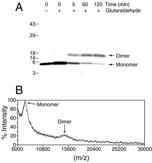
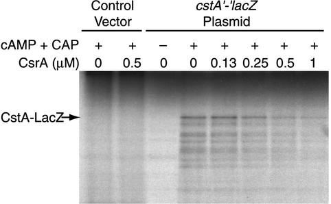
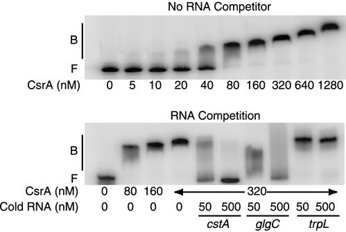
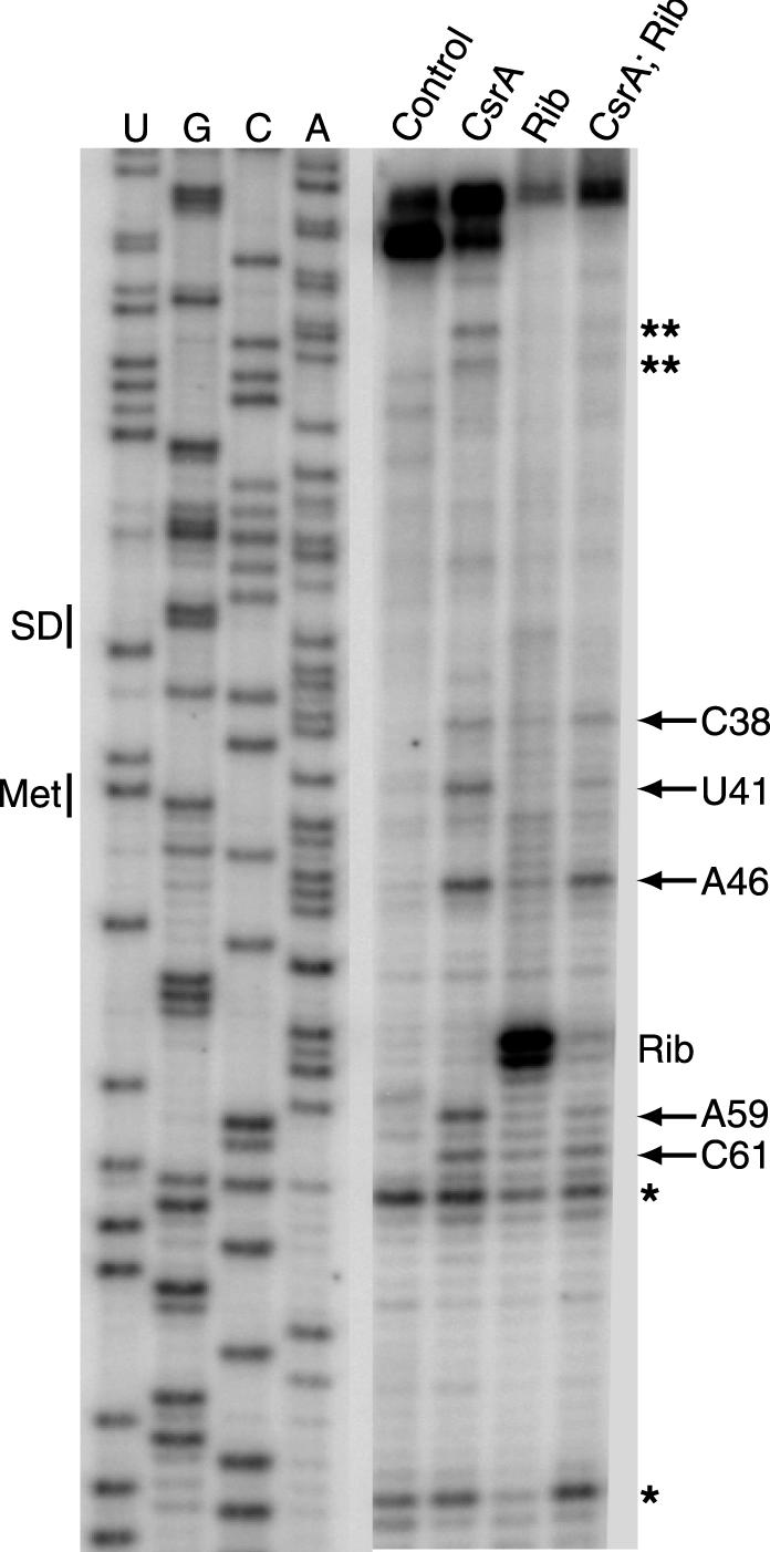
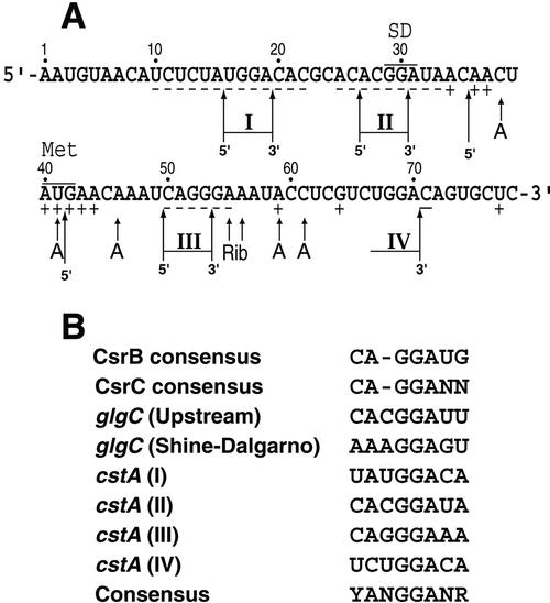
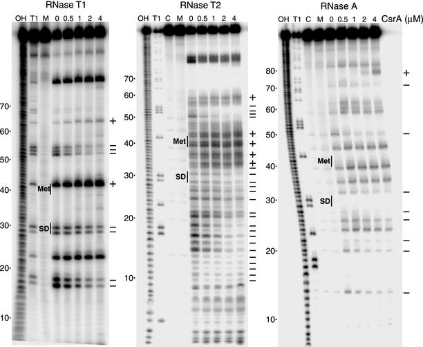
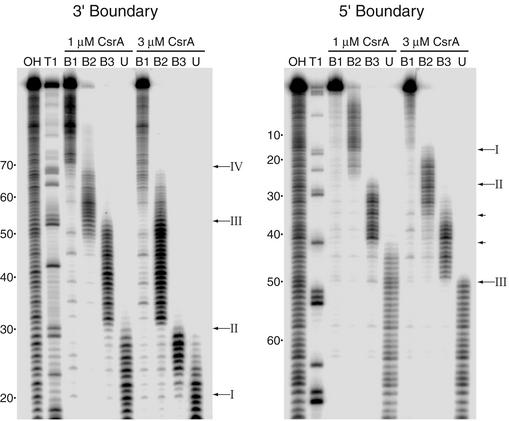
CsrA is a global regulator that binds to two sites in the glgCAP leader transcript, thereby blocking ribosome access to the glgC Shine-Dalgarno sequence. The upstream CsrA binding site (GCACACGGAU) was used to search the Escherichia coli genomic sequence for other genes that might be regulated by CsrA. cstA contained an exact match that overlapped its Shine-Dalgarno sequence. cstA was previously shown to be induced by carbon starvation and to encode a peptide transporter. Expression of a cstA'-'lacZ translational fusion in wild-type and csrA mutant strains was examined. Expression levels in the csrA mutant were approximately twofold higher when cells were grown in Luria broth (LB) and 5- to 10-fold higher when LB was supplemented with glucose. It was previously shown that cstA is regulated by the cyclic AMP (cAMP)-cAMP receptor protein complex and transcribed by Esigma(70). We investigated the influence of sigma(S) on cstA expression and found that a sigma(S) deficiency resulted in a threefold increase in cstA expression in wild-type and csrA mutant strains; however, CsrA-dependent regulation was retained. The mechanism of CsrA-mediated cstA regulation was also examined in vitro. Cross-linking studies demonstrated that CsrA is a homodimer. Gel mobility shift results showed that CsrA binds specifically to cstA RNA, while coupled-transcription-translation and toeprint studies demonstrated that CsrA regulates CstA synthesis by inhibiting ribosome binding to cstA transcripts. RNA footprint and boundary analyses revealed three or four CsrA binding sites, one of which overlaps the cstA Shine-Dalgarno sequence, as predicted. These results establish that CsrA regulates translation of cstA by sterically interfering with ribosome binding.


Figures

FIG. 1.
FIG. 1.
Subunit composition of CsrA. (A) A 4 to 20% gradient sodium dodecyl sulfate-polyacrylamide gel of glutaraldehyde-cross-linked and non-cross-linked CsrA is shown. The absence (−) or presence (+) of the glutaraldehyde cross-linking reagent and the time of the cross-linking reaction are shown at the top of each lane. The positions of the CsrA monomer and dimer are shown. Numbers at the left are kilodaltons. (B) MALDI-TOF mass spectrum of CsrA after cross-linking with glutaraldehyde for 60 min. Monomer and dimer peaks are indicated.
FIG. 2.
FIG. 2.
Effect of csrA and rpoS on expression of a cstA′-′lacZ translational fusion. (A) Effect of csrA on expression of the cstA′-′lacZ fusion carried on a low-copy-number plasmid when cells were grown in LB. (B) Effect of csrA on expression of the cstA′-′lacZ fusion as a single-copy chromosomal integrate when cells were grown in LB. (C) Effect of csrA on expression of the cstA′-′lacZ fusion as a single-copy chromosomal integrate when cells were grown in LB supplemented with 0.2% glucose. (D) Effect of rpoS on expression of the cstA′-′lacZ fusion as a single-copy chromosomal integrate when cells were grown in LB. (E) Effect of rpoS on expression of the cstA′-′lacZ fusion as a single-copy chromosomal integrate when cells were grown in LB supplemented with 0.2% glucose. (F) Effect of replacing the cAMP-CRP-dependent cstA promoter with a tac promoter on CsrA-mediated regulation of the cstA′-′lacZ fusion as a single-copy chromosomal integrate when cells were grown in LB supplemented with 0.2% glucose. Symbols for β-galactosidase activity: wild type, solid squares; csrA strain, open squares; rpoS strain, solid triangles; csrA rpoS strain, open triangles. Symbols for growth: wild type, solid diamonds. Time is hours of cell growth. These experiments were conducted three times with similar results. Results from representative experiments are shown.
FIG. 3.
FIG. 3.
Effects of CsrA on coupled transcription-translation of the cstA′-′lacZ translational fusion. Reaction mixtures contained pCSB36 (cstA′-′lacZ) or vector only (1.6 μg), as indicated. Reactions were carried out in the presence of various concentrations of purified CsrA in the absence (−) or presence (+) of cAMP (0.1 mM) and CRP (1.6 μg). The position of the full-length CstA-LacZ fusion polypeptide is shown.
FIG. 4.
FIG. 4.
Gel mobility shift analysis of CsrA-cstA RNA interaction. 5′-end-labeled cstA RNA (0.5 nM) was incubated with CsrA at the concentration indicated at the bottom of each lane. Gel shift assays were performed in the absence (top) or presence (bottom) of various competitor RNAs. The concentrations of specific (cstA and glgC) and nonspecific (trpL) competitor RNAs are shown at the bottom of each lane. Positions of free (F) and bound (B) RNAs are shown.
FIG. 5.
FIG. 5.
CsrA and 30S ribosomal subunit toeprints of cstA RNA. The presence, as well as the order of addition, of CsrA and/or 30S ribosomal subunits is shown at the top of each lane. Positions of bands corresponding to CsrA (arrows) and 30S ribosomal subunit (Rib) toeprints are shown. Two additional CsrA-dependent toeprints (∗∗) and bands observed in all lanes due to RNA secondary structure (∗) are indicated. The regions of the gel corresponding to the cstA Shine-Dalgarno (SD) sequence and start codon (Met) are shown. Sequencing lanes used to reveal U, G, A, and C residues are marked.
FIG. 6.
FIG. 6.
Summary of the in vitro results and a comparison of known CsrA binding sites. (A) Summary of the data presented in Fig. 5, 7, and 8. Short arrows, positions of the CsrA (A) and 30S ribosomal subunit (Rib) toeprints. Nucleotides in which bound CsrA increases (+) or decreases (−) cleavage by single-strand-specific ribonucleases are indicated. Long arrows, positions of the 5′ and 3′ boundaries. I to IV, the four CsrA binding sites. Positions of the cstA Shine-Dalgarno (SD) sequence and start codon (Met) are shown. Numbering is from the start of cstA transcription. (B) Sequence comparison of E. coli CsrA binding sites. The CsrA binding sites in the cstA transcript (I to IV) are compared with the CsrB RNA consensus (18 sites), the CsrC RNA consensus (9 sites), and two binding sites in the glgCAP leader transcript. The consensus sequence derived from this comparison is shown at the bottom. See text for details.
FIG. 7.
FIG. 7.
CsrA-cstA RNA footprint and RNA structure mapping. 5′-end-labeled cstA RNA was treated with RNase T1, RNase T2, or RNase A. Reactions were carried out in the absence or presence of CsrA at the concentrations indicated at the top of the lanes. Lanes corresponding to mock-treated RNA (M) and control RNA (C), as well as partial hydrolysis (OH) and RNase T1 digestion (T1) ladders are shown. The RNase T1 ladder was generated under denaturing conditions so that every G residues in the transcript could be visualized. The nucleotides that were protected from RNase cleavage (−), as well as those that were enhanced for cleavage (+), by bound CsrA are shown at the right of each panel. Positions of the cstA Shine-Dalgarno (SD) sequence and start codon (Met) are shown. Numbering is from the start of cstA transcription.
FIG. 8.
FIG. 8.
3′ and 5′ boundary analysis of CsrA-cstA RNA interaction. Limited alkaline hydrolysis ladders of cstA RNA were incubated with 1 or 3 μM CsrA. CsrA-RNA complexes were separated from unbound RNA on a native gel and subsequently fractionated through a 10% denaturing gel (shown). Lanes corresponding to distinct bound complexes (B1, B2, and B3) and unbound (U) RNA are shown. Lanes corresponding to limited base hydrolysis (OH) and RNase T1 digestion (T1) ladders are indicated. Arrows, positions of the boundaries. Roman numerals correspond to the CsrA binding sites depicted in Fig. 6. Short arrows, positions of 5′ boundaries that do not have corresponding 3′ boundaries. Numbering is from the start of cstA transcription.

References

    1. Allen, C. A., P. J. Fedorka-Cray, A. Vazquez-Torres, M. Suyemoto, C. Altier, L. R. Ryder, F. C. Fang, and S. J. Libby. 2001. In vitro and in vivo assessment of Salmonella enterica serovar Typhimurium DT104 virulence. Infect. Immun. 69:4673-4677. - PMC - PubMed
    1. Altier, C., M. Suyemoto, and S. D. Lawhon. 2000. Regulation of Salmonella enterica serovar Typhimurium invasion genes by csrA. Infect. Immun. 68:6790-6797. - PMC - PubMed
    1. Babitzke, P., J. T. Stults, S. J. Shire, and C. Yanofsky. 1994. TRAP, the trp RNA-binding attenuation protein of Bacillus subtilis, is a multisubunit complex that appears to recognize G/UAG repeats in the trpEDCFBA and trpG transcripts. J. Biol. Chem. 269:16597-16604. - PubMed
    1. Baker, C. S., I. Morozov, K. Suzuki, T. Romeo, and P. Babitzke. 2002. CsrA regulates glycogen biosynthesis by preventing translation of glgC in Escherichia coli. Mol. Microbiol. 44:1599-1610. - PubMed
    1. Bevilacqua, J. M., and P. C. Bevilacqua. 1998. Thermodynamic analysis of an RNA combinatorial library contained in a short hairpin. Biochemistry 37:15877-15884. - PubMed

Publication types

MeSH terms

LinkOut - more resources