. 2003 Aug;185(15):4585-92.
doi: 10.1128/JB.185.15.4585-4592.2003.

Cell death in Pseudomonas aeruginosa biofilm development

Affiliations

Cell death in Pseudomonas aeruginosa biofilm development

Jeremy S Webb et al. J Bacteriol. 2003 Aug.

Abstract

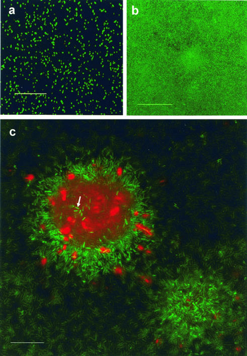
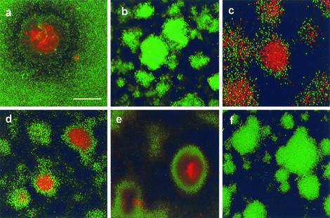
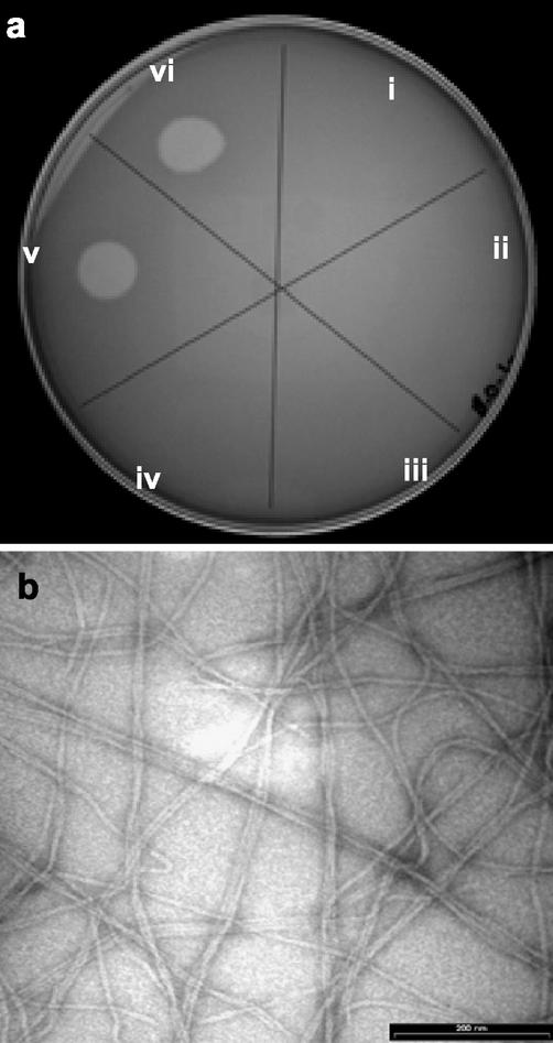
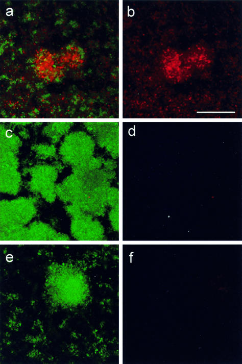
Bacteria growing in biofilms often develop multicellular, three-dimensional structures known as microcolonies. Complex differentiation within biofilms of Pseudomonas aeruginosa occurs, leading to the creation of voids inside microcolonies and to the dispersal of cells from within these voids. However, key developmental processes regulating these events are poorly understood. A normal component of multicellular development is cell death. Here we report that a repeatable pattern of cell death and lysis occurs in biofilms of P. aeruginosa during the normal course of development. Cell death occurred with temporal and spatial organization within biofilms, inside microcolonies, when the biofilms were allowed to develop in continuous-culture flow cells. A subpopulation of viable cells was always observed in these regions. During the onset of biofilm killing and during biofilm development thereafter, a bacteriophage capable of superinfecting and lysing the P. aeruginosa parent strain was detected in the fluid effluent from the biofilm. The bacteriophage implicated in biofilm killing was closely related to the filamentous phage Pf1 and existed as a prophage within the genome of P. aeruginosa. We propose that prophage-mediated cell death is an important mechanism of differentiation inside microcolonies that facilitates dispersal of a subpopulation of surviving cells.


Figures

FIG. 1.
FIG. 1.
Cell death occurs in localized regions in microcolonies of mature P. aeruginosa biofilms: confocal micrographs of P. aeruginosa biofilm development in glass flow cells visualized by using the BacLight LIVE/DEAD viability stain. Green fluorescent cells are viable, whereas red fluorescent cells have a compromised cell membrane and are dead. Images were obtained 3 h (a), 3 days (b), and 7 days (c) after the flow cells were inoculated. (a and b) Images taken at the biofilm-substratum interface. Scale bars = 50 μm. (c) x-y plane view through the center of a microcolony that was 50 μm high. A subpopulation of cells inside the microcolony was not killed (arrow). Scale bar = 25 μm. Similar results were obtained in five other experiments.
FIG. 2.
FIG. 2.
Confocal photomicrographs showing viability within microcolonies in 10-day-old biofilms containing the P. aeruginosa wild type (a), rpoN mutant (b), rpoN mutant with rpoN complemented in trans (c), lasI mutant (d), rhlI mutant (S. Beatson) (e), and an fliM pilA double mutant (f). Scale bar = 50 μm. The results are representative of the results of three experiments.
FIG. 3.
FIG. 3.
Analysis of bacteriophage activity in the fluid effluents from wild-type P. aeruginosa biofilms. (a) Droplets of biofilm effluent fluids were placed on a lawn of target cells of the P. aeruginosa strain. Fluids from 1-day (sector i), 2-day (sector ii), 3-day (sector iii), 5-day (sector iv), 7-day (sector v), and 9-day (sector vi) biofilms were used. Lysis of P. aeruginosa cells occurred after 1 week, which correlated with the onset of biofilm killing inside microcolonies. (b) Transmission electron microscopy examination of the purified phage revealed filamentous phage particles. Bar = 200 nm.
FIG. 4.
FIG. 4.
Add-back of purified phage to young (3-day) biofilms of P. aeruginosa strains before the onset of normal cell death. (a and b) Wild-type strain; (c and d) rpoN mutant; (e and f) pilA fliM double mutant. Biofilms were visualized by using the BacLight LIVE/DEAD stain after 2 days of growth in the presence of phage. (a, c, and e) Combined live (green) and dead (red) channels; (b, d, and f) corresponding dead channel only. Bar = 50 μm.
FIG. 5.
FIG. 5.
Microcolonies in mature P. aeruginosa biofilms can accumulate ROS: phase-contrast images of DHR-stained biofilms (a and c) and the corresponding rhodamine 123 fluorescence displays (b and d). Young (3-day) biofilms (a and b) and mature (10-day) biofilms (c and d) were used. Bar = 50 μm.

References

    1. Andersen, J. B., C. Sternberg, L. K. Poulsen, S. P. Bjorn, M. Givskov, and S. Molin. 1998. New unstable variants of green fluorescent protein for studies of transient gene expression in bacteria. Appl. Environ. Microbiol. 64:2240-2246. - PMC - PubMed
    1. Auschill, T. M., N. B. Arweiler, L. Netuschil, M. Brecx, E. Reich, A. Sculean, and N. B. Artweiler. 2001. Spatial distribution of vital and dead microorganisms in dental biofilms. Arch. Oral Biol. 46:471-476. - PubMed
    1. Ausubel, F. M., R. Brent, R. E. Kingston, D. D. Moore, J. G. Seidman, J. A. Smith, and K. Struhl. 1992. Short protocols in molecular biology. Greene Publishing and John Wiley & Sons, New York, N.Y.
    1. Beatson, S. A., C. B. Whitchurch, A. B. Semmler, and J. S. Mattick. 2002. Quorum sensing is not required for twitching motility in Pseudomonas aeruginosa. J. Bacteriol. 184:3598-3604. - PMC - PubMed
    1. Berk, R. S. 1965. Effect of antibacterial agents on the autoplaque phenomenon of Pseudomonas aeruginosa. Can. J. Microbiol. 11:213-219. - PubMed

Publication types

MeSH terms