. 2003 Jul 16;23(15):6163-70.
doi: 10.1523/JNEUROSCI.23-15-06163.2003.

CNS glucagon-like peptide-1 receptors mediate endocrine and anxiety responses to interoceptive and psychogenic stressors

Affiliations

CNS glucagon-like peptide-1 receptors mediate endocrine and anxiety responses to interoceptive and psychogenic stressors

Kimberly P Kinzig et al. J Neurosci. .

Erratum in

  • J Neurosci. 2003 Sep 3;23(22):following 8158. Figueredo, HF [corrected to Figueiredo, HF]

Abstract

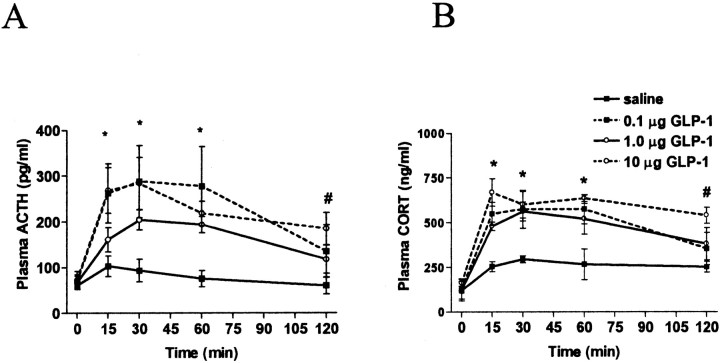
Responses to stressors serve to adjust physiology and behavior to increase short-term survival at the potential expense of increasing susceptibility to disease over the long term. We show that glucagon-like peptide-1 (7-36) amide (GLP-1) increases levels of the stress-activated hormones ACTH and corticosterone when administered directly into the rat brain and increases levels of anxiety as measured by the elevated plus maze. The endocrine response is preferentially activated by GLP-1 administration in the paraventricular nucleus of the hypothalamus, whereas the anxiety response is preferentially activated by administration in the central nucleus of the amygdala. Furthermore, GLP-1 antagonists block increases in stress hormones associated with the toxin LiCl and both the endocrine and anxiety responses to vertical heights. Although diverse neural circuits must necessarily process disparate stressors, the current data implicate a role for the GLP-1 system as a critical mediator of multiple stress responses.


Figures

Figure 1.
Figure 1.
Central nucleus of the amygdala injection sites. After the completion of the experiments involving CeA-cannulated rats, the animals were killed and given injections of cresyl violet via the CeA cannula. Brains were subsequently sectioned (50 μm/section), and cannula placement was verified. Black dots within the CeA of each bottom diagram represent injection sites for the rats with correctly placed cannulas. Rats whose cannulas were not in the CeA were excluded from the data analyses (n = 6 of 25 removed).
Figure 2.
Figure 2.
Paraventricular nucleus of the hypothalamus injection sites. After the completion of the experiments involving PVN-cannulated rats, the animals were killed and given injections of cresyl violet via the PVN cannula. Brains were subsequently sectioned (50 μm/section), and cannula placement was verified. Black dots within the PVN of each bottom diagram represent injection sites for the rats with correctly placed cannulas. Rats whose cannulas were not in the PVN were excluded from the data analyses (n = 5 of 22 removed).
Figure 3.
Figure 3.
A, Plasma ACTH levels after i3vt administration of GLP-1 (0.1, 1.0, or 10 μg) or saline. Plasma ACTH was significantly elevated in response to all three doses of GLP-1 at 15, 30, and 60 min. At 120 min after injection, only 10 μg of GLP-1 resulted in significantly elevated ACTH. B, Plasma CORT after i3vt administration of GLP-1 (0.1, 1.0, or 10 μg). All three doses resulted in significant elevations of CORT at 15, 30, and 60 min after injection. When animals received the highest dose, plasma CORT remained significantly elevated at 120 min. *All three treatment groups significantly different from saline; p < 0.05. #Highest dose (10 μg) significantly different from saline; p < 0.05.
Figure 4.
Figure 4.
A, Plasma ACTH after intraperitoneal administration of GLP-1. Fifteen minutes after injection (10, 100, and 500 μg/kg or saline), the highest dose of GLP-1 resulted in a significant increase in plasma ACTH (p < 0.05). There were no other differences between GLP-1 and saline treatment. B, Plasma CORT after intraperitoneal administration of GLP-1. Intraperitoneal administration of GLP-1 (10, 100, and 500 μg/kg) did not elevate plasma CORT. *Highest dose (500 μg/kg) significantly different from saline; p < 0.05.
Figure 5.
Figure 5.
A, Plasma ACTH after CRH receptor antagonism and i3vt GLP-1. CRH receptor antagonism attenuated GLP-1-induced elevations in plasma ACTH. Plasma ACTH was significantly lower in rats treated with the CRH receptor antagonist astressin before 1 μg of GLP-1 administered i3vt. B, Plasma CORT after CRH receptor antagonism and i3vt GLP-1. CRH receptor antagonism attenuated GLP-1-induced elevations in plasma CORT. Plasma CORT was significantly lower in rats treated with the CRH receptor antagonist astressin before 1 μg of GLP-1 administered i3vt (p < 0.01 at 15, 30, and 60 min).
Figure 6.
Figure 6.
A, Plasma ACTH after GLP-1 receptor antagonism and intraperitoneal LiCl. Plasma ACTH levels were significantly elevated after intraperitoneal administration of LiCl. This effect was attenuated by pretreatment with the GLP-1 receptor antagonist des-His1,Glu9-exendin-4 (exendin). At 15, 30, 60, and 120 min, saline plus LiCl resulted in significantly elevated ACTH compared with saline plus saline (p < 0.001; p < 0.001; p < 0.01; p < 0.01, respectively), compared with exendin plus saline (p < 0.001 at 15 min; p < 0.01 at 30 and 60 min; not significantly different at 120 min), and compared with exendin plus LiCl (p < 0.001 at 15 min; p < 0.01 at 30 min; p < 0.05 at 60 min; no difference at 120 min). B, Plasma CORT after GLP-1 receptor antagonism and intraperitoneal LiCl. At 15, 30, and 60 min, saline plus LiCl resulted in significantly elevated CORT compared with saline plus saline (p < 0.001; p < 0.001; p < 0.01, respectively), compared with exendin plus saline (p < 0.001 at 15 and 30 min; p < 0.01 at 60 min), and compared with exendin plus LiCl (p < 0.001 at 15 and 30 min; p < 0.05 at 60 min). There were no significant differences in CORT levels at 120 min.
Figure 7.
Figure 7.
A, Plasma ACTH after GLP-1 receptor antagonism before placement on an isolated open arm of the elevated plus maze. At 15, 30, and 60 min after EPM stress, rats that were pretreated with exendin had significantly lower plasma ACTH levels than did saline-treated rats. B, Plasma CORT after GLP-1 receptor antagonism before placement on an isolated open arm of the elevated plus maze. At 15, 30, and 60 min after EPM stress, rats that were pretreated with exendin had significantly lower plasma CORT levels than did saline-treated rats. *Significantly different from saline; p < 0.01.
Figure 8.
Figure 8.
A, Plasma CORT levels in response to administration of GLP-1 directly into the CeA. There was no significant effect of either dose (0.2 or 1.0 μg) on plasma CORT at 60 min after infusion compared with saline. B, Plasma CORT levels in response to administration of GLP-1 directly into the PVN. Plasma CORT (0.2 or 1.0 μg) was significantly elevated at 60 min after infusion of either 0.2 or 1.0 μg of GLP-1 compared with saline; *p < 0.05.

References

    1. Bittencourt JC, Sawchenko PE 2000. Do centrally administered neuropeptides access cognate receptors? An analysis in the central corticotropin-releasing factor system. J Neurosci 20: 1142–1156. - PMC - PubMed
    1. Charney DS, Deutch AY, Krystal JH, Southwick SM, Davis M ( 1993) Psychobiologic mechanisms of posttraumatic stress disorder. Arch Gen Psychiatry 50: 295–305. - PubMed
    1. Drucker DJ ( 1990) Glucagon and the glucagon-like peptides. Pancreas 5: 484–488. - PubMed
    1. Goke R, Larsen PJ, Mikkelsen JD, Skeikh SP ( 1995) Distribution of GLP-1 binding sites in the rat brain: evidence that exendin is a ligand of brain GLP-1 sites. Eur J Neurosci 7: 2294–2300. - PubMed
    1. Grillon C, Southwick SM, Charney DS ( 1996) The psychobiological basis of posttraumatic stress disorder: association between an agouti-related protein gene polymorphism and anorexia nervosa. Mol Psychiatry 1: 278–297. - PubMed

Publication types

MeSH terms