. 2003 Jul 16;23(15):6181-7.
doi: 10.1523/JNEUROSCI.23-15-06181.2003.

Critical role for microglial NADPH oxidase in rotenone-induced degeneration of dopaminergic neurons

Affiliations

Critical role for microglial NADPH oxidase in rotenone-induced degeneration of dopaminergic neurons

Hui-Ming Gao et al. J Neurosci. .

Abstract

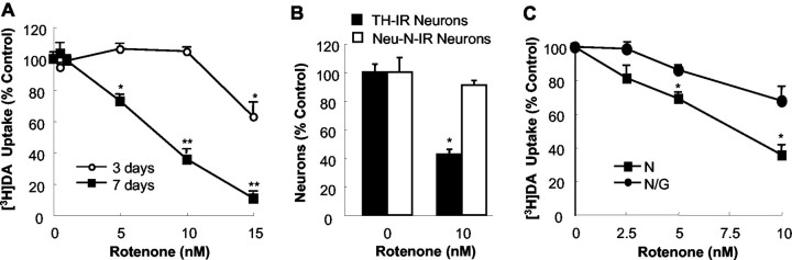
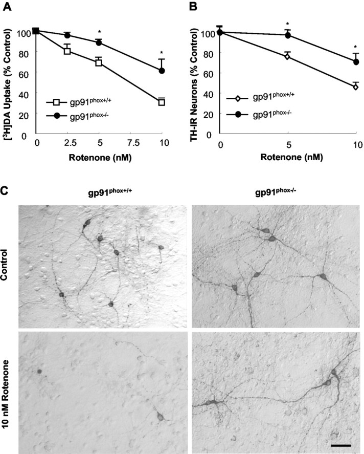
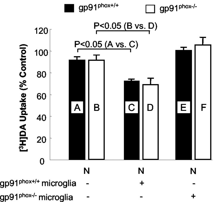
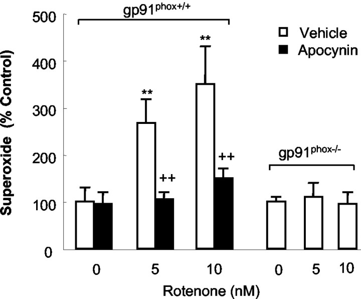
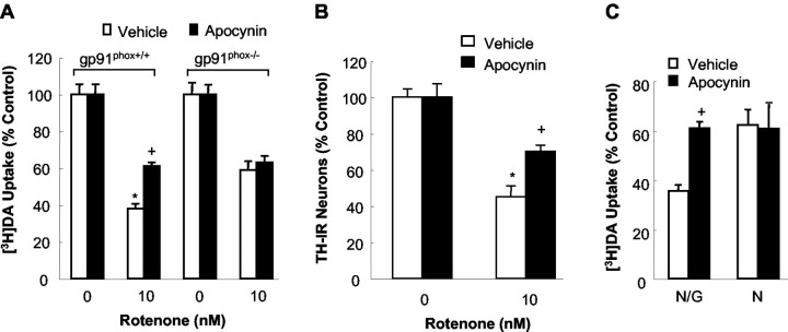
Increasing evidence has suggested an important role for environmental toxins such as pesticides in the pathogenesis of Parkinson's disease (PD). Chronic exposure to rotenone, a common herbicide, reproduces features of Parkinsonism in rats. Mechanistically, rotenone-induced dopaminergic neurodegeneration has been associated with both its inhibition of neuronal mitochondrial complex I and the enhancement of activated microglia. Our previous studies with NADPH oxidase inhibitors, diphenylene iodonium and apocynin, suggested that NADPH oxidase-derived superoxide might be a major factor in mediating the microglia-enhanced rotenone neurotoxicity. However, because of the relatively low specificity of these inhibitors, the exact source of superoxide induced by rotenone remains to be further determined. In this study, using primary mesencephalic cultures from NADPH oxidase--null (gp91phox-/-) or wild-type (gp91phox+/+) mice, we demonstrated a critical role for microglial NADPH oxidase in mediating microglia-enhanced rotenone neurotoxicity. In neuron--glia cultures, dopaminergic neurons from gp91phox-/- mice were more resistant to rotenone neurotoxicity than those from gp91phox+/+ mice. However, in neuron-enriched cultures, the neurotoxicity of rotenone was not different between the two types of mice. More importantly, the addition of microglia prepared from gp91phox+/+ mice but not from gp91phox-/- mice to neuron-enriched cultures markedly increased rotenone-induced degeneration of dopaminergic neurons. Furthermore, apocynin attenuated rotenone neurotoxicity only in the presence of microglia from gp91phox+/+ mice. These results indicated that the greatly enhanced neurotoxicity of rotenone was attributed to the release of NADPH oxidase-derived superoxide from activated microglia. This study also suggested that microglial NADPH oxidase may be a promising target for PD treatment.


Figures

Figure 1.
Figure 1.
Glia enhanced dopaminergic neurodegeneration induced by rotenone. A, Primary neuron—glia cultures were treated for 3 or 7 d with vehicle or 0.5—15 nm rotenone, and the capability of cultures to uptake DA was evaluated. B, Neuron—glia cultures were treated for 7 d with vehicle or 10 nm rotenone, and the selectivity of rotenone-induced neurodegeneration was determined by quantification of TH-IR and NeuN-IR neurons after immunocytochemical staining. C, Neuron—glia (N/G; •) or neuron-enriched (N; ▪) cultures were treated for 7 d with vehicle or 2.5—10 nm rotenone. [3H]DA uptake was measured to assess neurotoxicity to dopaminergic neurons. The results for DA uptake (A, C) and cell counts (B) are expressed as a percentage of the control cultures and are means ± SEM of three experiments performed in triplicate. *p < 0.05, **p < 0.005 compared with the corresponding control (A, B). *p < 0.05 compared with the corresponding rotenone-treated neuron-enriched cultures (C).
Figure 2.
Figure 2.
Resistance of dopaminergic neurons from gp91 phox-/- mice to rotenone neurotoxicity. Neuron—glia cultures were treated for 7 d with vehicle or desired concentrations of rotenone. Afterward, cultures were assayed for uptake of [3H]DA (A), quantification of the TH-IR neurons (B), or immunocytochemical analysis of TH-IR neurons (C). Results are expressed as a percentage of the control cultures and are means ± SEM of three experiments performed in triplicate. *p < 0.05 compared with the corresponding rotenone-treated cultures from gp91 phox+/+ mice. Scale bar, 50 μm.
Figure 3.
Figure 3.
Effect of the addition of microglia to neuron-enriched (N) cultures on rotenone-induced degeneration of dopaminergic neurons. Mesencephalic neuron-enriched cultures were supplemented with 5 × 10⁴ microglia per well prepared from either gp91 phox+/+ or gp91 phox-/- mice. After 24 hr, the cultures were treated with vehicle or 5 nm rotenone, and [3H]DA uptake was determined 7 d after the treatment. Results are expressed as a percentage of the control cultures and are means ± SEM of three experiments performed in triplicate. p < 0.05 was considered statistically significant. All other comparisons were not significant.
Figure 4.
Figure 4.
Rotenone stimulated release of superoxide in microglia from gp91 phox+/+ mice but not gp 91 phox-/- mice. Mouse primary microglia were seeded to 96 well plates. After 24 hr, microglia were pretreated with vehicle or 0.5 mm apocynin for 30 min. Afterward, microglia were stimulated with rotenone (5 or 10 nm) or corresponding vehicle. Superoxide production, measured as SOD-inhibitable cytochrome c reduction, was determined as described in Materials and Methods. There was no significant difference in the basal levels of superoxide production (control) between the cultures from gp91 phox-/- mice and those of their wild-type litter-mates. Results are a percentage of the control cultures and are expressed as means ± SEM of three experiments performed in triplicate. **p < 0.005 compared with the control. ++p < 0.005 compared with corresponding rotenone-treated cultures.
Figure 5.
Figure 5.
Effect of apocynin on rotenone-induced degeneration of dopaminergic neurons. A, B, Neuron—glia cultures were pretreated for 30 min with vehicle or 0.5 mm apocynin before treatment with 10 nm rotenone. Seven days later, degeneration of dopaminergic neurons was determined by [3H]DA uptake (A) and counts of TH-IR neurons (B). C, Neuron—glia (N/G) or neuron-enriched (N) cultures were pretreated for 30 min with the vehicle or 0.5 mm apocynin before treatment with 10 nm rotenone. [3H]DA uptake was measured 7 d later. Results are means ± SEM of three to four experiments performed in triplicate. *p < 0.05 compared with the corresponding control. +p < 0.05 compared with rotenone-treated cultures.

References

    1. Alam M, Schmidt WJ ( 2002) Rotenone destroys dopaminergic neurons and induces parkinsonian symptoms in rats. Behav Brain Res 136: 317—324. - PubMed
    1. Aloisi F ( 1999) The role of microglia and astrocytes in CNS immune surveillance and immunopathology. Adv Exp Med Biol 468: 123—133. - PubMed
    1. Babior BM ( 1999) NADPH oxidase: an update. Blood 93: 1464 —1476. - PubMed
    1. Betarbet R, Sherer TB, MacKenzie G, Garcia-Osuna M, Panov AV, Greenamyre JT ( 2000) Chronic systemic pesticide exposure reproduces features of Parkinson's disease. Nat Neurosci 3: 1301—1306. - PubMed
    1. Cassarino DS, Fall CP, Swerdlow RH, Smith TS, Halvorsen EM, Miller SW, Parks JP, Parker Jr WD, Bennett Jr JP ( 1997) Elevated reactive oxygen species and antioxidant enzyme activities in animal and cellular models of Parkinson's disease. Biochim Biophys Acta 1362: 77—86. - PubMed

MeSH terms

LinkOut - more resources