. 2003 Jul 16;23(15):6232-7.
doi: 10.1523/JNEUROSCI.23-15-06232.2003.

Tenascin-R as a repellent guidance molecule for developing optic axons in zebrafish

Affiliations

Tenascin-R as a repellent guidance molecule for developing optic axons in zebrafish

Catherina G Becker et al. J Neurosci. .

Abstract

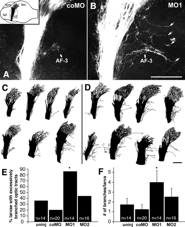
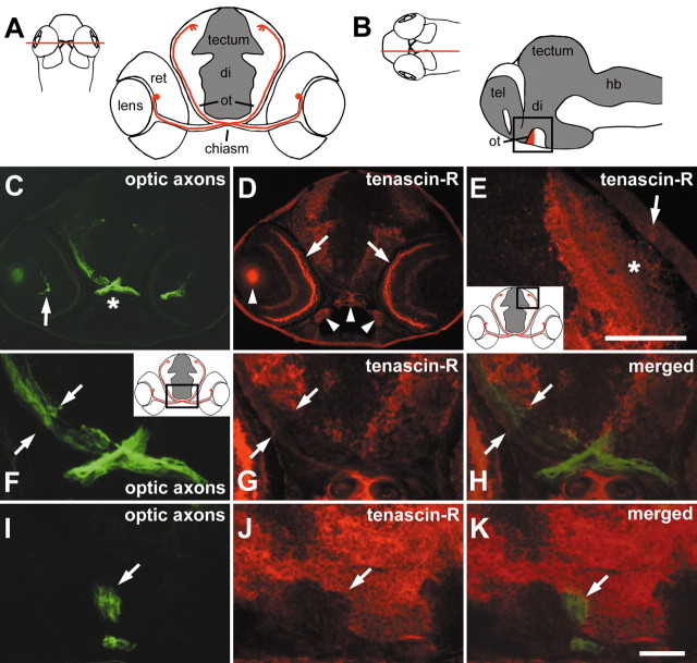
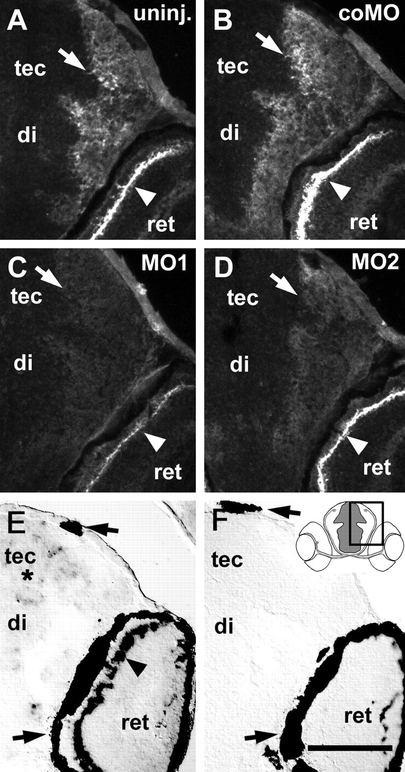
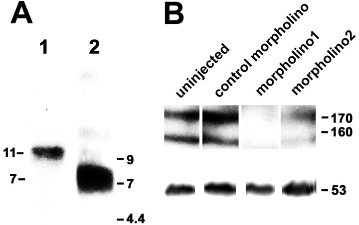
To investigate the role of tenascin-R in nervous system development, we studied axon pathfinding in the developing optic system of zebrafish. Zebrafish tenascin-R has the same domain structure as tenascin-R in amniotes. Amino acid sequence identity with human tenascin-R is 60%. In 3-d-old larvae, tenascin-R mRNA is expressed in scattered cells throughout the periventricular cell layer of the diencephalon and tectum. Tenascin-R immunoreactivity is not detectable in the optic nerve, optic tract, or tectal optic neuropil but immediately borders the optic tract caudally. Reducing expression of tenascin-R in 3-d-old larvae in vivo by injecting morpholinos into fertilized eggs led to excessive branching of the optic tract in 86% of all injected larvae compared with 20-37% in controls. Branches were almost exclusively caudal, where tenascin-R immunoreactivity normally borders the optic tract, suggesting a role for tenascin-R in guiding optic axons in the ventral diencephalon by a contact-repellent mechanism.


Figures

Figure 4.
Figure 4.
Morpholino treatment leads to excessive caudal branching of the optic tract in 3-d-old larvae. A, B, Lateral views of confocal image stacks of the optic tract in whole-mounted brains, after labeling of the temporal retina with DiI, correspond to the boxed area in the inset in A. The inset gives the orientation of the brain with the labeled optic projection indicated in black (tel, telencephalon; ot, optic tract; tec, tectum; eye, contralateral eye; the ipsilateral eye was removed before mounting); dorsal is up, and rostral is left. In a control morpholino-treated larva (A, coMO), axons are found only in arborization field 3 (AF-3) caudal to the optic tract, whereas in a morpholino1-treated larva (B, MO1), multiple excessive branches are seen (arrows). C, D, Examples of entire DiI-labeled optic projections from one set of experiments are shown for control morpholino- (C) and morpholino1-treated larvae (D). E, F, The percentages of larvae with excessively branched optic tracts (E); the mean numbers of excessive axon branches per larva are given (F) (uninj, uninjected; coMO, control morpholino; MO1, morpholino1, MO2, morpholino2). Significant differences from controls are indicated by asterisks (E, Fisher's exact test, p ≤ 0.001; F, Mann–Whitney U test, p ≤ 0.019). Scale bars: (in B) A, B, 210 μm; (in D) C, D, 210 μm.
Figure 2.
Figure 2.
Tenascin-R immunoreactivity borders the optic projection in 3-d-old larvae. A, B, Schematic presentations of the optic projection are shown in a cross-section in A. The optic projection is represented by one retinal ganglion cell in red with its soma located in the retina (ret) and its axon entering the diencephalon (di) through the chiasm, where it crosses the midline and reaches the superficial optic neuropil of the optic tectum (tectum) through the optic tract (ot). InB, the ventral optic tract (red) is indicated in a sagittal section (tel, telencephalon; hb, hindbrain). Cell-dense areas are depicted in gray. Red lines in dorsal views of whole heads indicate approximate levels of sectioning. C, D, A cross-section through a head at the level of the optic chiasm as indicated in A, double immunolabeled for optic axons (C, green) and tenascin-R (D, red), is shown. Optic axons are labeled from the optic fiber layer of the retina (arrow in C) through the chiasm (asterisk in C) into the ventral diencephalon. Tenascin-R immunoreactivity is present in the retinal outer plexiform layer (arrows in D) and the brain. Arrowheads in D indicate nonspecific fluorescence of the lens and cartilage. E, In a cross-section through the tectum, corresponding to the boxed area in the inset, tenascin-R immunoreactivity is found primarily in deep fiber layers. The asterisk indicates the superficial optic neuropil that is low in tenascin-R immunoreactivity. The arrow indicates the epidermis overlaying the tectum. F–H, A higher magnification of the section shown in C and D corresponds to the boxed area in the inset ofF. The optic tract is indicated by opposing arrows, showing that tenascin-R (G) directly borders optic axons (F) medially. In H, signals are merged. I–K, In a double-labeled sagittal section through the brain (oriented as in the boxed area in B), the ventral optic tract is indicated by an arrow. Tenascin-R immunoreactivity (J) directly borders optic axons (I) caudally but not rostrally. In K, signals are merged. Arrows in I–K point to the ventral optic tract. Scale bars: E,50 μm; (in K) C, D, 100 μm and F–K, 33 μm.
Figure 3.
Figure 3.
Effect of morpholinos on tenascin-R immunoreactivity and tenascin-R mRNA expression in 3-d-old larvae. Cross-sections including the diencephalon (di), tectum (tec), and part of the retina (ret) correspond to the boxed area in the inset in F. A–D, Arrows depict deep tectal fiber layers, and arrowheads indicate the outer plexiform layer of the retina. In uninjected (A, uninj.) and control morpholino-injected (B, coMO) larvae, tenascin-R immunoreactivity is most intense in the outer plexiform layer of the retina and high in deep fiber layers of the tectum and diencephalon. Injection of morpholino1 (C, MO1) more strongly reduces immunoreactivity than morpholino2 (D, MO2). E, F, Tenascin-R mRNA is detected by in situ hybridization in scattered cells throughout the periventricular cell layer of the brain (asterisk) and in a row of cells in the retina (arrowhead in E) but not after incubation with a sense RNA probe (F). Arrows in E and F indicate the retinal pigment epithelium and pigment cells in the epidermis, which always appear black. Scale bar (in F): A–F, 100 μm.
Figure 1.
Figure 1.
Tenascin-R mRNA and protein are expressed in zebrafish. A, Northern blot analysis of total RNA isolated from adult zebrafish brains yielded a band at 11 kb (lane 1). A probe to tenascin-C labeled a band at 7 kb (lane 2). Lengths of transcripts are indicated on the left; positions of molecular markers are indicated on the right. B, Western blot analysis using anti-EGF-L antibody on detergent lysates of heads of 3-d-old larvae, separated on the same gel, indicate reduction of immunoreactivity by morpholino1 and morpholino2 but not by control morpholino. Reprobing of the blot with an antibody to tubulin is depicted in the bottom row. Molecular weights of tenascin-R and tubulin are indicated on the right.

Similar articles

Cited by

References

    1. Bartsch U, Pesheva P, Raff M, Schachner M ( 1993) Expression of janusin (J1–160/180) in the retina and optic nerve of the developing and adult mouse. Glia 9: 57–69. - PubMed
    1. Becker T, Anliker B, Becker CG, Taylor J, Schachner M, Meyer RL, Bartsch U ( 2000) Tenascin-R inhibits regrowth of optic fibers in vitro and persists in the optic nerve of mice after injury. Glia 29: 330–346. - PubMed
    1. Burrill JD, Easter SS ( 1994) Development of the retinofugal projections in the embryonic and larval zebrafish (Brachydanio rerio). J Comp Neurol 346: 583–600. - PubMed
    1. Campbell DS, Regan AG, Lopez JS, Tannahill D, Harris WA, Holt CE ( 2001) Semaphorin 3a elicits stage-dependent collapse, turning, and branching in Xenopus retinal growth cones. J Neurosci 21: 8538–8547. - PMC - PubMed
    1. Dickson BJ ( 2002) Molecular mechanisms of axon guidance. Science 298: 1959–1964. - PubMed

Publication types

MeSH terms

Associated data