. 2003 Aug 5;100(16):9464-9.
doi: 10.1073/pnas.1630663100. Epub 2003 Jul 16.

Alpha-galactosylceramide (KRN7000) suppression of chemical- and oncogene-dependent carcinogenesis

Affiliations

Alpha-galactosylceramide (KRN7000) suppression of chemical- and oncogene-dependent carcinogenesis

Yoshihiro Hayakawa et al. Proc Natl Acad Sci U S A. .

Abstract

Recent studies have revealed significant efficacy of the marine sponge glycolipid, alpha-galactosylceramide (alpha-GalCer), in treatment of experimental metastatic cancers, infections, and autoimmune diseases. However, the capacity of alpha-GalCer to prevent tumor development had never, to our knowledge, been evaluated in mouse models of chemical- and oncogene-dependent carcinogenesis. In this study, we demonstrate that long-term administration of soluble alpha-GalCer, spanning the time of tumor initiation, inhibits primary tumor formation in three different models: methylcholanthrene-induced sarcomas, mammary carcinomas in Her-2/neu transgenic mice, and spontaneous sarcomas in p53-/- mice. Weekly treatment of mice with alpha-GalCer maintained lymphoid tissue natural killer cell and T cell activation and elevated serum IFN-gamma and IL-4 concentrations. Consistent with the antimetastatic activity of alpha-GalCer, prevention of methylcholanthrene-induced sarcoma was IFN-gammaand tumor necrosis factor-related apoptosis-inducing ligand dependent, but not perforin-dependent. Taken together, our results demonstrate that NK1.1+alphabetaTCR+ cell-based immune therapy can inhibit primary tumorigenesis.


Figures
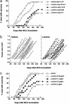
Fig. 1.
Fig. 1.
α-GalCer-mediated control of MCA-induced fibrosarcoma. (a) Groups of 20 B6 WT mice were injected s.c. in the hind flank with 400 μg of MCA diluted in 0.1 ml of corn oil. Mice received 2 μg of α-GalCer i.p. as follows: (i) weekly from day –28 to day 0 (where day 0 = day of MCA inoculation); (ii) weekly from day 0 to day 28; (iii) weekly from day 0 to day 70; (iv) weekly from day 28 to day 98. Mice were observed weekly for tumor development over the course of 60–200 days. Tumors >4 mm in diameter and demonstrating progressive growth over 2 weeks were counted as positive. (b) Representative individual fibrosarcomas from the group of 20 B6 WT mice receiving 400 μgof MCA and vehicle (Left) were compared for growth with a similar group of B6 WT mice inoculated with 400 μg of MCA and receiving α-GalCer treatment (2μg of α-GalCer weekly; Right), commencing in each mouse when sarcoma was palpated for the second successive week. Tumor size was measured weekly with a caliper square as the product of two diameters, and results were recorded as the tumor size (cm2). The lines of best fit of each group demonstrate growth rate, are represented by the solid lines, and are not significantly different (P = 0.1649, Mann–Whitney U test). (c) Groups of 15 B6 WT mice were injected s.c. in the hind flank with 400 μg of MCA diluted in 0.1 ml of corn oil and received 0.02–2.0 μg of α-GalCer i.p. (as indicated) weekly from day 0 to day 70.
Fig. 2.
Fig. 2.
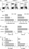
α-GalCer-mediated activation, cytokine production, and expansion of lymphocyte subsets. Groups of three B6 WT mice received either vehicle or 2 μg of α-GalCer i.p. on day 0 (α-GalCer ×1) or weekly from day 0 to day 28 (α-GalCer ×5). (a) Liver MNC were collected 7 days after each α-GalCer or vehicle injection, and the surface phenotype of the MNC was characterized by flow cytometry as indicated, NK1.1 vs. CD3 vs. CD69. (b) The percentage of CD8+ T cells and NK cells in the liver MNC were plotted. Cell numbers (×105) were as follows: CD8+ T cells; vehicle, 2.5 ± 0.3; α-GalCer × 1, 18.7 ± 1.5; α-GalCer × 5, 13.5 ± 1.1; CD3 NK1.1+ cells; vehicle, 1.2 ± 0.2; α-GalCer × 1, 16.1 ± 1.2; α-GalCer × 5, 4.8 ± 0.7; CD3+ NK1.1+ cells; vehicle, 4.1 ± 0.7; α-GalCer × 1, 15.4 ± 1.2; and α-GalCer × 5, 4.8 ± 0.7. (c) Sera were collected before (0 h), or4hor24h after a single (×1) or multiple (×5) i.p. injections of 2 μgof α-GalCer or vehicle. IFN-γ and IL-4 levels in the serum were evaluated by using ELISA, and statistical differences were determined by comparing levels with the 0-h time point for each treatment using a Mann–Whitney U test (*, P < 0.05). Levels of IFN-γ and IL-4 were below detection (<50 pg/ml) in vehicle-treated mice.
Fig. 3.
Fig. 3.
α-GalCer-mediated control of MCA-induced fibrosarcoma by innate immunity and IFN-γ activity. Groups of 15–20 B6 WT, B6 IFN-γ/, B6 TRAIL/,B6 pfp/,orB6pfp/ IFN-γ/ mice were injected s.c. in the hind flank with 400 μg of MCA diluted in 0.1 ml of corn oil. Mice received either vehicle or 2 μg of α-GalCer i.p. weekly from day 0 to day 70. All mice were observed weekly for tumor development over the course of 60–200 days. Tumors >4 mm in diameter and demonstrating progressive growth over 2 weeks were counted as positive.
Fig. 4.
Fig. 4.
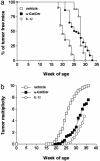
Appearance of the first tumor (a) and tumor multiplicity (b) in CBf1-neuT mice treated with α-GalCer and IL-12. Mice received 2 μg of α-GalCer (n = 13) or vehicle only (n = 20) i.p. at weeks 5, 9, 13, 17, 21, 25, and 29. Alternatively, a group of mice (n = 8) received 100 ng in 0.2 ml of IL-12/day for 5 days at weeks 5, 9, 13, 17, 21, 25, and 29. Progressively growing masses (>1-mm mean diameter) were regarded as tumors. Differences in tumor incidence at 25 weeks were statistically different between vehicle-treated and IL-12 or α-GalCer-treated mice, as evaluated by a Fisher's exact test (P < 0.0004). Tumor multiplicity was statistically different between vehicle-treated and IL-12- or α-GalCer-treated mice at week 25 and beyond, as demonstrated by using a Mann–Whitney U test (P < 0.004).
Fig. 5.
Fig. 5.
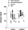
α-GalCer protects mice from spontaneous sarcoma. The appearance of tumors was recorded in groups of 34–35 B6 p53/ mice receiving either vehicle or 2 μgof α-GalCer i.p. weekly from 4 weeks of age. Mice were evaluated on a weekly basis. When mice were moribund, tumor type (open squares, thymic lymphoma; filled triangles, disseminated lymphoma; open circles, sarcoma; filled diamonds, other tumors) was recorded against the age (in days) at the time of death/autopsy. The mean age of p53/ mice developing spontaneous sarcoma is depicted by the cross bars, and the statistically later incidences of sarcomas in the α-GalCer-treated group were noted (192.6 ± 6.1 days), compared with vehicle-treated mice (147.1 ± 8.3 days; P = 0.0005, Mann–Whitney U test).

Similar articles

Cited by

References

    1. Morita, M., Motoki, K., Akimoto, K., Natori, T., Sakai, T., Sawa, E., Yamaji, K., Koezuka, Y., Kobayashi, E. & Fukushima, H. (1995) J. Med. Chem. 38, 2176–2187. - PubMed
    1. Nakagawa, R., Motoki, K., Ueno, H., Iijima, R., Nakamura, H., Kobayashi, E., Shimosaka, A. & Koezuka, Y. (1998) Cancer Res. 58, 1202–1207. - PubMed
    1. Nakagawa, R., Serizawa, I., Motoki, K., Sato, M., Ueno, H., Iijima, R., Nakamura, H., Shimosaka, A. & Koezuka, Y. (2000) Oncol. Res. 12, 51–58. - PubMed
    1. Fuji, N., Ueda, Y., Fujiwara, H., Toh, T., Yoshimura, T. & Yamagishi, H. (2000) Clin. Cancer Res. 6, 3380–3387. - PubMed
    1. Kobayashi, E., Motoki, K., Uchida, T., Fukushima, H. & Koezuka, Y. (1995) Oncol. Res. 7, 529–534. - PubMed

Publication types

MeSH terms