. 2003 Aug;133(2):200-7.
doi: 10.1046/j.1365-2249.2003.02208.x.

Immune complex-FcgammaR interaction modulates monocyte/macrophage molecules involved in inflammation and immune response

Affiliations

Immune complex-FcgammaR interaction modulates monocyte/macrophage molecules involved in inflammation and immune response

P Barrionuevo et al. Clin Exp Immunol. 2003 Aug.

Abstract

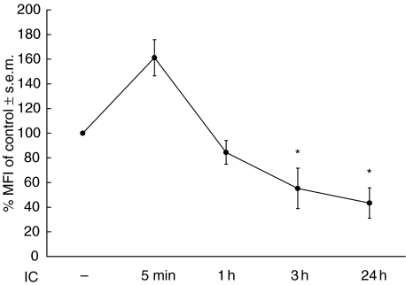
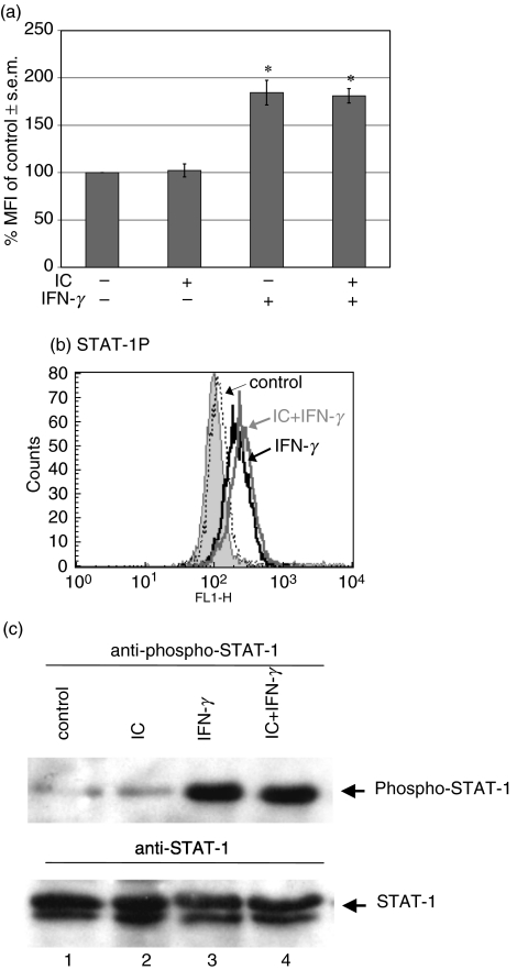
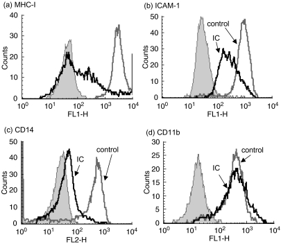
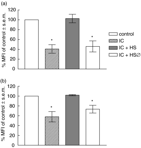
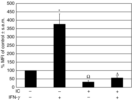
The interaction between receptors for the Fc portion of IgG (FcgammaRs) from monocytes/macrophages and immune complexes (IC) triggers regulatory and effector functions. Recently, we have demonstrated that IC exert a drastic inhibition of basal and IFN-gamma-induced expression of MHC class II on human monocytes. Taking into account that the regulation of MHC class II molecules is a crucial event in the immune response, in this report we extend our previous studies analysing the effect of STAT-1 phosphorylation in the down-regulatory process, the fate of the intracellular pool of MHC class II molecules and the effect of complement on MHC class II down-regulation induced by IC. We also studied the effect of IC on the expression of MHC class II (I-A(d)) in macrophages using a mouse model of chronic inflammation. We demonstrate that IC induce a depletion not only on surface expressed but also on intracellular MHC class II content and that IC-induced down-regulation of MHC class II is not mediated by the inhibition of STAT-1 phosphorylation. On the other hand, the effect of IC is not specific for the down-regulation of MHC class II, for it could be restricted to other molecules involved in inflammatory processes. Our experiments also show that the activation of the complement system could be a crucial step on the regulation of the effect of IC on MHC class II expression. In agreement with our in vitro experiments using human monocytes, IC treatment reduces the expression of MHC class II in a mouse model of chronic inflammation.


Figures

Fig. 1
Fig. 1
Kinetics of the effect of IC on basal MHC class II expression. Human monocytes (0·5 × 106 cells/ml) were incubated with medium (control) or IC for 5 min, 1 h, 3 h or 24 h. After these periods, the cells were stained with an anti-HLA-DR antibody and the expression of this molecule was evaluated by flow cytometry. Data are expressed as % MFI ± s.e.m. of control cells. Statistical significance was calculated using the Mann–Whitney test, two-tailed; n= 5. *P < 0·05 significantly different from control.
Fig. 2
Fig. 2
Effect of IC on phosphorylation of STAT-1. Human monocytes (2·5 × 106 cells/ml) were allowed to adhere onto γ-globulin-coated or uncoated plastic dishes for 1 h at 37°C. Then, the cells were treated with medium or IFN-γ for 15 min After that period, the cells were removed, permeabilized and the phosphorylation of STAT-1 was evaluated by flow cytometry as indicated in Materials and methods. (a) Data are expressed as % MFI ± s.e.m. of control cells. Statistical significance was calculated using the Mann–Whitney test, two-tailed; n= 5. *P < 0·05 significantly different from control. (b) The histogram corresponds to a representative experiment of five. Non-specific binding (filled peak) was determined using normal rabbit IgG as control; x axis: fluorescence intensity (arbitrary units), y axis: cell number. (c) Human monocytes (2 × 10⁶ cells/ml) were allowed to adhere onto γ-globulin-coated or uncoated plastic dishes for 1 h at 37°C. Then, the cells were treated with medium or IFN-γ for 5 min. After that period, the cells were lysed and the extracts immunoprecipitated with anti-STAT-1. They were then analysed by SDS-PAGE, transferred to PVDF membranes and probed with antiphospho-STAT-1 MoAb (upper panel). The lower panel represents the same membrane reprobed with anti-STAT-1 MoAb to show the amount of STAT-1 protein loaded in each lane. Untreated cells, lane 1; IC, lane 2; IFN-γ, lane 3; IC + IFN-γ, lane 4.
Fig. 3
Fig. 3
Effect of IC on the surface expression of other molecules. Human monocytes (0·5 × 106 cells/ml) were incubated with medium (control) or IC (100 µg/ml) for 24 h. After that period, the cells were stained with: (a) anti-HLA-ABC (MHC class I); (b) anti-CD54 (ICAM-1); (c) anti-CD14; (d) anti-CD11b or anti-CD11a (n = 5, data not shown), and the expression of these molecules was evaluated by flow cytometry. The histograms correspond to a representative experiment of five [for (a, b and d)] or 17 (for (c). Non-specific binding (filled peak) was determined using control isotype antibodies; x axis: fluorescence intensity (arbitrary units), y axis: cell number.
Fig. 4
Fig. 4
Effect of complement on IC-induced down-regulation of MHC class II. (a) IC were formed and then incubated with medium, normal or heat-inactivated (56°C, 30 min) human serum, for 1 h at 37°C. (b) IC were formed in the presence of medium, normal or heat-inactivated (56°C, 30 min) human serum, for 1 h at 37°C. Then, for both experimental conditions, human monocytes (0·5 × 106 cells/ml) were incubated with medium (control) or the IC preparations indicated above for 18 h. After this period, human monocytes were stained with an anti-HLA-DR antibody and the level of expression of MHC class II was evaluated by flow cytometry. Data are expressed as % MFI ± s.e.m. of control cells. Statistical significance was calculated using the Mann–Whitney test, two-tailed; n= 8. *P < 0·01 significantly different from control.
Fig. 5
Fig. 5
Effect of IC on basal and IFN-γ-induced MHC class II (I-Ad) expression in macrophages in a mouse model of chronic inflammation. A glass cylinder was introduced subcutaneously into BALB/c mice. After 20 days, the cylinders caused a chronic inflammatory process and pyrogen-free saline or 50 µl of IC were injected into the cylinder. Simultaneously, pyrogen-free saline or 50 µl of murine IFN-γ (3000 U/ml) were added to the cylinder. After 24 h, the cylinders were extracted from the mice and the cells that had been recruited were stained with an antibody against murine MHC class II (anti-I-Ad). Flow cytometry was performed and the macrophage population was analysed using macrophage-specific forward light scatter (FSC) and side light scatter (SSC) gates. Data are expressed as % MFI ± s.e.m. of control cells. Statistical significance was calculated using the unpaired t-test, two-tailed; n= 5. *P < 0·0001 significantly different from control. Ω, P < 0·0001 significantly different from control and IFN-γ-treated cells. Δ, P < 0·01 significantly different from control and P < 0·0001 significantly different from IFN-γ-treated cells.

References

    1. Schifferli JA, Taylor RP. Physiological and pathological aspects of circulating immune complexes. Kidney Int. 1989;35:993–1003. - PubMed
    1. Mannik M. Pathophysiology of circulating immune complexes. Arthritis Rheumat. 1982;25:783–7. - PubMed
    1. Kasai S, Akaike T, Kunimoto T, Nitta K. Superoxide anion production from mouse peritoneal macrophages stimulated with surface-bound IgG and immune complexes: adsorption characteristics of IgG in relationship to biological activity. Eur J Immunol. 1982;12:1054–7. - PubMed
    1. Bonney RJ, Naruns P, Davies P, Humes JL. Antigen–antibody complexes stimulate the synthesis and release of prostaglandins by mouse peritoneal macrophages. Prostaglandins. 1979;18:605–16. - PubMed
    1. Rouzer CA, Scott WA, Hamill AL, Liu FT, Katz DH, Cohn ZA. Secretion of leukotriene C and other arachidonic acid metabolites by macrophages challenged with immunoglobulin E immune complexes. J Exp Med. 1982;156:1077–86. - PMC - PubMed

Publication types