Comparative Study
. 2003 May-Jun;5(3):255-66.
doi: 10.1016/S1476-5586(03)80057-X.

4-Hydroxy-3-methoxybenzoic acid methyl ester: a curcumin derivative targets Akt/NF kappa B cell survival signaling pathway: potential for prostate cancer management

Affiliations
Comparative Study

4-Hydroxy-3-methoxybenzoic acid methyl ester: a curcumin derivative targets Akt/NF kappa B cell survival signaling pathway: potential for prostate cancer management

Addanki P Kumar et al. Neoplasia. 2003 May-Jun.

Abstract

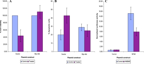
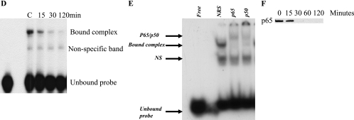
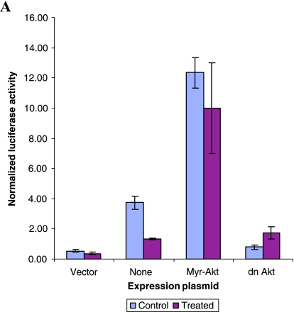
Transcription factor NFkappaB and the serine/threonine kinase Akt play critical roles in mammalian cell survival signaling and have been shown to be activated in various malignancies including prostate cancer (PCA). We have developed an analogue of curcumin called 4-hydroxy-3-methoxybenzoic acid methyl ester (HMBME) that targets the Akt/NFkappaB signaling pathway. Here, we demonstrate the ability of this novel compound to inhibit the proliferation of human and mouse PCA cells. HMBME-induced apoptosis in these cells was tested by using multiple biochemical approaches, in addition to morphologic analysis. Overexpression of constitutively active Akt reversed the HMBME-induced growth inhibition and apoptosis, illustrating the direct role of Akt signaling in HMBME-mediated growth inhibition and apoptosis. Further, investigation of the molecular events associated with its action in LNCaP cells shows that: 1) HMBME reduces the level of activated form of Akt (phosphorylated Akt); and 2) inhibits the Akt kinase activity. Further, the transcriptional activity of NFkappaB, the DNA-binding activity of NFkappaB, and levels of p65 were all significantly reduced following treatment with HMBME. Overexpression of constitutively active Akt, but not the kinase dead mutant of Akt, activated the basal NFkappaB transcriptional activity. HMBME treatment had no influence on this constitutively active Akt-augmented NFkappaB transcriptional activity. These data indicate that HMBME-mediated inhibition of Akt kinase activity may have a potential in suppressing/decreasing the activity of major survival/antiapoptotic pathways. The potential use of HMBME as an agent that targets survival mechanisms in PCA cells is discussed.


Figures

Figure 1
Figure 1
Structure of HMBME.
Figure 2
Figure 2
(A) Effect of HMBME on proliferation of androgen-responsive (LNCaP), androgen-independent (DU145), benign prostatic epithelial (BPH-1), and nontumorigenic fibroblasts (GM0637). Cells were plated in 96-well plates as described in Materials and Methods section and treated with indicated concentrations of either HMBME or solvent control. Cell proliferation was measured by CellTiter96 Aqueous One solution assay at a 72-hour time point by determining the absorbance at 570 nm using SpectraMaxPlus plate reader (Molecular Devices, Sunnyvale, CA). The data shown here are an average ± SD of five replicate wells and are representative of four independent experiments. (B) Effect of HMBME on the proliferation of TRAMP cell lines. The experimental procedure was the same as described in legends for (A). (C) Effect of HMBME on colonyforming ability of LNCaP cells. Cells were plated in triplicate in 35-mm dishes on 0.5% agarose-containing media as described in Materials and Methods section. Following 14-day incubation, cells were stained with 0.5 ml of 0.02% p-iodonitrotetrazolium and colonies were counted in 10 different fields from each plate. The results are expressed as mean ± SD of five replicate wells and are representative of two independent experiments.
Figure 3
Figure 3
Effect of HMBME on cell cycle distribution in LNCaP cells. LNCaP cells were treated with either DMSO alone or with 25 µM HMBME for 24 hours as described in Materials and Methods section. Following treatment, cells were harvested, washed with PBS, and then resuspended in 1 ml of Krishan stain containing 1.1 mg/ml sodium citrate, 46 µg/ml propidium iodide, 0.01% of NP40, and 10 µg/ml RNase. Data were analyzed using Modfit LT (panel B). Alterations in the distribution of cells in different phases are also shown as a graph (panel A). This is an average ± SD of three independent experiments.
Figure 3
Figure 3
Effect of HMBME on cell cycle distribution in LNCaP cells. LNCaP cells were treated with either DMSO alone or with 25 µM HMBME for 24 hours as described in Materials and Methods section. Following treatment, cells were harvested, washed with PBS, and then resuspended in 1 ml of Krishan stain containing 1.1 mg/ml sodium citrate, 46 µg/ml propidium iodide, 0.01% of NP40, and 10 µg/ml RNase. Data were analyzed using Modfit LT (panel B). Alterations in the distribution of cells in different phases are also shown as a graph (panel A). This is an average ± SD of three independent experiments.
Figure 4
Figure 4
(A) Morphologic alterations of LNCaP cells following HMBME treatment. LNCaP cells were treated with either DMSO or with different concentrations of HMBME (5, 25, and 50 µM) for 24 hours. Photomicrographs of these cells were taken by phase contrast microscopy using a Nikon Microscope with a digital camera system Coolpix 995 at a magnification of 20 x (Nikon, Tokyo, Japan). The picture shown here shows cells treated with 25 µM HMBME for 24 hours. Arrows indicate apoptotic cells. (B and C) Induction of apoptosis following treatment with HMBME. Cells were treated with HMBME (25 µM for 24 hours) and induction of apoptosis was detected by FITC-Annexin staining through flow cytometry and acridine/orange staining. Both the adherent and floating cells were collected by trypsinization for quantification of apoptosis as described in Materials and Methods section. A representative graph of FITC-Annexin is shown (B). Data shown for acridine orange/ethidium bromide staining are average ± SD of two independent experiments conducted in triplicate. Cells were counted in four different fields for each sample (panel C).
Figure 4
Figure 4
(A) Morphologic alterations of LNCaP cells following HMBME treatment. LNCaP cells were treated with either DMSO or with different concentrations of HMBME (5, 25, and 50 µM) for 24 hours. Photomicrographs of these cells were taken by phase contrast microscopy using a Nikon Microscope with a digital camera system Coolpix 995 at a magnification of 20 x (Nikon, Tokyo, Japan). The picture shown here shows cells treated with 25 µM HMBME for 24 hours. Arrows indicate apoptotic cells. (B and C) Induction of apoptosis following treatment with HMBME. Cells were treated with HMBME (25 µM for 24 hours) and induction of apoptosis was detected by FITC-Annexin staining through flow cytometry and acridine/orange staining. Both the adherent and floating cells were collected by trypsinization for quantification of apoptosis as described in Materials and Methods section. A representative graph of FITC-Annexin is shown (B). Data shown for acridine orange/ethidium bromide staining are average ± SD of two independent experiments conducted in triplicate. Cells were counted in four different fields for each sample (panel C).
Figure 5
Figure 5
(A) Overexpression of constitutively active Akt protects LNCaP cells from HMBME-induced cell growth inhibition. Subconfluent LNCaP cells were transfected with either pCMVMyrAkt (an activated form of Akt with the Src myristoylation signal fused in-frame to the c-Akt coding sequence) or control vector (pCMV) using Lipofectamin (Invitrogen) in triplicate dishes. Forty-eight hours following transfection, cells were treated either with 25 µM HMBME or solvent control. Both floating and adherent cells were collected after 2 hours of treatment and assessed for cell viability by trypan blue exclusion assay. The data shown here are average ± SD of three independent experiments. (B) Overexpression of constitutively active Akt protects LNCaP cells from undergoing apoptosis following treatment with HMBME. Experiments were conducted essentially as described in (A). Both floating and adherent cells were collected after 2 hours of treatment and assessed for apoptosis as described in Materials and Methods section. The data shown here are average ± SD of two independent transfections. (C) Influence of HMBME on transcriptional activity of the NFκB promoter. Transient transfections were performed with pNFκB reporter plasmid (1 µg/well) and pRL-TK plasmid (50 ng/well; renilla luciferase for normalization) as described in Materials and Methods section using Lipofectin reagent. Forty-eight hours after transfection, cells were treated with HMBME (25 µM) for 2 hours. Firefly and renilla luciferase activity was measured in the extracts prepared from these using Dual Luciferase Reporter Assay System (Promega) in duplicate samples containing equal amounts of protein. Renilla luciferase activity was used to normalize for transfection efficiency. Results are expressed as the ratio of firefly luciferase/renilla luciferase at equal amounts of protein. The data shown here are a representative experiment that was performed for four times with two different preparations of plasmid. For cotransfection experiments, indicated expression plasmids (1 µg/well) were included along with the pNFκB reporter plasmid. The data shown here are a representative experiment that was performed for four times with two different preparations of plasmid. (D) EMSA of nuclear extracts prepared from control and HMBME-treated cells. Nuclear extract (5 µg) was incubated with approximately 0.2 ng of NFκB consensus oligonucleotide as radiolabeled probe as described in Materials and Methods section and the DNA-protein complexes were resolved on a 4% nondenaturing gel by electrophoresis and subject to autoradiography. (E) Identification of protein components of NFκB DNA-binding activity. Nuclear extracts prepared from LNCaP cells were preincubated with p65, p50, or NRS for 30 minutes on ice. These extracts were used in EMSA with NFkB probe as described in legends for (C). Supershifted complex is indicated as p65/p50 complex and NS indicates nonspecific band. (F) Western blot analysis of whole cell extracts from LNCaP cells following treatment with HMBME. Twenty-five micrograms of extract from control or HMBME-treated cells was fractionated on 10% SDS-PAGE and transferred to a nitrocellulose membrane. After blocking, the membrane was incubated for 2 or 3 hours with the antibody p65. This was followed by incubation with secondary horseradish peroxidase-conjugated antirabbit IgG antibody (Sigma) in blocking solution. Bound antibody was detected by Supersignal West Pico Chemiluminescent Substrate, following the manufacturer's directions (Pierce). The blot shown here is a representative blot of three independent experiments.
Figure 5
Figure 5
(A) Overexpression of constitutively active Akt protects LNCaP cells from HMBME-induced cell growth inhibition. Subconfluent LNCaP cells were transfected with either pCMVMyrAkt (an activated form of Akt with the Src myristoylation signal fused in-frame to the c-Akt coding sequence) or control vector (pCMV) using Lipofectamin (Invitrogen) in triplicate dishes. Forty-eight hours following transfection, cells were treated either with 25 µM HMBME or solvent control. Both floating and adherent cells were collected after 2 hours of treatment and assessed for cell viability by trypan blue exclusion assay. The data shown here are average ± SD of three independent experiments. (B) Overexpression of constitutively active Akt protects LNCaP cells from undergoing apoptosis following treatment with HMBME. Experiments were conducted essentially as described in (A). Both floating and adherent cells were collected after 2 hours of treatment and assessed for apoptosis as described in Materials and Methods section. The data shown here are average ± SD of two independent transfections. (C) Influence of HMBME on transcriptional activity of the NFκB promoter. Transient transfections were performed with pNFκB reporter plasmid (1 µg/well) and pRL-TK plasmid (50 ng/well; renilla luciferase for normalization) as described in Materials and Methods section using Lipofectin reagent. Forty-eight hours after transfection, cells were treated with HMBME (25 µM) for 2 hours. Firefly and renilla luciferase activity was measured in the extracts prepared from these using Dual Luciferase Reporter Assay System (Promega) in duplicate samples containing equal amounts of protein. Renilla luciferase activity was used to normalize for transfection efficiency. Results are expressed as the ratio of firefly luciferase/renilla luciferase at equal amounts of protein. The data shown here are a representative experiment that was performed for four times with two different preparations of plasmid. For cotransfection experiments, indicated expression plasmids (1 µg/well) were included along with the pNFκB reporter plasmid. The data shown here are a representative experiment that was performed for four times with two different preparations of plasmid. (D) EMSA of nuclear extracts prepared from control and HMBME-treated cells. Nuclear extract (5 µg) was incubated with approximately 0.2 ng of NFκB consensus oligonucleotide as radiolabeled probe as described in Materials and Methods section and the DNA-protein complexes were resolved on a 4% nondenaturing gel by electrophoresis and subject to autoradiography. (E) Identification of protein components of NFκB DNA-binding activity. Nuclear extracts prepared from LNCaP cells were preincubated with p65, p50, or NRS for 30 minutes on ice. These extracts were used in EMSA with NFkB probe as described in legends for (C). Supershifted complex is indicated as p65/p50 complex and NS indicates nonspecific band. (F) Western blot analysis of whole cell extracts from LNCaP cells following treatment with HMBME. Twenty-five micrograms of extract from control or HMBME-treated cells was fractionated on 10% SDS-PAGE and transferred to a nitrocellulose membrane. After blocking, the membrane was incubated for 2 or 3 hours with the antibody p65. This was followed by incubation with secondary horseradish peroxidase-conjugated antirabbit IgG antibody (Sigma) in blocking solution. Bound antibody was detected by Supersignal West Pico Chemiluminescent Substrate, following the manufacturer's directions (Pierce). The blot shown here is a representative blot of three independent experiments.
Figure 6
Figure 6
(A) Akt activates NFκB promoter activity. Cotransfection experiments were performed essentially as described above for Figure 5B with 1 µg/well of the indicated expression plasmids (pCMVMyrAkt, an activated form of Akt with the Src myristoylation signal fused in-frame to the c-Akt coding sequence; dn Akt, construct expressing inactive Akt due to mutation in the kinase domain; or control vector, pCDNA-3, along with the pNFκB reporter plasmid). Luciferase activity was determined as described above for Figure 5C. (B) EMSA using transfected extracts. Cell extracts prepared from the transfected cells were used in gel shift experiments as described in Figure 5D. “C” indicates control extract and “T” indicates treated extract (25 µM HMBME for 2 hours). “F” denotes free probe line without the protein extract. (C) Akt kinase activity and levels of p-Akt following treatment with HMBME. Actively growing LNCaP cells were treated with HMBME (25 µM) for 24 hours or for different time points as described in Materials and Methods section. Endogenous Akt was immunoprecipitated using an Akt monoclonal antibody. Following extensive washes, kinase reaction was performed in the presence of 200 µM cold ATP and GSK-3 substrate. Phosphorylation of GSK-3 was measured by Western blotting using an antiphospho-GSK-3 antibody. Western blotting using pAkt was performed essentially as described in legends for Figure 5F.
Figure 6
Figure 6
(A) Akt activates NFκB promoter activity. Cotransfection experiments were performed essentially as described above for Figure 5B with 1 µg/well of the indicated expression plasmids (pCMVMyrAkt, an activated form of Akt with the Src myristoylation signal fused in-frame to the c-Akt coding sequence; dn Akt, construct expressing inactive Akt due to mutation in the kinase domain; or control vector, pCDNA-3, along with the pNFκB reporter plasmid). Luciferase activity was determined as described above for Figure 5C. (B) EMSA using transfected extracts. Cell extracts prepared from the transfected cells were used in gel shift experiments as described in Figure 5D. “C” indicates control extract and “T” indicates treated extract (25 µM HMBME for 2 hours). “F” denotes free probe line without the protein extract. (C) Akt kinase activity and levels of p-Akt following treatment with HMBME. Actively growing LNCaP cells were treated with HMBME (25 µM) for 24 hours or for different time points as described in Materials and Methods section. Endogenous Akt was immunoprecipitated using an Akt monoclonal antibody. Following extensive washes, kinase reaction was performed in the presence of 200 µM cold ATP and GSK-3 substrate. Phosphorylation of GSK-3 was measured by Western blotting using an antiphospho-GSK-3 antibody. Western blotting using pAkt was performed essentially as described in legends for Figure 5F.

Similar articles

Cited by

References

    1. American Cancer Society, author. Cancer Facts and Figures. 2002. ( http://www.cancer.org)
    1. Garnick MB. Prostate cancer: screening, diagnosis, and management. Ann Intern Med. 1993;118:804–818. - PubMed
    1. Huggins C, Hodges CV. Studies on prostate cancer: I.The effect of estrogen and of androgen injection on serum phosphatase in metastatic prostate cancer. Cancer Res. 1941;1:293–297.
    1. Scher HI, Steineck G, Kelly WK. Hormone refractory (D3) prostate cancer: refining the concept. Urology. 1995;46:142–148. - PubMed
    1. Pratt DE. Antioxidants and cancer prevention. In: M-T Huang, C-T Ho, CY Lee., editors. Phenolic Compounds in Food and their Effect on Health II ACS Symposium Series 507. Washington, DC: American Chemical Society; 1992. pp. 54–71. (Chapter 5)

Publication types

MeSH terms