

The loss of TGF-beta signaling promotes prostate cancer metastasis
- PMID: 12869309
- PMCID: PMC1502411
- DOI: 10.1016/S1476-5586(03)80058-1
The loss of TGF-beta signaling promotes prostate cancer metastasis
Abstract
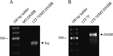
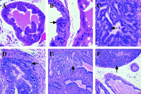
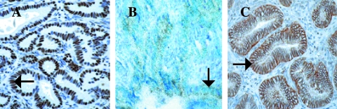
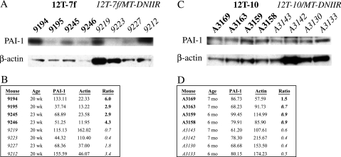
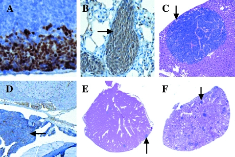
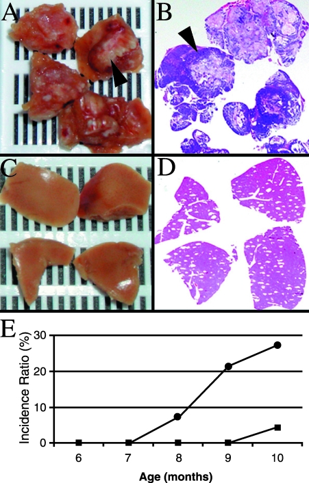
In breast and colon cancers, transforming growth factor (TGF)-beta signaling initially has an antineoplastic effect, inhibiting tumor growth, but eventually exerts a proneoplastic effect, increasing motility and cancer spread. In prostate cancer, studies using human samples have correlated the loss of the TGF-beta type II receptor (T beta R II) with higher tumor grade. To determine the effect of an inhibited TGF-beta pathway on prostate cancer, we bred transgenic mice expressing the tumorigenic SV40 large T antigen in the prostate with transgenic mice expressing a dominant negative T beta R II mutant (DN II R) in the prostate. Transgene(s) and TGF-beta 1 expression were identified in the prostate and decreased protein levels of plasminogen activator inhibitor type I, as a marker for TGF-beta signaling, correlated with expression of the DN II R. Although the sizes of the neoplastic prostates were not enlarged, increased amounts of metastasis were observed in mice expressing both transgenes compared to age-matched control mice expressing only the large T antigen transgene. Our study demonstrates for the first time that a disruption of TGF-beta signaling in prostate cancer plays a causal role in promoting tumor metastasis.
Figures
Similar articles
-
Stromal transforming growth factor-beta signaling mediates prostatic response to androgen ablation by paracrine Wnt activity.Cancer Res. 2008 Jun 15;68(12):4709-18. doi: 10.1158/0008-5472.CAN-07-6289. Cancer Res. 2008. PMID: 18559517 Free PMC article.
-
Blockade of transforming growth factor-beta signaling suppresses progression of androgen-independent human prostate cancer in nude mice.Clin Cancer Res. 2005 Jun 15;11(12):4512-20. doi: 10.1158/1078-0432.CCR-04-2571. Clin Cancer Res. 2005. PMID: 15958637
-
Restoration of transforming growth factor beta signaling pathway in human prostate cancer cells suppresses tumorigenicity via induction of caspase-1-mediated apoptosis.Cancer Res. 1999 Mar 15;59(6):1366-71. Cancer Res. 1999. PMID: 10096572
-
TGF beta in prostate cancer: a growth inhibitor that can enhance tumorigenicity.Prostate. 1997 Apr 1;31(1):61-70. doi: 10.1002/(sici)1097-0045(19970401)31:1<61::aid-pros10>3.0.co;2-m. Prostate. 1997. PMID: 9108888 Review.
-
TGF-BETA IN THE NATURAL HISTORY OF PROSTATE CANCER.Acta Clin Croat. 2019 Mar;58(1):128-138. doi: 10.20471/acc.2019.58.01.17. Acta Clin Croat. 2019. PMID: 31363335 Free PMC article. Review.
Cited by
-
SFMBT2 (Scm-like with four mbt domains 2) negatively regulates cell migration and invasion in prostate cancer cells.Oncotarget. 2016 Jul 26;7(30):48250-48264. doi: 10.18632/oncotarget.10198. Oncotarget. 2016. PMID: 27340776 Free PMC article.
-
Endoglin phosphorylation by ALK2 contributes to the regulation of prostate cancer cell migration.Carcinogenesis. 2010 Mar;31(3):359-66. doi: 10.1093/carcin/bgp217. Epub 2009 Sep 7. Carcinogenesis. 2010. PMID: 19736306 Free PMC article.
-
Mouse models of prostate cancer: picking the best model for the question.Cancer Metastasis Rev. 2014 Sep;33(2-3):377-97. doi: 10.1007/s10555-013-9487-8. Cancer Metastasis Rev. 2014. PMID: 24452759 Free PMC article. Review.
-
Matriptase-2/NR4A3 axis switches TGF-β action toward suppression of prostate cancer cell invasion, tumor growth, and metastasis.Oncogene. 2022 May;41(20):2833-2845. doi: 10.1038/s41388-022-02303-z. Epub 2022 Apr 13. Oncogene. 2022. PMID: 35418692
-
Network-based prediction for sources of transcriptional dysregulation using latent pathway identification analysis.Proc Natl Acad Sci U S A. 2011 Aug 9;108(32):13347-52. doi: 10.1073/pnas.1100891108. Epub 2011 Jul 25. Proc Natl Acad Sci U S A. 2011. PMID: 21788508 Free PMC article.
References
-
- Ohori M, Goad JR, Wheeler TM, Eastham JA, Thompson TC, Scardino PT. Can radical prostatectomy alter the progression of poorly differentiated prostate cancer? J Urol. 1994;152:1843–1849. - PubMed
-
- Quinn DI, Henshall SM, Head DR, Golovsky D, Wilson JD, Brenner PC, Turner JJ, Delprado W, Finlayson JF, Stricker PD, Grygiel JJ, Sutherland RL. Prognostic significance of p53 nuclear accumulation in localized prostate cancer treated with radical prostatectomy. Cancer Res. 2000;60:1585–1594. - PubMed
-
- Kim IY, Ahn HJ, Lang S, Oefelein MG, Oyasu R, Kozlowski JM, Lee C. Loss of expression of transforming growth factor-beta receptors is associated with poor prognosis in prostate cancer patients. Clin Cancer Res. 1998;4:1625–1630. - PubMed
-
- Guo Y, Jacobs SC, Kyprianou N. Down-regulation of protein and mRNA expression for transforming growth factor-beta (TGF-beta1) type I and type II receptors in human prostate cancer. Int J Cancer. 1997;71:573–579. - PubMed
Publication types
MeSH terms
Substances
Grants and funding
LinkOut - more resources
Full Text Sources
Medical
Molecular Biology Databases
