. 2003 Aug 1;17(15):1882-93.
doi: 10.1101/gad.1107803. Epub 2003 Jul 17.

SKN-1 links C. elegans mesendodermal specification to a conserved oxidative stress response

Affiliations

SKN-1 links C. elegans mesendodermal specification to a conserved oxidative stress response

Jae Hyung An et al. Genes Dev. .

Abstract

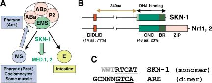
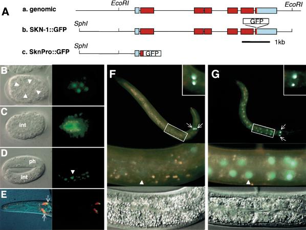
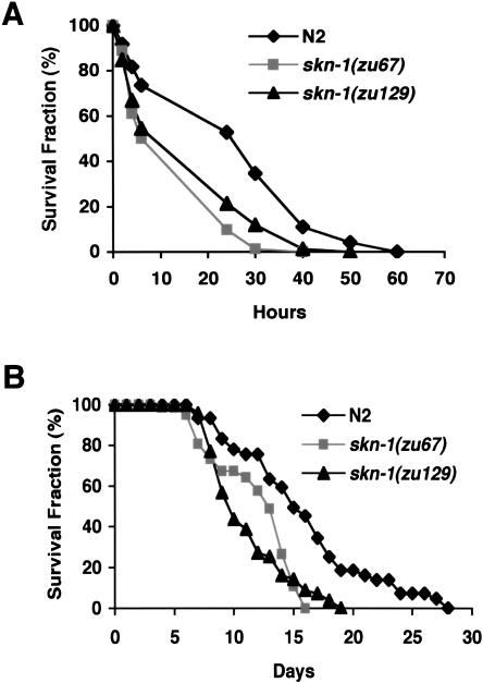
During the earliest stages of Caenorhabditis elegans embryogenesis, the transcription factor SKN-1 initiates development of the digestive system and other mesendodermal tissues. Postembryonic SKN-1 functions have not been elucidated. SKN-1 binds to DNA through a unique mechanism, but is distantly related to basic leucine-zipper proteins that orchestrate the major oxidative stress response in vertebrates and yeast. Here we show that despite its distinct mode of target gene recognition, SKN-1 functions similarly to resist oxidative stress in C. elegans. During postembryonic stages, SKN-1 regulates a key Phase II detoxification gene through constitutive and stress-inducible mechanisms in the ASI chemosensory neurons and intestine, respectively. SKN-1 is present in ASI nuclei under normal conditions, and accumulates in intestinal nuclei in response to oxidative stress. skn-1 mutants are sensitive to oxidative stress and have shortened lifespans. SKN-1 represents a connection between developmental specification of the digestive system and one of its most basic functions, resistance to oxidative and xenobiotic stress. This oxidative stress response thus appears to be both widely conserved and ancient, suggesting that the mesendodermal specification role of SKN-1 was predated by its function in these detoxification mechanisms.


Figures

Figure 1.
Figure 1.
SKN-1 embryonic functions and comparison to Nrf proteins. (A) Cell fate specification. In four-cell embryos, SKN-1 initiates mesendodermal development by establishing the EMS blastomere fate (Bowerman et al. 1992). Anterior is to the left, and ventral at the bottom. (B) SKN-1 compared to Nrf proteins. The SKN-1 minor groove-binding arm is shown in light green. The percent identity between SKN-1 and mouse Nrf2 regions is indicated. (C) Consensus sequences for SKN-1 binding and the ARE. The SKN-1 BR recognizes a consensus bZIP half-site (underlined) adjacent to an AT-rich motif (gray) that is specified by the arm (B). Nrf proteins bind to the ARE as obligate heterodimers with Maf or other bZIP proteins (Hayes and McMahon 2001). (R) G/A; (W) T/A.
Figure 2.
Figure 2.
skn-1-dependent GCS-1::GFP expression in the intestine and ASI neurons. (A-F) GCS-1::GFP expression in wild-type animals. A gcs-1 genomic fragment containing its 17 N-terminal codons and 1840 upstream base pairs was fused to the N terminus of GFP, which contained a nuclear localization signal. The expression patterns shown are each representative of more than two independent transgenic lines, and of all postembryonic stages examined (L2-adult; data not shown). (A,B) Nomarski (A) and fluorescent (B) views of an L2 larva. (B) A line demarcates the approximate boundary between the anterior intestine (I) and posterior pharynx (P). (C,D) Combined Nomarski/fluorescent (C) and fluorescent (D) views of the head of a typical L4-stage animal that had been exposed to DiI. (D) One of the two ASI neurons is indicated with an arrow. (E,F) An L2 larva in which GCS-1::GFP expression was induced to high levels in the intestine by heat. A similar induction occurred in response to paraquat (Table 2). The boundary between the anterior intestine and posterior pharynx is indicated as in B. (G-L) GCS-1::GFP was not detectable outside of the pharynx in skn-1 homozygotes. Typical animals are shown from experiments that parallel those displayed to the left in A-F. Note the absence of GCS-1::GFP in the intestine and ASI neurons under normal conditions (G-J), and after treatment with heat (K,L) or paraquat (data not shown). In two independent transgenic lines, in a homozygous skn-1 background GCS-1::GFP expression was not detected in these tissues in any animals under either normal or induction conditions.
Figure 3.
Figure 3.
Specific elements required for skn-1-independent and -dependent GCS-1::GFP expression. (A) Analysis of the gcs-1 promoter. The expression of the indicated constructs from transgenic extrachromosomal arrays was assayed in two to three independent transgenic lines under normal conditions, and after induction by paraquat and heat. The relative expression levels in the tissues designated to the right (data not shown) are indicated by plus signs, with ++ indicating a reproducible reduction and + indicating barely detectable expression. Within each set of transgenic lines that carried promoter mutations, levels of normal and induced expression were affected in parallel. Mutations that were created in predicted SKN-1 sites 1, 2, and 3 are described in Materials and Methods, and are not compatible with SKN-1 binding (Blackwell et al. 1994; see text). Red ovals indicate predicted SKN-1 binding sites and a green bar indicates the 5′-end of the gcs-1::gfp coding region. Map numbers refer to the predicted translation start. (B) Uncoupling pharyngeal GCS-1::GFP expression from intestinal and ASI neuron expression. The gcsΔ2 mutation eliminated pharyngeal GCS-1::GFP expression, but allowed near-wild-type levels of ASI and intestinal expression. Concurrent ablation of SKN-1 binding site 3 (gcsΔ2, mut3) eliminated transgene expression in all tissues. Paraquat-treated worms are shown in the GFP column. (C) Composite gcs-1 promoter element that includes SKN-1 site 3, and is also present in the med-1 and med-2 promoters. SKN-1 binding sites are red, and identical sequences are boxed.
Figure 4.
Figure 4.
Specific binding of SKN-1 to an essential gcs-1 promoter sequence. (A) Binding of full-length SKN-1 to site 3 within the gcs-1 composite element, assayed by EMSA. (Lanes 2-5) Binding of increasing amounts of in vitro translated SKN-1 protein (0, 0.25, 0.5, and 3 μL translation lysate; indicated by a triangle) to the wild-type site. (Lane 1) Binding to 3 μL of un-programmed lysate. A background species is labeled. (Lanes 6-10) The same assay performed with the mutant probe. (Lanes 11-20) SKN-1 DNA binding is assayed in the presence of the indicated unlabeled competitor oligonucleotides. Lanes 12-15 and 17-20 correspond to addition of a 20-, 50-, 150-, and 400-fold molar excess of competitor over the labeled wild-type DNA. (B) The in vitro translated SKN-1 DNA-binding domain (Fig. 1B) binds specifically to the gcs-1 composite element. Binding was assayed as in A.
Figure 5.
Figure 5.
Expression and stress-induced nuclear accumulation of SKN-1::GFP. (A) SKN-1::GFP transgenes. (a) skn-1 gene. Transcribed coding and untranslated regions are indicated in red and blue, respectively. (b) SKN-1::GFP translational fusion construct, which includes an EcoRI fragment that previously rescued maternal skn-1 lethality (Bowerman et al. 1992). C. elegans DNA is indicated by a black line. (c) SknPro::GFP promoter fusion, in which the 38 N-terminal SKN-1 amino acids are fused to GFP containing a nuclear localization signal. (B-D) Embryonic expression of SKN-1::GFP. Nomarski (left) and fluorescent (right) views of 100-cell (B), 280-min (C), and threefold (D) embryos. Endogenous intestinal autofluorescence is visible as yellow or orange (see Materials and Methods). White triangles, intestine precursor nuclei; int, intestine; ph, pharynx. (E) SKN-1::GFP expression in ASI neurons (arrows). Nomarski/fluorescent (left) and fluorescent (right) views are shown of a typical DiI-exposed L4 larva. (F) Larval SKN-1::GFP expression under normal conditions. (Bottom) Fluorescent and Nomarski closeups of the boxed central region of this L2. An intestinal nucleus is indicated by a white triangle, and the ASI neurons (arrows) are shown in detail in the top right box. (G) SKN-1::GFP localization under oxidative stress. A heat-shocked L2 is shown, but similar results were obtained after exposure to other oxidative stress inducers (Table 3). The integrated strain Is007 is shown, but two extrachromosomal lines and a different integrated line exhibited similar patterns.
Figure 6.
Figure 6.
skn-1 mutants are sensitive to oxidative stress and have reduced lifespans. (A) Paraquat sensitivity. Individual worms were scored for survival at the times shown after they had been placed in M9 that contained 100 mM paraquat. An average of three experiments involving 24 worms each is graphed. Among these experiments, for skn-1(zu67) and skn-1(zu129) animals, the mean survival times expressed as percentage of wild type were 52.9% ± 9.5% and 60.7% ± 16.1%, respectively. All wild-type and skn-1 mutant worms survived a parallel control 72-h incubation in M9 alone (data not shown). (B) Lifespan assay. Worms were maintained at 20°C and scored for survival at the indicated time after the L4 stage. An average of three consecutive experiments involving 25-28 worms each is plotted (Table 4).

Similar articles

Cited by

References

    1. Blackwell T.K., Bowerman, B., Priess, J., and Weintraub, H. 1994. Formation of a monomeric DNA binding domain by Skn-1 bZIP and homeodomain elements. Science 266: 621-628. - PubMed
    1. Bluher M., Kahn, B.B., and Kahn, C.R. 2003. Extended longevity in mice lacking the insulin receptor in adipose tissue. Science 299: 572-574. - PubMed
    1. Bowerman B., Eaton, B.A., and Priess, J.R. 1992. skn-1, a maternally expressed gene required to specify the fate of ventral blastomeres in the early C. elegans embryo. Cell 68: 1061-1075. - PubMed
    1. Bowerman B., Draper, B.W., Mello, C., and Priess, J. 1993. The maternal gene skn-1 encodes a protein that is distributed unequally in early C. elegans embryos. Cell 74: 443-452. - PubMed
    1. Brenner S. 1974. The genetics of Caenorhabditis elegans. Genetics 77: 71-94. - PMC - PubMed

Publication types

MeSH terms