. 2003 Aug;9(8):1007-18.
doi: 10.1261/rna.5590603.

A structural linkage between the dimerization and encapsidation signals in HIV-2 leader RNA

Affiliations

A structural linkage between the dimerization and encapsidation signals in HIV-2 leader RNA

Jean-Marc Lanchy et al. RNA. 2003 Aug.

Abstract

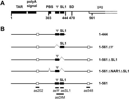
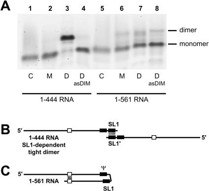
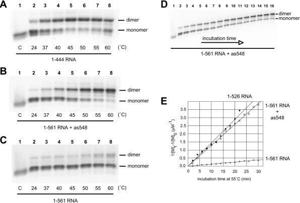
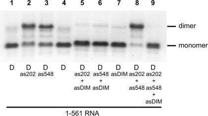
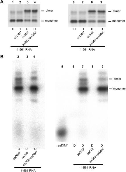
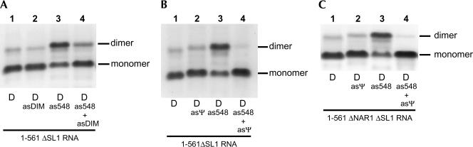
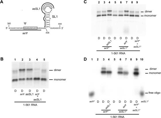
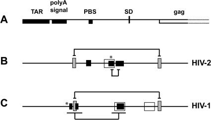
The 5' untranslated leader region of retroviral RNAs contains noncoding information that is essential for viral replication, including signals for transcriptional transactivation, splicing, primer binding for reverse transcription, dimerization of the genomic RNA, and encapsidation of the viral RNA into virions. These RNA motifs have considerable structural and functional overlap. In this study, we investigate the conformational dynamics associated with the use and silencing of a sequence in HIV-2 RNA that is involved in genomic RNA dimerization called stem-loop 1 (SL1) and its relationship with a flanking sequence that is known to be important for encapsidation of viral RNAs. We demonstrate that a long-distance intramolecular interaction between nucleotides located upstream of the primer-binding site domain and nucleotides encompassing the Gag translation start codon functionally silences SL1 as a dimerization element. This silencing can be relieved by mutation or by hybridization of an oligonucleotide that disrupts the long-distance interaction. Furthermore, we identify a palindrome within the packaging/encapsidation signal Psi (just 5' of SL1) that can either serve as an efficient dimerization signal itself, or can mediate SL1 silencing through base pairing with SL1. These results provide a tangible link between the functions of genomic RNA dimerization and encapsidation, which are known to be related, but whose physical relationship has been unclear. A model is proposed that accounts for observations of dimerization, packaging, and translation of viral RNAs during different phases of the viral replication cycle.


Figures

FIGURE 1.
FIGURE 1.
5′ leader region of HIV-2 ROD genomic RNA. (A) The landmark sequences with known functions are indicated by boxes with the name indicated above. TAR, polyA signal, PBS, Ψ, SL1, SD, and gag represent the trans-activation region, the poly(A) signal domain, the primer binding site, the encapsidation signal, the stem–loop 1, the major splice donor site, and the 5′ end of the Gag protein coding region, respectively. (B) RNAs used in this study. The closed boxes represent the in vivo characterized encapsidation signal Ψ (Griffin et al. 2001) and the in vitro characterized dimerization element SL1 (in HIV-2; Dirac et al. 2001; Lanchy and Lodmell 2002). The open boxes represent the cores of the two dimer-interfering elements previously characterized 189–196 and 543–550 (Lanchy et al. 2003). The name of each RNA construct is indicated at the right. The short thick lines below some elements represent the binding sites of antisense DNA oligonucleotides used in this study.
FIGURE 2.
FIGURE 2.
TBE-resistant dimerization of 1–444 and 1–561 RNAs. (A) 1–444 and 1–561 RNAs were assayed for dimerization at 55°C with monomer (M) or dimer (D) buffer. Dimerization was also assayed in the presence of a 20-fold excess of antisense DNA oligonucleotide asDIM, which is complementary to nucleotides 397–426. After incubation for 30 min, samples were subjected to electrophoresis on a TBE agarose gel at 28°C. Only tight dimers withstand the warm TBE electrophoresis. (Control lanes C) Monomeric RNA that was denatured at 90°C, then quenched on ice immediately prior to loading. (B) Schematic representation of the tight dimer of 1–444 RNA from A, lane 3. The two molecules interact through an extended base pairing of the SL1 elements. (C) Schematic representation of the proposed monomeric form of 1–561 RNA from A, lane 7. Upon incubation of 1–561 RNA at high temperature, there is an intramolecular folding of SL1 that competes with the intermolecular interaction (i.e., dimerization). The SL1 structure is shown partially base paired to the upstream encapsidation signal Ψ, as described in Lanchy et al. (2003).
FIGURE 3.
FIGURE 3.
Influence of incubation temperature and time on the as548 oligonucleotide-induced dimerization of 1–561 RNA. The 1–444 RNA (A), 1–561 RNA with the as548 oligonucleotide (B), or 1–561 RNA without oligonucleotide (C) were incubated in dimer buffer for 30 min at the indicated temperatures and subjected to electrophoresis on TBE agarose gel run at 28°C. In B, the 1–561 RNA was incubated with a 20-fold excess of the antisense DNA oligonucleotide as548, which is complementary to nt 528–548. The band below the indicated monomer band in A and the two faint bands between monomer and dimer bands in B probably represent low-abundance conformers of monomer (in A) or dimer (in B) species. Control lanes C are described in the previous figure. (D) The time-dependent dimerization of 1–561 RNA at 55°C in the presence of the as548 oligonucleotide was monitored on a TBE agarose gel at 28°C. (Lanes 1–16) Incubation times of 0, 2, 4, 6, 8, 10, 12, 14, 16, 18, 20, 22, 24, 26, 28, 30 min, respectively. (E) Plots of the kinetic data for 1–526 RNA and 1–561 RNA with or without as548 (represented by closed circles, open squares, and open diamonds, respectively). [M]t is the concentration of monomer at time t, and [M]0 is the initial concentration of dimerization-competent monomer.
FIGURE 4.
FIGURE 4.
Antisense oligonucleotide-mediated activation and suppression of 1–561 RNA tight dimerization. 1–561 RNA was incubated for 30 min at 55°C in dimer buffer with a 20-fold excess of as202, as548, asDIM, or a combination of these oligonucleotides and subjected to electrophoresis on TBE agarose gel at 28°C. Antisense DNA oligonucleotides as202 and as548 target the upstream and downstream dimer-interfering elements, respectively, previously characterized in our laboratory (Lanchy et al. 2003). asDIM is complementary to nt 397–426 and inhibits SL1-dependent tight dimerization of the 1–444 model RNA (Fig. 2 ▶).
FIGURE 5.
FIGURE 5.
Partitioning of asDIM oligonucleotide with monomer and dimer species of 1–561 RNA upon dimer induction by as202 or as548 oligonucleotides. 1–561 RNA and oligonucleotides were mixed, heated to 90°C, quench cooled, then incubated at 55°C. Reactions shown in lanes 2,4,7,9 included a small amount of radioactively labeled asDIM oligonucleotide. Reactions shown in lanes 4 and 9 additionally contained a 20-fold excess of as202 or as548 oligonucleotides, respectively. The monomer and dimer species were separated on TBE/28°C gel as described in the previous figures. (A) The gel was stained with ethidium bromide and visualized by fluorescence scanning. (B) The same gel was then fixed and dried and the radioactivity associated with the free and bound oligonucleotide was visualized by phosphorimager analysis. Lanes 2,7 suggest that a different interacting region than SL1or Ψ mediated the low level of residual dimers. The exclusion of asDIM* from the dimer bands in lanes 4,9 suggests that nt 397–426 are required for dimerization. Lane 5 corresponds to free radioactively labeled asDIM oligonucleotide loaded without RNA.
FIGURE 6.
FIGURE 6.
as548-Mediated tight dimerization of 1–561RNA lacking SL1and PBS dimerization sites. (A) 1–561 ΔSL1RNA was incubated at 55°C in dimer buffer with a 20-fold excess of as548, asDIM oligonucleotides, or both and subjected to electrophoresis on TBE gel. The 1–561ΔSL1RNA lacks the SL1structure (deletion of nt 409–436). (B) The experiment described in A was reproduced using asΨ instead of asDIM. The asΨ oligonucleotide is complementary to nt 380–404 (Table 1 ▶) and binds to the encapsidation signal Ψ. (C) The experiment described in B was reproduced with the 1–561ΔNAR1ΔSL1RNA. This construct lacks both SL1and the 5′-304-GGCGCC-309-3′ sequence located at the 5′ end of the primer-binding site shown previously to mediate loose dimerization of 1–561 RNA (Jossinet et al. 2001; Lanchy and Lodmell 2002). Loose dimers are not expected to withstand the TBE electrophoresis.
FIGURE 7.
FIGURE 7.
Antisense oligonucleotide directed against SL1induces Ψ-dependent tight dimerization of 1–561 RNA and vice versa. (A) Representation of the Ψ –SL1region and antisense oligonucleotides used in this experiment. The two palindromic sequences involved in dimerization are boxed. asΨ and asSL1antisense oligonucleotides binding sites are represented by thick lines. (B) 1–561 RNA was incubated in dimer buffer at 55°C without or with a 20-fold molar excess of asΨ, asSL1, or both oligonucleotides, and subjected to TBE electrophoresis. (C, D) Partitioning of asΨ or asSL1oligonucleotides in monomer or dimer species of 1–561RNA. The experiment described in B was repeated with one of the oligonucleotides radioactively labeled and present in a small amount and the other present in a 20-fold molar excess. In C, the gel is visualized with ethidium bromide and in D, the same dried gel is visualized by phosphorimager analysis to visualize the radioactivity associated with the labeled oligonucleotide.
FIGURE 8.
FIGURE 8.
Structural analysis of the Ψ–SL1region. (A) 1–561 RNA was probed with RNase T1 in the presence (lane 2) or absence (lane 3) of as548. The lanes labeled G, U, A, and C are sequencing lanes. (Lane 1) Control (1–561RNA primer extended without RNase T1 modification); (lane 2) 1–561RNA activated with oligonucleotide as548 during RNase T1 probing; (and lane 3) RNase T1 probing of 1–561RNA without as548 present during modification. Theregion labeled Ψ represents the palindromic sequence GGAGUGCUCC present in this region.The region labeled SL1represents the palindromic sequence GGUACC present in this region.(B) A model representing HIV-2 RNA SL1in an extended duplex conformation. (Open triangles) Cleavages observed in 1–561RNA by RNase T1 in the absence of as548. (Solid circles) RNase T1 cleavages observed in 1–561RNA with as548 present. An increase in nucleotide reactivity is represented by an increase in the symbol present at that location.
FIGURE 9.
FIGURE 9.
Structural and functional elements involved in encapsidation and dimerization of HIV-1and HIV-2 genomic RNAs. (A) The landmark sequences with known functions are indicated by boxes (see Fig. 1A ▶). (B, C) The encapsidation, dimerization, and regulatory elements in HIV-2 (Griffin et al. 2001; Dirac et al. 2002; Lanchy et al. 2003) and HIV-1(McBride and Panganiban 1996; Abbink and Berkhout 2002; Berkhout et al. 2002; Clever et al. 2002; Russell et al. 2002) are indicated by open, closed, and hatched boxes, respectively. The described short- or long-range interactions between structural elements are indicated with brackets. The asterisk indicates palindrome (GAGUGCUU/C) locations in HIV-1and HIV-2.

Similar articles

Cited by

References

    1. Abbink, T.E. and Berkhout, B. 2002. A novel long distance base pairing interaction in human immunodeficiency virus type 1 RNA occludes the Gag start codon. J. Biol. Chem. 278: 11601–11611. - PubMed
    1. Bender, W. and Davidson, N. 1976. Mapping of poly(A) sequences in the electron microscope reveals unusual structure of type C oncornavirus RNA molecules. Cell 7: 595–607. - PubMed
    1. Berglund, J.A., Charpentier, B., and Rosbash, M. 1997. A high affinity binding site for the HIV-1 nucleocapsid protein. Nucleic Acids Res. 25: 1042–1049. - PMC - PubMed
    1. Berkhout, B. 1996. Structure and function of the human immunodeficiency virus leader RNA. Prog. Nucleic Acid Res. Mol. Biol. 54: 1–34. - PubMed
    1. Berkhout, B., Ooms, M., Beerens, N., Huthoff, H., Southern, E., and Verhoef, K. 2002. In vitro evidence that the untranslated leader of the HIV-1 genome is an RNA checkpoint that regulates multiple functions through conformational changes. J. Biol. Chem. 277: 19967–19975. - PubMed

Publication types

LinkOut - more resources