

doi: 10.1562/0031-8655(2003)077<0604:iioydo>2.0.co;2.
Increased intensities of YOYO-1-labeled DNA oligomers near silver particles
Affiliations
- PMID: 12870845
- PMCID: PMC2753835
- DOI: 10.1562/0031-8655(2003)077<0604:iioydo>2.0.co;2
Item in Clipboard
Increased intensities of YOYO-1-labeled DNA oligomers near silver particles
Photochem Photobiol.
2003 Jun.
Abstract
DNA detection is usually performed using fluorescence probes. Using a DNA oligomer stained with the widely used dye 1,1'-[1,3-propanediylbis[(dimethylimino)-3,1-propanediyl]]bis[4-[(3-methyl-2(3H)-benzoxazolylidene)methyl]]-quinolinum tetraiodide (YOYO-1), we show that a substrate containing silver particles can lead to a greater than 10-fold increase in the fluorescence intensity. Proximity to silver particles also increases the photostability of YOYO-1-DNA. These results suggest that substrates or gels containing silver particles may be used for increased sensitivity in DNA detection.
Figures

Emission spectra of the DNA oligomers labeled with increasing concentrations of YOYO-1. The inserts show the anisotropies and intensities with increasing concentrations of YOYO-1.

Absorption spectra of an SIF and emission spectra of the protein-coated surfaces treated with YOYO-1 but without DNA.
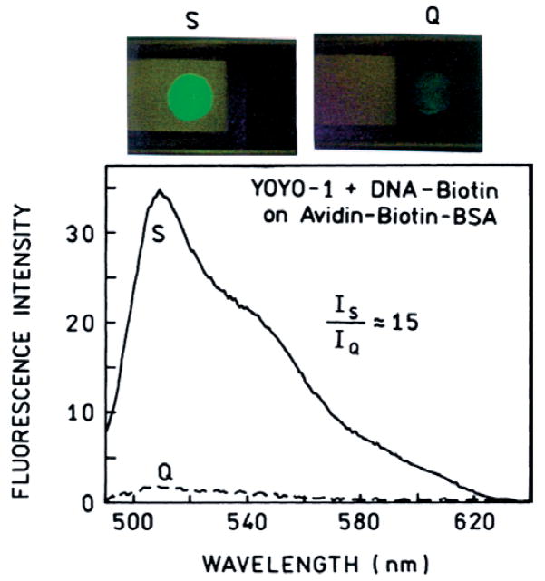
Emission spectra of YOYO-1–labeled DNA bound to the quartz (Q) and silver (S) surfaces. The upper panels show a real-color photograph of labeled DNA spotted on the silver (left) and quartz (right) surfaces.

FD (top) and reconstructed time-domain (bottom) intensity decays of YOYO-1–DNA on quartz (Q) and silver (S).

Photostabilities of YOYO-1–DNA on quartz and silver with the same incident power (top) and at the same incident power but normalized at time zero (bottom).

Schematic of surface, sequence of the DNA oligomers, structures of YOYO-1 and biotinylated oligonucleotide and experimental geometry. Note that sizes in the surface schematic are not in scale. The BSA–avidin protein layer is about 80 Å thick, and the silver particles are about 400 Å high.
Similar articles
-
Fluorescence images of DNA-bound YOYO between coupled silver particles.Langmuir. 2007 Nov 6;23(23):11734-9. doi: 10.1021/la702064v. Epub 2007 Oct 3. Langmuir. 2007. PMID: 17914851 Free PMC article.
-
Stable fluorescent complexes of double-stranded DNA with bis-intercalating asymmetric cyanine dyes: properties and applications.Nucleic Acids Res. 1992 Jun 11;20(11):2803-12. doi: 10.1093/nar/20.11.2803. Nucleic Acids Res. 1992. PMID: 1614866 Free PMC article.
-
1H NMR studies of the bis-intercalation of a homodimeric oxazole yellow dye in DNA oligonucleotides.J Biomol Struct Dyn. 1998 Oct;16(2):205-22. doi: 10.1080/07391102.1998.10508240. J Biomol Struct Dyn. 1998. PMID: 9833661
-
Triplet fraction buildup effect of the DNA-YOYO complex studied with fluorescence correlation spectroscopy.Anal Biochem. 2007 Jul 1;366(1):87-92. doi: 10.1016/j.ab.2007.03.040. Epub 2007 Apr 24. Anal Biochem. 2007. PMID: 17490596
-
Structure and dynamics of condensed DNA probed by 1,1'-(4,4,8,8-tetramethyl-4,8-diazaundecamethylene)bis[4-[[3- methylbenz-1,3-oxazol-2-yl]methylidine]-1,4-dihydroquinolinium] tetraiodide fluorescence.Biochemistry. 2002 Dec 24;41(51):15277-87. doi: 10.1021/bi020440y. Biochemistry. 2002. PMID: 12484766
Cited by
-
Advances in surface-enhanced fluorescence.J Fluoresc. 2004 Jul;14(4):425-41. doi: 10.1023/b:jofl.0000031824.48401.5c. J Fluoresc. 2004. PMID: 15617385 Free PMC article. Review.
-
Fluorescence spectral properties of labeled thiolated oligonucleotides bound to silver particles.Biopolymers. 2004 Jun 15;74(3):263-71. doi: 10.1002/bip.20071. Biopolymers. 2004. PMID: 15150802 Free PMC article.
-
Increasing the sensitivity of DNA microarrays by metal-enhanced fluorescence using surface-bound silver nanoparticles.Nucleic Acids Res. 2007;35(2):e13. doi: 10.1093/nar/gkl1054. Epub 2006 Dec 14. Nucleic Acids Res. 2007. PMID: 17169999 Free PMC article.
-
Dansyl-Labelled Ag@SiO2 Core-Shell Nanostructures-Synthesis, Characterization, and Metal-Enhanced Fluorescence.Materials (Basel). 2020 Nov 16;13(22):5168. doi: 10.3390/ma13225168. Materials (Basel). 2020. PMID: 33207805 Free PMC article.
-
Strategy for photostable proximity bioassays using lanthanides.Appl Opt. 2007 Apr 1;46(10):1918-23. doi: 10.1364/ao.46.001918. Appl Opt. 2007. PMID: 17356638 Free PMC article.
References
-
- Benson SC, Zeng Z, Glazer AN. Fluorescence energy transfer cyanine heterodimers with high affinity for double-stranded DNA. Anal Biochem. 1995;231:247–255. - PubMed
Publication types
MeSH terms
Substances
Grants and funding
LinkOut - more resources
Full Text Sources
Other Literature Sources
Miscellaneous
