. 2003 Aug;109(4):580-7.
doi: 10.1046/j.1365-2567.2003.01700.x.

Human secretory immunoglobulin A may contribute to biofilm formation in the gut

Affiliations

Human secretory immunoglobulin A may contribute to biofilm formation in the gut

R Randal Bollinger et al. Immunology. 2003 Aug.

Abstract

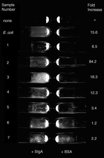
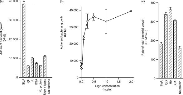
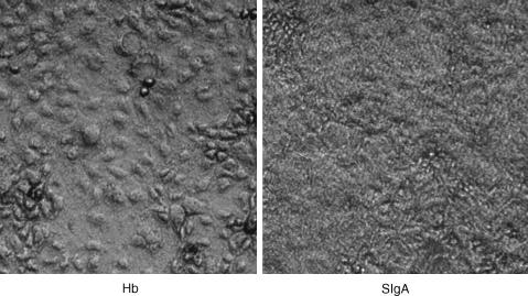
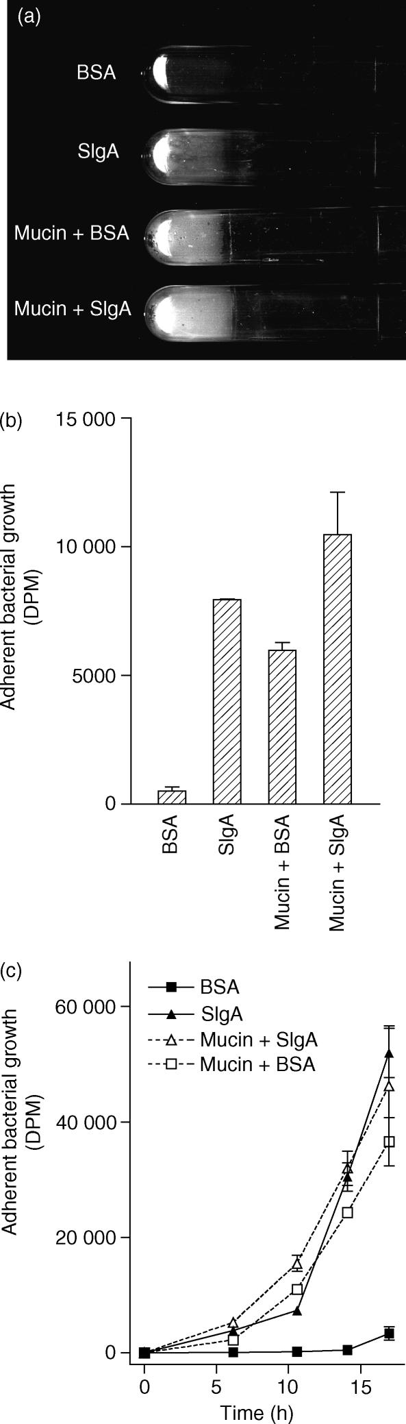
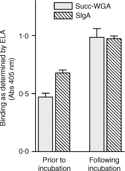
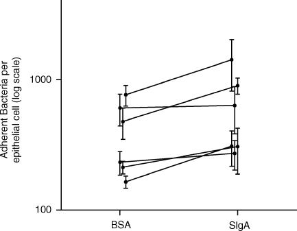
It is critical, both for the host and for the long-term benefit of the bacteria that colonize the gut, that bacterial overgrowth with subsequent bacterial translocation, which may lead to sepsis and death of the host, be avoided. Secretory IgA (sIgA) is known to be a key factor in this process, agglutinating bacteria and preventing their translocation in a process termed 'immune exclusion'. To determine whether human sIgA might facilitate the growth of normal enteric bacteria under some conditions, the growth of human enteric bacteria on cultured, fixed human epithelial cells was evaluated in the presence of sIgA or various other proteins. Human sIgA was found to facilitate biofilm formation by normal human gut flora and by Escherichia coli on cultured human epithelial cell surfaces under conditions in which non-adherent bacteria were repeatedly washed away. In addition, the presence of sIgA resulted in a 64% increase in adherence of E. coli to live cultured epithelial cells over a 45-min period. Mucin, another defence factor thought to play a key role in immune exclusion, was found to facilitate biofilm formation by E. coli. Our findings suggest that sIgA may contribute to biofilm formation in the gut.


Figures

Figure 1
Figure 1
Biofilms on cultured human epithelial cells formed by E. coli or by human faecal bacteria obtained from seven healthy volunteers. The sample identifications on the left refer to either the bacterial species (E. coli) or to the number of the donor. The ‘fold increase’ on the right is the ratio of bacterial growth in the presence of sIgA over the growth in the presence of BSA. Biofilms were grown with the tubes in a vertical position so that the biofilms formed on the side of the tube. Culture tubes were washed three times with saline and the tubes were emptied of all liquid before the photograph was taken. The quantitative bacteria growth is shown in Table 1 and the conditions of the experiment are as described for Table 1. Representative tubes are shown in the photograph.
Figure 2
Figure 2
Effect of sIgA on bacterial growth. Bacteria were grown in the presence of uniformly labelled [14C]glucose on cultured human epithelial cells and the uptake of 14C was used as a measure of bacterial growth. (a) Bacterial growth was measured in the presence of 0·5 mg/ml of various proteins following four changes of growth medium with three saline washes between each change. (b) Using the same washing procedure, adherent bacterial growth was measured as a function of sIgA concentration. (c) The total (non-adherent + adherent) growth rate of enteric bacteria in the presence of 0·5 mg/ml of various proteins was measured. In this case, bacteria were grown in 15-ml conical tubes (no epithelial cells), vortexed for 2 min to dislodge adherent growth, and quantified by 14C-incorporation. All experiments were run in duplicate, and the standard errors are shown. Results comparing sIgA and BSA are representative of eight experiments.
Figure 3
Figure 3
A biofilm as viewed under the light microscope. Phase contrast microscopy revealed the formation of bacterial plaques covering fixed, cultured epithelial cells in the presence of 0·5 mg/ml sIgA (right panel) but not 0·5 mg/ml of other proteins, including hamoglobin (Hb, left panel). In the left panel, the gross topology of the epithelial cells is evident, but is obscured by the bacterial plaque in the right panel.
Figure 4
Figure 4
Biofilms on cultured human epithelial cells formed by E. coli in the presence of 0·5 mg/ml sIgA, 50 mg/ml mucin, or both 0·5 mg/ml sIgA and 50 mg/ml mucin. (a,b) Biofilms were grown for 2·5 days as described in the Materials and methods section with the tubes in a vertical position so that the biofilms formed on the side of the tube. Tubes were washed three times with saline and the tubes were emptied of all liquid before the photograph was taken. Representative tubes are shown in (a), and the amount of adherent growth as quantified by 14C-incorporation is shown in (b). The experiment was reproducible. (c) The rate of adherent bacterial growth is shown. Cultured human epithelial cells were incubated with E. coli in the position described above. The ‘standard protocol’, which entails removal of non-adherent growth four times over a period of 2·5 days (see Materials and Methods section), was not utilized. For this experiment, the frequency with which non-adherent bacteria were removed and fresh media were added to cultures was increased as described in the text. All experiments were run in duplicate, and the standard errors are shown.
Figure 5
Figure 5
Exposure of ‘neoepitopes’ on cultured human epithelial cells following incubation with human enteric bacteria. The binding of succinylated wheat germ agglutinin (succ-WGA) and sIgA were evaluated using ELA as described in the Materials and Methods section. The binding of succ-WGA was used to evaluate the exposure of saccharides normally buried on the cell surface, as described in the text. Twelve wells were used for each condition, and the standard errors are shown. The experiment shown is representative of three experiments performed.
Figure 6
Figure 6
Adherence of E. coli to live human epithelial cells in the presence or absence of sIgA. The number of bacteria adherent to live epithelial cells was determined in the presence of 0·5 mg/ml sIgA or 0·5 mg/ml BSA. Six trials were conducted, with the results of a given trial represented by two points connected by a solid line in the graph. Each trial was conducted in triplicate and the standard errors are shown. In all trials, the number of adherent bacteria in the presence of sIgA was greater than the number adherent in the presence of BSA. However, there was considerable variability in the number of adherent bacteria between trials, perhaps because of day-to-day variability in the cultured epithelial cells. The number of bacteria was determined by measuring the number of plaque forming units.

References

    1. Biancone L, Monteleone I, Del Vecchio Blanco G, Vavassori P, Pallone F. Resident bacterial flora and immune system. Digest Liver Dis. 2002;34:S37–43. - PubMed
    1. Schiffrin EJ, Blum S. Interactions between the microbiota and the intestinal mucosa. Eur J Clin Nutr. 2002;56:S60–4. - PubMed
    1. Williams RC, Gibbons RJ. Inhibition of bacterial adherence by secretory immunoglobulin A. a mechanism of antigen disposal. Science. 1972;177:697–9. - PubMed
    1. Brandtzaeg P. Development and basic mechanisms of human gut immunity. Nutr Rev. 1998;56:S5–18. - PubMed
    1. Mestecky J, Russell MW, Elson CO. Intestinal IgA: novel views on its function in the defence of the largest mucosal surface. Gut. 1999;44:2–5. - PMC - PubMed

Publication types

LinkOut - more resources