. 2003 Aug 5;100(16):9494-9.
doi: 10.1073/pnas.1531901100. Epub 2003 Jul 18.

Angiopoietin-related growth factor (AGF) promotes epidermal proliferation, remodeling, and regeneration

Affiliations

Angiopoietin-related growth factor (AGF) promotes epidermal proliferation, remodeling, and regeneration

Yuichi Oike et al. Proc Natl Acad Sci U S A. .

Abstract

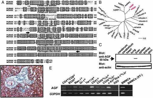
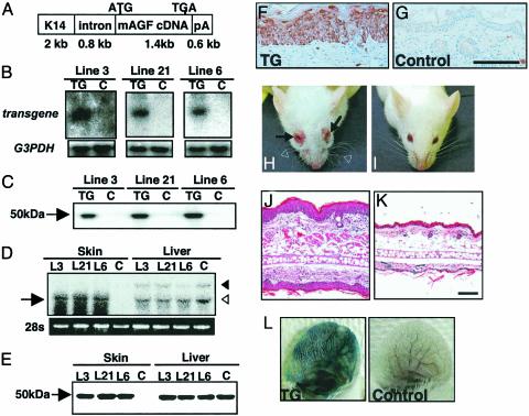
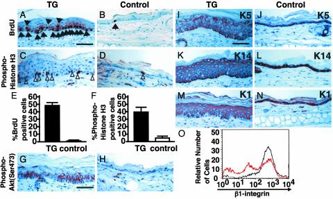
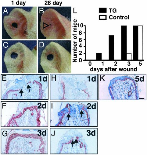
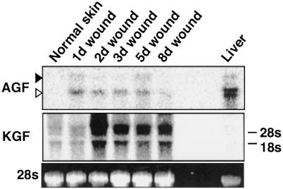
We report here the identification of an angiopoietin-related growth factor (AGF). To examine the biological function of AGF in vivo, we created transgenic mice expressing AGF in epidermal keratinocytes (K14-AGF). K14-AGF mice exhibited swollen and reddish ears, nose and eyelids. Histological analyses of K14-AGF mice revealed significantly thickened epidermis and a marked increase in proliferating epidermal cells as well as vascular cells in the skin compared with nontransgenic controls. In addition, we found rapid wound closure in the healing process and an unusual closure of holes punched in the ears of K14-AGF mice. Furthermore, we observed that AGF is expressed in platelets and mast cells, and detected at wounded skin, whereas there was no expression of AGF detected in normal skin tissues, suggesting that AGF derived from these infiltrated cells affects epidermal proliferation and thereby plays a role in the wound healing process. These findings demonstrate that biological functions of AGF in epidermal keratinocytes could lead to novel therapeutic strategies for wound care and epidermal regenerative medicine.


Figures

Fig. 1.
Fig. 1.
Sequence and expression analyses of AGF. (A) Deduced amino acid sequences of human and mouse AGF. Open and filled arrows indicate the limits of the coiled-coil and fibrinogen-like domains, respectively. (B) The evolutionary relationship of AGF (red) to the angiopoietin superfamily was derived by using dnasis for windows v. 2.1 (Hitachi Software, Tokyo). The length of each horizontal line is proportional to the degree of amino acid sequence divergence. (C) Western blot analysis of various mouse tissues by using the anti-AGF (Upper) and anti-actin (Lower) antibodies. (D) Immunoreaction using the anti-AGF antibody shows AGF specifically expressed in hepatic parenchymal cells, not in the Glisson region. (Scale bar = 100 μm.) (E) Analysis of AGF mRNA expression in hematopoietic cells from adult bone marrow or BMMCs by RT-PCR. CD4+/CD8+, T cell; B220, B cell; Mac-1, macrophage and monocyte; Gr-1, granulocyte; Ter119, erythrocyte; CD41, megakaryocyte/platelet; CD45+Lin+, mature HCs; CD45+Lin-, immature HCs; c-Kit+Sca-1+Lin-, hematopoietic stem cell-enriched population. A mixture of anti-Mac-1, -Gr-1, -B220, -CD4, -CD8, and -Ly-6 antibodies was used as a lineage marker (Lin). GAPDH mRNA served as a loading control. All RNA without RT treatment (RT-) show no transcript by PCR. BMMCs (RT-) is one representative data.
Fig. 2.
Fig. 2.
Markedly thickened epidermal layers in K14-AGF mice. (A) Schematic representation of the transgene used to generate K14-AGF mice. K14, intron, and pA indicate the human K14 promoter, rabbit β-globin intron, and a polyadenylation signal derived from the K14 gene, respectively. (B and C) Expression of the transgene was detected in the whole skin of F1 mice (TG) 3 days after birth by Northern (B) and Western (C) blotting analysis. No expression of the transgene was detected in controls (C). Blotting analysis for GAPDH was performed as an internal control experiment. (D and E) Comparison of the mRNA (D) and protein (E) level of AGF from skin and liver between K14-AGF mice and controls. Arrow in D indicates the transcription of the transgene. Open and filled arrowheads in D indicate 1.8- and 4.0-kb endogeneous AGF transcripts, respectively. Five micrograms of protein was loaded in each lane in E.(F and G) Immunohistochemical analysis of AGF detects expression of the transgene in the epidermis of skin from the ears of F1 K14-AGF mice (F) and their controls (G). (Scale bar = 100 μm.) (H and I) Front view of the K14-AGF mouse and controls. Swelling of the eyelid (arrows in H), ears, and nose, and wavy whiskers (open arrowheads in H) were detected in K14-AGF mice. (J and K) Hematoxylin/eosin histology of swollen ear of the K14-AGF mouse (J) and control (K). (Scale bar = 100 μm.) (L) Photographs of ears injected intravenously with Evans blue dye to visualize plasma leakage. The ear of a K14-AGF mouse was strongly blue, whereas the control ear was not changed. One representative experiment is shown.
Fig. 3.
Fig. 3.
Actively cycling expression of keratin proteins in epidermal cells of K14-AGF mice. (AF) Comparison of levels of DNA synthesis in the epidermis from K14-AGF mice and controls. Skin sections from both mice were stained immunohistochemically for BrdUrd (A and B) and anti-phospho-histone H3 (C and D)by using peroxidase-based detection. Sections were counterstained with hematoxylin. Arrows and arrowheads indicate examples (brown-stained nuclei) of BrdUrd-positive and phospho-histone H3-positive cells, respectively. (E and F) The average numbers of labeled cells with BrdUrd and anti-phospho-histone H3 immunoreactivity from five sections each from three mice, respectively. (G and H) Immunoreactivity against anti-phospho-Akt antibody was seen in the thickened epidermis from the K14-AGF mouse (G), whereas no immunoreactivity was seen in epidermis from controls (H). (IN)K5(J) and K14 (L) were detected in the basal layer of the epidermis in controls. Similar sections obtained from K14-AGF mice show positive staining in the suprabasal layer of the epidermis as well(I and K). K1 staining was similar for both the K14-AGF mouse (M) and controls (N). (Scale bar = 50 μm.) (O) Surface levels of β1-integrins in basal keratinocytes of K14-AGF (red line) and controls (black line). Three peaks for intensity of β1-integrin expression are detected in K14-AGF, whereas one peak with high intensity is seen in controls.
Fig. 4.
Fig. 4.
Reepithelialization of wounds in K14-AGF mice. (AD) Representative photograph ear wound healing. K14-AGF and control ears were punched in the center creating a 2-mm open hole and followed for 28 days. (A and B) One can see the progression of hole closure from day 1 (A) to day 28 (B). Open arrowhead in B indicates shortened hole in K14-AGF. (EK) Wounding was accomplished by ear segment excisions. Shown are representative data of skin sections with staining for anti-K14 antibody from the exposed portion of the remaining ear of K14-AGF mice (EG) and control littermates (HK)at1,2,3, and 5 days after the initial wounds. All sections were photographed at the same magnification. (Scale bar = 100 μm.) Arrowheads indicate migrating and proliferating epidermal keratinocytes, indicating that keratinocytes overlapped the injury site rapidly in K14-AGF mice. (L) Analysis of frequencies of wound closure by histological examination at 1, 2, 3, and 5 days after the initial wounding. Filled (controls) and open (K14-AGF mice) columns represent the number of mice in which the wound was completely covered with keratinocytes. Ten mice were examined on each day of the experiment.
Fig. 5.
Fig. 5.
Expression of AGF mRNA in normal and wounded skin. The total RNA (10 μg) from normal and wounded ear skin was analyzed by Northern blotting analysis with cDNA probes for AGF and KGF. The relative amount of each mRNA was quantified with normalization to 28S rRNA levels. The time after injury is indicated on top of each lane: 1, 2, 3, 5, and 8 days.

References

    1. Vassar, R. & Fuchs, E. (1991) Genes Dev. 5, 714-727. - PubMed
    1. Guo, L., Yu, Q-C. & Fuchs, E. (1993) EMBO J. 12, 973-986. - PMC - PubMed
    1. Werner, S., Smola, H., Liao, X., Longaker, M. T., Krieg, T., Hofschneider, P. H. & Williams, L. T. (1994) Science 266, 819-822. - PubMed
    1. Mann, G. B., Fowler, K. J., Gabriel, A., Nice, E. C., Williams, R. L. & Dunn, A. R. (1993) Cell 73, 249-261. - PubMed
    1. Guo, L., Degensteim, L. & Fuchs, E. (1996) Genes Dev. 10, 165-175. - PubMed

Publication types

MeSH terms