. 2003 Aug 19;100(17):9980-5.
doi: 10.1073/pnas.1533448100. Epub 2003 Jul 18.

Neurodegeneration and defective neurotransmission in a Caenorhabditis elegans model of tauopathy

Affiliations

Neurodegeneration and defective neurotransmission in a Caenorhabditis elegans model of tauopathy

Brian C Kraemer et al. Proc Natl Acad Sci U S A. .

Abstract

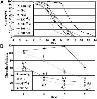
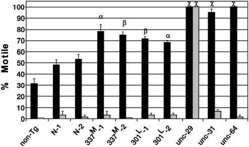
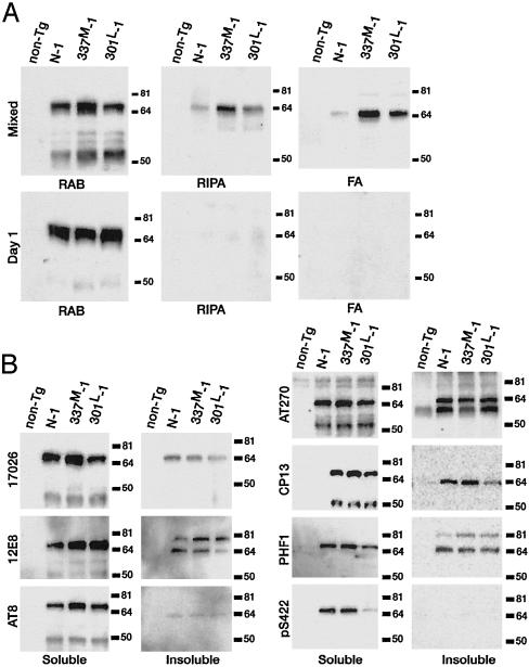
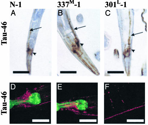
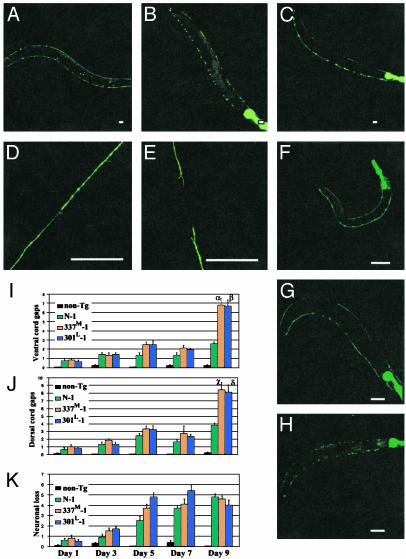
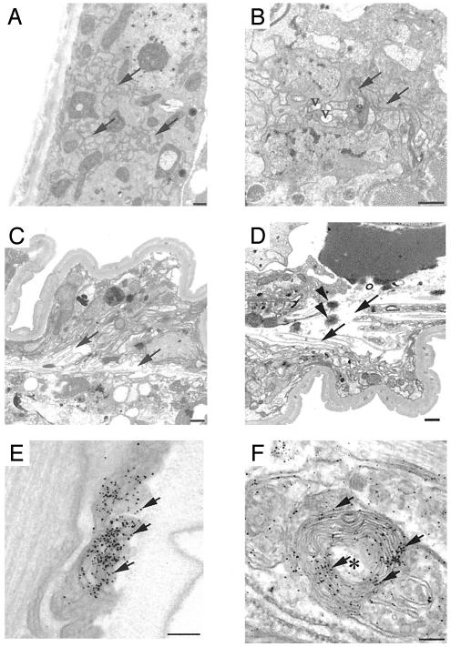
Frontotemporal dementia with parkinsonism chromosome 17 type (FTDP-17) is caused by mutations in MAPT, the gene encoding tau. FTDP-17 begins with executive function deficits and other abnormal behaviors, which progress to dementia. Neurodegenerative changes include accumulation of aggregated tau as neuronal and glial fibrillary tangles. Aggregated tau is seen in numerous other neurodegenerative diseases, including Alzheimer's disease (AD). We expressed normal and FTDP-17 mutant human tau (mutations P301L and V337M) in Caenorhabditis elegans to model tauopathy disorders. Tau pan-neuronal expression caused progressive uncoordinated locomotion (Unc), characteristic of nervous system defects in worms. Subsequently, insoluble tau accumulates and both soluble and insoluble tau is phosphorylated at many of the sites hyperphosphorylated in FTDP-17, AD, and other tauopathies. Substantial neurodegeneration, seen as bulges and gaps in nerve cords followed by loss of neurons, occurs after insoluble tau begins to accumulate. Axons show vacuoles, membranous infoldings, and whorls with associated amorphous tau accumulations and abnormal tau-positive aggregates. FTDP-17 mutation lines had a more severe Unc phenotype, accumulated more insoluble tau at a younger age, were more resistant to cholinergic inhibitors, and had more severe axonal degeneration when compared with lines expressing normal tau. The Unc phenotype is caused by a presynaptic defect. Postsynaptic transmission is intact. This transgenic model will enable mechanistic dissection of tau-induced neurodegeneration and identification of genes and compounds that inhibit pathological tau formation.


Figures

Fig. 1.
Fig. 1.
(A) Tg worm lifespans. Average lifespans in days (±SD) are: N-1, 11.1 ± 0.8; N-2, 12.6 ± 0.8; 337M-1, 10.9 ± 0.4; 337M-2, 12.8 ± 0.3; 301P-1, 12.3 ± 0.3; and 301P-2, 13.31 ± 0.5. Lifespans for each Tg line was different from the non-Tg line (P < 104 to P < 1012; two-tailed t test) but not different from other Tg lines. (B) Unc phenotype and age. Each point is the mean thrashing rate for 10 developmentally staged non-Tg, N-1, 337M-1, or 301L-1 animals. Error bars are the SEM. For N-1, symbols are for comparison to non-Tg values. For 301L and 337M-1, the first symbol is for comparisons to non-Tg and the second symbol is for comparisons to N-1. Symbols are as follows: Φ, P < 0.2; χ, P < 1 × 10²; α, P < 1 × 10³; β, P < 1 × 10⁴; γ, P < 1 × 10⁵; δ, P < 1 × 10⁶; ε, P < 1 × 10⁹; η, P < 1 × 10¹⁰; θ, P < 1 × 10¹⁵.
Fig. 2.
Fig. 2.
Cholinergic neuronal transmission. Worms incubated in aldicarb (dark bars) or levamisole (light bars) were assessed for motility (36). Each bar is the average of three independent experiments with 20 animals each. Symbols are for comparisons to N-1 (α, P = 0.01; β, P < 102; χ, P < 103). Strain unc-29 is deficient for AChR, unc-31 is deficient for Ca2+-dependent activator protein for secretion (CAPS), and unc-64 is deficient for syntaxin.
Fig. 3.
Fig. 3.
(A) Tau solubility. RAB, RIPA, and FA fractions from equivalent amounts of packed mixed stage or 1-day-old worms were analyzed by immunoblotting using the phosphorylation-independent antibody 17026. (B) Tau phosphorylation. The soluble and insoluble proteins are the RAB supernatant and pellet, respectively. Fractions were analyzed by immunoblotting using antibodies that recognize the following tau phospho-epitopes: 12E8, S262; AT-8, S199, S202, and T205; AT-270, T181; CP13, S202; PHF-1, S396 and S404; pS422, S422.
Fig. 4.
Fig. 4.
Immunostaining for Tau. (AC) Paraffin-embedded sections were stained with antibody Tau-46. (DF) Whole mount animals were immunostained (37) by using Tau-46 as the primary antibody and AlexA 568 as the secondary antibody (red). GFP in the pharynx is green. Arrows point to the ventral nerve cord, and arrowheads are head neurons. No staining was seen in non-Tg animals (not shown). D and E, head; F, commissures, dorsal, and ventral nerve cords (see also Fig. 10, which is published as supporting information on the PNAS web site). (Scale bars = 50 μm.)
Fig. 5.
Fig. 5.
Degeneration of γ-aminobutyric acid (GABA)ergic neurons. (AH) GFP fluorescence in anesthetized living worms singly Tg for unc-25::GFP (A,9 days old; D, mid-body dorsal nerve cord, 5 days old), doubly Tg for unc-25::GFP and 337M-1 (B, 9 days old; E, mid-body dorsal cord, 5 days old), or doubly Tg for unc-25::GFP and 301L-1 (C, 9 days old). Dorsal is up, and anterior is left. (Scale bar = 40 μm.) (FH) GABAergic neuronal degeneration in a single 337M-1/unc-25::GFP animal examined at age 1 day (F), 5 days (G), and 7 days (H). (IK) Quantitation of dorsal cord (I) and ventral cord (J) gaps and neuronal loss (K) at different ages. Each measurement is the mean from 10 animals ± SEM. Symbols are for comparisons to N-1 (α, P = 1 × 10³; β, P = 2 × 10²; χ, P = 3 × 10⁴; δ, P = 3 × 10³).
Fig. 6.
Fig. 6.
Ultrastructural of degenerating axons. (AD) Transmission EM. Transverse sections of 9-day-old N-1 (A), 301L-1 (B), and 337M-1 (C and D) lines. Arrows show normal axons in A, mildly degenerative axons in B, and severely degenerating disoriented axons in C and D. V, vacuolar clearing of axoplasm. Spherical, osmophilic protein aggregates (arrowheads) were evident (D). (E and F) Preembedding immuno-EM of 337M-1 animals by using antibody 17026. The arrow shows immuno-silver-enhanced particles that identify tau-labeled structures. Degenerating axons with collapsed membranous profiles (onion skin lesions) are indicated by an asterisk and contain amorphous aggregates labeled by anti-tau antibodies. (Scale bars = 500 nm in AD and 100 nm in E and F.)

Comment in

  • Neurodegenerative tauopathy in the worm.
    Goedert M. Goedert M. Proc Natl Acad Sci U S A. 2003 Aug 19;100(17):9653-5. doi: 10.1073/pnas.1834191100. Epub 2003 Aug 11. Proc Natl Acad Sci U S A. 2003. PMID: 12913116 Free PMC article. No abstract available.

References

    1. Hutton, M., Lendon, C. L., Rizzu, P., Baker, M., Froelich, S., Houlden, H., Pickering-Brown, S., Chakraverty, S., Isaacs, A., Grover, A., et al. (1998) Nature 393, 702–705. - PubMed
    1. Poorkaj, P., Bird, T. D., Wijsman, E., Nemens, E., Garruto, R. M., Anderson, L., Andreadis, A., Wiederholt, W. C., Raskind, M. & Schellenberg, C. D. (1998) Ann. Neurol. 43, 815–825. - PubMed
    1. Spillantini, M. G., Murrell, J. R., Goedert, M., Farlow, M. R., Klug, A. & Ghetti, B. (1998) Proc. Natl. Acad. Sci. USA 95, 7737–7741. - PMC - PubMed
    1. Hong, M., Zhukareva, V., Vogelsberg-Ragaglia, V., Wszolek, Z., Reed, L., Miller, B. I., Geschwind, D. H., Bird, T. D., McKeel, D. Goate, A., et al. (1998) Science 282, 1914–1917. - PubMed
    1. Goedert, M., Jakes, R. & Crowther, R. A. (1999) FEBS Lett. 450, 306–311. - PubMed

Publication types

MeSH terms

LinkOut - more resources