. 2003 Aug;34(4):413-20.
doi: 10.1038/ng1217.

Mutations in INVS encoding inversin cause nephronophthisis type 2, linking renal cystic disease to the function of primary cilia and left-right axis determination

Affiliations

Mutations in INVS encoding inversin cause nephronophthisis type 2, linking renal cystic disease to the function of primary cilia and left-right axis determination

Edgar A Otto et al. Nat Genet. 2003 Aug.

Abstract

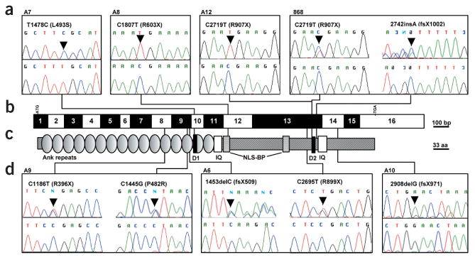
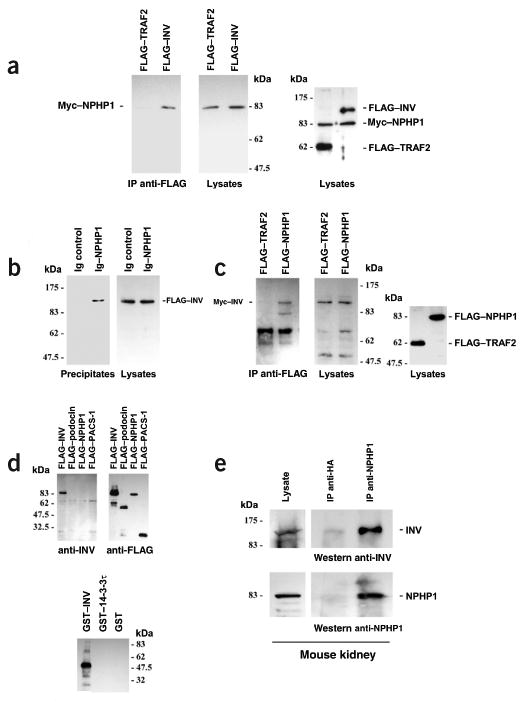
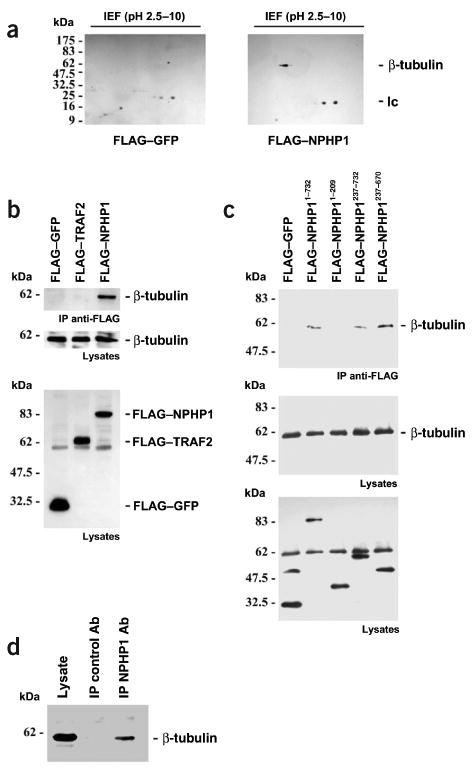
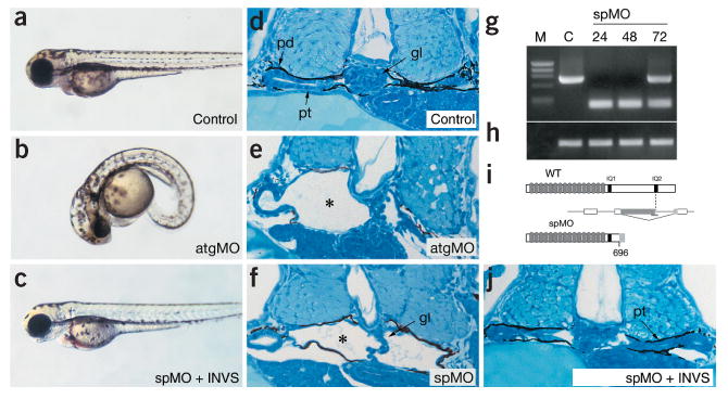
Nephronophthisis (NPHP), an autosomal recessive cystic kidney disease, leads to chronic renal failure in children. The genes mutated in NPHP1 and NPHP4 have been identified, and a gene locus associated with infantile nephronophthisis (NPHP2) was mapped. The kidney phenotype of NPHP2 combines clinical features of NPHP and polycystic kidney disease (PKD). Here, we identify inversin (INVS) as the gene mutated in NPHP2 with and without situs inversus. We show molecular interaction of inversin with nephrocystin, the product of the gene mutated in NPHP1 and interaction of nephrocystin with beta-tubulin, a main component of primary cilia. We show that nephrocystin, inversin and beta-tubulin colocalize to primary cilia of renal tubular cells. Furthermore, we produce a PKD-like renal cystic phenotype and randomization of heart looping by knockdown of invs expression in zebrafish. The interaction and colocalization in cilia of inversin, nephrocystin and beta-tubulin connect pathogenetic aspects of NPHP to PKD, to primary cilia function and to left-right axis determination.


Figures

Figure 1
Figure 1
Mutations in INVS in individuals with NPHP2. (a,d) Mutations in INVS (nucleotide exchange and amino acid exchange) are shown together with sequence traces for mutated sequence (top) and sequence from healthy controls (bottom). Family numbers are given above boxes. If only one mutation is shown, it occurred in the homozygous state, except in individual A10, in whom only one mutation in the heterozygous state was detected. In individual 868, the 2742insA mutation is shown in the flipped version of the reverse strand. (b) Exon structure of INVS. Lines indicate relative positions and connect to mutations detected in INVS. Open and filled boxes represent INVS exons drawn relative to scale bar. Positions of start codon (ATG) at nucleotide +1 and of stop codon (TGA) are indicated. (c) Representation of protein motifs is drawn to scale parallel to exon structure. Lines connect to point mutations detected, as shown in boxes (a,d). aa, amino acid residues; Ank, ankyrin/swi6 motif; D1, D box1 (Apc2-binding); D2, D box2; IQ, calmodulin binding domains.
Figure 2
Figure 2
Nephrocystin associates with inversin in HEK293T cells and mouse tissue. (a) Myc-tagged nephrocystin (Myc–NPHP1) was coexpressed with N-terminally FLAG-tagged full-length inversin (FLAG–INV) or FLAG-tagged TRAF2 (FLAG–TRAF2) protein as a negative control. After immunoprecipitation with anti-FLAG antibody, coprecipitating nephrocystin was detected with nephrocystin-specific antiserum (left panel). We controlled for protein expression levels in cellular lysates by immunoblotting using a nephrocystin antibody (middle panel) or FLAG-specific and nephrocystin-specific antibodies (right panel). Molecular weight markers are shown in kDa. (b) Full-length nephrocystin was fused to the CH2 and CH3 domains of human IgG1 and precipitated with protein G sepharose beads. FLAG-tagged inversin specifically coprecipitated with nephrocystin but not with control protein (CH2 and CH3 domains of human IgG1 without nephrocystin fusion) as shown with FLAG-specific antibody. (c) FLAG-tagged nephrocystin or FLAG-tagged TRAF2 protein as a negative control was coexpressed with N-terminally Myc-tagged full-length inversin (Myc–INV). After immunoprecipitation with anti-FLAG antibody, coprecipitating inversin was detected with inversin-specific antiserum (left and middle panels). Right panel shows lysate control developed with FLAG-specific antibody. (d) A rabbit antiserum to an MBP-inversin fusion protein (amino acids 561–716 of mouse inversin) specifically recognized inversin (amino acids 1–716) expressed in HEK293T cells (left panel) but not the FLAG-tagged control proteins podocin (FLAG–podocin), nephrocystin (FLAG-NPHP1) or PACS-1 (FLAG–PACS–1, amino acids 85–280; left panel). It also specifically recognized recombinant GST-inversin (amino acids 561–716) but not two other control GST fusion proteins (lower panel). (e) To show endogenous nephrocystin-inversin interaction in vivo in mouse kidney, half of mouse kidney tissue lysates was immunoprecipitated with a control antibody to hemagglutinin (anti-HA), and the other half was precipitated with anti-nephrocystin antisera. Immobilized inversin was detected with the inversin-specific antisera (right upper panel). Precipitation of endogenous nephrocystin was confirmed by reprobing the blot for nephrocystin (right lower panel). Left panels show lysate control.
Figure 3
Figure 3
Molecular interaction of nephrocystin with β-tubulin. (a) To identify nephrocystin-interacting proteins, HEK 293T cells were transfected with the FLAG-tagged control protein GFP or FLAG-tagged nephrocystin. Specific association of β-tubulin with nephrocystin was confirmed by immunoblotting of 2D gels using anti-tubulin antibody. lc, immunoglobulin light chain. (b) Several FLAG-tagged nephrocystin truncations were generated to analyze the interaction of nephrocystin with β-tubulin. Endogenous β-tubulin precipitated with transfected full-length nephrocystin but not with the control proteins GFP or TRAF2 (upper panel). Middle panel, expression of native β-tubulin in lysates. Lower panel shows a reprobe of the membrane depicted in middle panel with anti-FLAG antibody (β-tubulin is still detected below the 62 kDa marker), confirming comparable expression levels of the FLAG-tagged proteins. (c) The interaction was mapped to a region of nephrocystin involving amino acids 237–670 (c, upper panel). Expression levels of β-tubulin are shown (c, bottom panel). The blot was reprobed with anti-FLAG antibody to confirm expression of the FLAG-tagged proteins in the lysates (c, lower panel). (d) Coprecipitation of endogenous β-tubulin with native nephrocystin in ciliated mCcd-K1 cells. mCcd-K1 cells were grown for 7 d until cilia were recognized (data not shown). Cell lysates were precleared extensively and immunoprecipitated with a control antibody (Ab) or nephrocystin-specific antiserum and separated by 10% SDS–PAGE. We detected endogenous β-tubulin in the nephrocystin-containing precipitates using a β-tubulin-specific antibody, indicating that the nephrocystin–β-tubulin interaction occurs in vivo.
Figure 4
Figure 4
Nephrocystin and inversin localize to primary cilia in renal tubular epithelial cells. (a) Colocalization of nephrocystin and β-tubulin-4 in primary cilia of MDCK cells. Two examples of cilia are shown in the upper and lower panels. Wild-type MDCK cells (clone II) were grown on coverslips at 100% confluence and cultivated for 7 d before the experiment to allow full polarization and cilia formation. Localization of nephrocystin was determined by immunofluorescence using nephrocystin-specific antibody with confocal images captured at the level of the apical membrane. Cells were costained with rabbit antibody to nephrocystin (left panels) and mouse antibody to β-tubulin-4 (middle panels) followed by the respective secondary antibodies. (b) Specific localization of nephrocystin in primary cilia was confirmed by the use of blocking recombinant nephrocystin protein. (c) Inversin localizes to primary cilia in MDCK cells. Localization of endogenous inversin was determined by immunofluorescence using inversin-specific antibody with confocal images captured at the level of the apical membrane. Cells were costained with mouse antibody to β-tubulin-4 and rabbit antibody to inversin followed by the respective secondary antibodies (lower panel). In additional stainings, the antibody to β-tubulin-4 was omitted to reduce potential spectral overlap between the inversin and β-tubulin-4 signals (upper panel). (d) Partial colocalization of nephrocystin and inversin in primary cilia. Localization of nephrocystin was determined by immunofluorescence using nephrocystin-specific antibody with confocal images captured at the level of the apical membrane. Cells were costained with goat antibody to inversin (left panel) and rabbit antibody to nephrocystin (middle panel) followed by the respective secondary antibodies. Partial colocalization is shown in the right panel.
Figure 5
Figure 5
Disruption of zebrafish invs function results in renal cyst formation. (a) Embryos injected with a control, non-specific oligonucleotide have normal morphology. (b) Embryos injected with atgMO and spMO (not shown) have a pronounced ventral axis curvature at 3 d.p.f. (combined totals for atgMO and spMO: 432 of 479 injected embryos; 90%). (c) Coinjection of 100 pg mouse Invs mRNA with spMO completely rescued axis curvature defects (combined totals for atgMO and spMO: 363 of 381 mRNA+MO injected embryos were rescued; 95%). (d) Histological section of a 2.5-d.p.f. control embryo pronephros showing the midline glomerulus (Gl), pronephric tubule (Pt) and pronephric duct (Pd). (e) atgMO-injected 3-d.p.f. embryo showing cystic dilatation of pronephric tubules and glomerulus (indicated with an asterisk) lined with squamous epithelium. (f) spMO similarly causes cystic maldevelopment of the pronephric tubules (marked with an asterisk). (gi) Molecular analysis of morpholino targeted invs splicing defects: RT–PCR analysis of invs expression in 24-h.p.f. control injected embryos generates a 746-bp invs fragment encoding the C-terminal domain (g; lane C, nucleotides 2,233–2,979 of GenBank AF465261; lane M, ΦX174 markers). spMO-injected embryos (g; lanes spMO; 24, 48 and 72 h.p.f.) analyzed with the same RT–PCR primers generate a 189-bp RT–PCR product representing a C-terminal invs deletion allele. Some recovery of wild-type (WT) mRNA is observed at 72 h.p.f. (h) RT–PCR of ACTB mRNA on the same RNA samples as in g shows no effect of morpholino injection at any time point. (i) Diagram of effect of spMO on invs mRNA processing. Preventing normal splicing in the IQ2 domain recruits a cryptic splice donor in upstream invs coding sequence; the resulting out-of-frame fusion generates a C-terminally truncated invs mRNA at amino acid 696 with an altered 21 amino acid C terminus. (j) Rescue of normal morphology by coinjected spMO and mouse Invs mRNA shows a normal pronephric duct structure (Pt). When injected alone, mouse Invs mRNA had no effect.

Comment in

  • From cilia to cyst.
    Watnick T, Germino G. Watnick T, et al. Nat Genet. 2003 Aug;34(4):355-6. doi: 10.1038/ng0803-355. Nat Genet. 2003. PMID: 12923538 No abstract available.

References

    1. Smith C, Graham J. Congenital medullary cysts of the kidneys with severe refractory anemia. Am J Dis Child. 1945;69:369–377.
    1. Fanconi G, Hanhart E, Albertini A. Die familiäre juvenile Nephronophthise. Hel Pediatr Acta. 1951;6:1–49. - PubMed
    1. Hildebrandt F. Juvenile nephronophthisis. In: Barratt TM, Avner ED, Harmon WE, editors. Pediatric nephrology. Williams & Wilkins; Baltimore: 1999.
    1. Hildebrandt F, et al. A novel gene encoding an SH3 domain protein is mutated in nephronophthisis type 1. Nat Genet. 1997;17:149–153. - PubMed
    1. Saunier S, et al. A novel gene that encodes a protein with a putative src homology 3 domain is a candidate gene for familial juvenile nephronophthisis. Hum Mol Genet. 1997;6:2317–2323. - PubMed

Publication types

MeSH terms