. 2003 Sep;130(17):4123-33.
doi: 10.1242/dev.00559.

Altered fracture repair in the absence of MMP9

Affiliations

Altered fracture repair in the absence of MMP9

Céline Colnot et al. Development. 2003 Sep.

Abstract

The regeneration of adult skeletal tissues requires the timely recruitment of skeletal progenitor cells to an injury site, the differentiation of these cells into bone or cartilage, and the re-establishment of a vascular network to maintain cell viability. Disturbances in any of these cellular events can have a detrimental effect on the process of skeletal repair. Although fracture repair has been compared with fetal skeletal development, the extent to which the reparative process actually recapitulates the fetal program remains uncertain. Here, we provide the first genetic evidence that matrix metalloproteinase 9 (MMP9) regulates crucial events during adult fracture repair. We demonstrate that MMP9 mediates vascular invasion of the hypertrophic cartilage callus, and that Mmp9(-/-) mice have non-unions and delayed unions of their fractures caused by persistent cartilage at the injury site. This MMP9- dependent delay in skeletal healing is not due to a lack of vascular endothelial growth factor (VEGF) or VEGF receptor expression, but may instead be due to the lack of VEGF bioavailability in the mutant because recombinant VEGF can rescue Mmp9(-/-) non-unions. We also found that Mmp9(-/-) mice generate a large cartilage callus even when fractured bones are stabilized, which implicates MMP9 in the regulation of chondrogenic and osteogenic cell differentiation during early stages of repair. In conclusion, the resemblance between Mmp9(-/-) fetal skeletal defects and those that emerge during Mmp9(-/-) adult repair offer the strongest evidence to date that similar mechanisms are employed to achieve bone formation, regardless of age.


Figures
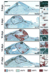
Fig. 1
Fig. 1
MMP9 expression during non-stable fracture healing. (Left column) Sagittal sections through the wild-type callus at 3, 6, 10, 14 and 21 days post-fracture, stained with Safranin-O/Fast Green (SO-FG), illustrates the abundant proteoglycan-containing cartilage (red, c) that appears during the soft callus phase of healing (4–10 days post-fracture) and is resorbed during the hard callus phase of healing (10–21 days post-fracture). The borders of the fracture callus are delimited with a dotted line. The proximal half of the broken tibia is on the left side, showing the growth plate, and the distal part of the tibia is on the right side. (Right column) In situ hybridization and immunostaining analyses at 3, 6, 10, 14 and 21 days post-fracture. At 3 days, analyses show Mmp9 mRNA (green) and MMP9 protein (brown) in inflammatory cells around the fracture site. At 6 days, MMP9 protein is detected in TRAP-positive cells at the fracture site. At 10 days, Mmp9 mRNA is localized to cells at the boundary between the cartilage callus (c) and newly forming bone (b). This cartilage-bone boundary, demarcated by a dotted line, is visualized with SO-FG staining on an adjacent section. At 14 days, Mmp9-expressing cells are detected at the site of hypertrophic cartilage degradation. At 21 days, MMP9- and TRAP-positive osteoclasts are localized in the callus at the site of new bone remodeling. Higher magnifications in the right column correspond to the boxed areas in the left column. gp, growth plate; bm, bone marrow; b, bone. Scale bars: SO-FG staining (low magnification), 2 mm; SO-FG staining (high magnification) and Mmp9 in situ hybridization, 250 μm; MMP9 immunostaining and TRAP-MMP9 double staining, 10 μm.
Fig. 2
Fig. 2
Mmp9−/− mice exhibit an accumulation of cartilage during non-stable fracture healing. (A) SO-FG staining of wild-type (top, +/+) and Mmp9−/− callus tissues (bottom, −/−) at 10, 14 and 21 days indicates that cartilage persists in the Mmp9−/− callus up to 21 days. (B) Histomorphometric measurements of total callus and cartilage volumes in wild-type (n=12) and Mmp9−/− (n=17) mice at 10 and 14 days. There is a statistically significant increase in the volume of cartilage in Mmp9−/− calluses compared with wild-type calluses at 14 days (asterisks, Student’s t-test, P=0.02). Bars represent mean±s.e.m. Scale bar: 1 mm.
Fig. 3
Fig. 3
Hypertrophic callus remodeling and bone formation are delayed in Mmp9−/− mice. Transcripts for collagen type X (ColX, yellow) and osteocalcin (Oc, black) are superimposed onto adjacent sections stained with SO-FG to illustrate the location of hypertrophic cartilage and new bone (same sections showing only SO-FG staining are also shown in Fig. 2B). By 14 days, ColX (yellow) is downregulated in the wild-type callus but remains strongly expressed throughout the Mmp9−/− callus. Conversely, Oc is strongly expressed in the wild-type callus by 14 days but is expressed at much lower levels in the Mmp9−/− callus. Blue dotted lines represent the boundary of the callus. Scale bar: 1 mm.
Fig. 4
Fig. 4
Mmp9−/− mice exhibit an ossification defect during stabilized fracture repair. In A and B, wild-type is left, Mmp9−/− is right. (A) SO-FG stained sections through the stabilized fracture site at 10 days confirm the presence of abundant new bone (arrows indicate Oc expression) and the absence of cartilage in the wild-type callus (no ColIIa signal is evident despite hybridization with this RNA probe). By sharp contrast, abundant cartilage forms in the Mmp9−/− callus regardless of stabilization of the bone segments, as shown by the expression of ColIIa (pink). Oc expression (black) is also detected in the periosteum, adjacent to the fracture site (arrows) and in the endosteum. (B) SO-FG staining of sections at a pin implant (*) at 10 days shows that although no cartilage is detected in wild-type animals, abundant cartilage (red) is present in Mmp9−/− animals. This cartilage is restricted to the periosteal surface (po). Oc expression (black) is localized both at the periosteal and endosteal (en) surfaces (arrows) in wild-type and Mmp9−/− animals. (C) SO-FG stained sections through the wild-type callus illustrate that if fractures are left unstable for 24 hours (left, 24h) and subsequently stabilized, they heal without evidence of cartilage (10 days; arrow indicates the healing fracture). However, fractures that are unstable for 48 hours (right, 48h) and subsequently stabilized tend to heal with abundant cartilage (red). (D) MMP9 immunostaining and double-staining with TRAP illustrate that MMP9 protein (brown) is detected by day 3 in the endosteal matrix (box 1), in inflammatory cells (box 2), in mesenchymal cells within the fracture gap and surrounding soft tissues (box 3), and in the periosteum (box 4). These MMP9-positive cells are TRAP-negative. TRAP-positive osteoclasts/chondroclasts are present at the epiphyseal growth plates of the fractured bone (not shown), and in the cortical bone. bm, bone marrow; c, cortex. Scale bars: in A,C, 1 mm; in B,D (low magnification), 500 μm; in D (high magnification), 10 μm.
Fig. 5
Fig. 5
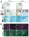
Mmp9−/− fracture healing is hindered by the reduction in chondroclasts/osteoclasts and a delay in vascular invasion. 10 days, left half; 14 days right half. (A) Top row, left panels show that in wild-type mice at 10 days post-fracture, Mmp9-expressing chondroclasts/osteoclasts (inset shows cells with characteristic ruffled borders) begin to accumulate at the wild-type cartilage/bone boundary (dotted red line). MMP9 is not expressed in null-mutant mice. In an adjacent section (middle row, left panels), TRAP activity highlights the location of osteoclasts in the wild-type callus (arrowheads), which are largely absent from the Mmp9−/− callus. b, bone; c, cartilage. In near-adjacent sections (bottom row, left panels), PECAM-expressing endothelial cells (arrow) have accumulated at the border between wild-type hypertrophic cartilage (c) and newly forming bone (b; arrow). Few PECAM-positive cells are detected in the Mmp9−/− callus (arrow). By 14 days post-fracture, abundant MMP9 protein (upper right panels, brown) and TRAP activity (middle right panels, arrowheads) indicate that osteoclasts-mediated degradation of the cartilage callus is well underway (dotted red line indicates the cartilage-bone junction). This degradation activity is associated with increased vascular invasion as illustrated by PECAM-expressing endothelial cells (arrows). Although more PECAM-positive cells are present at 14 days in the Mmp9−/−callus (arrow), they are restricted to the edge of the cartilage-bone junction (dotted red line). (B) Tissue sections adjacent to those analyzed in A were examined for the expression of Vegf (top, fuchsia) and one of its receptors, Flk1 (bottom, green). Vegf transcripts were detected in late hypertrophic chondrocytes in both wild-type and Mmp9−/−calluses at 10 and 14 days post-fracture. Transcripts for Flk1 were detected in endothelial cells surrounding the cartilage callus of both wild-type and Mmp9−/− calluses. b, bone; c, cartilage. The dotted lines in B delimit the boundary of the cartilage in the callus. Scale bars: in A (low magnification) and in B, 200 μm; in A (high magnification), 20 μm.
Fig. 6
Fig. 6
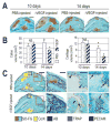
rVEGF rescues the Mmp9−/− fracture repair defect. (A) Tissue sections from PBS- and rVEGF-injected Mmp9−/− calluses were stained with SO-FG at 10 and 14 days post-fracture to illustrate that, during the maturation phase of fracture healing (10 days), there is no difference in either the amount of cartilage (red) or the size of the callus (indicated by dotted black line). However, by 14 days post-fracture, the amount of cartilage was substantially reduced in Mmp9−/−mice that received rVEGF compared with those that received PBS. These histological observations were confirmed by histomorphometric measurements of the total callus volume and the cartilage volume (B). By 14 days post-fracture, there was a statistically significant decrease in both the total callus volume (left graft, asterisks) and in the cartilage volume (right graft, asterisks) of Mmp9−/− mice that received rVEGF (n=10) versus PBS (n=10), as assessed by ANOVA (P=0.03 and P=0.02, respectively; bars represent mean±s.e.m.). (C) By 14 days post-fracture, substantially less hypertrophic cartilage was detected in the Mmp9−/− calluses that had been treated with rVEGF compared with their PBS counterparts. Left panels show the yellow ColX hybridization signal superimposed upon a tissue section stained with SO-FG. Higher magnification of area boxed in red illustrates that ossification was also accelerated by rVEGF when compared with PBS controls [Analine Blue, (AB) stain superimposed upon an adjacent tissue section stained with SO-FG]. Higher magnification of the area boxed in black illustrates that rVEGF induced substantially greater osteoclast-mediated degradation of the callus compared with PBS controls (arrowheads in upper and lower panels indicate TRAP immunostaining, red dotted line demarcates the boundary between the remaining hypertrophic cartilage and the newly forming bone). Higher magnification of this same region demonstrates that rVEGF resulted in an increased vascular invasion of the callus, as shown by the presence of PECAM-positive endothelial cells penetrating the Mmp9−/−hypertrophic cartilage callus (arrows, bottom right panel). Conversely, endothelial cells remain at the periphery of the PBS-injected Mmp9−/−callus. Scale bars: in A,C (SO/ColX panel), 1 mm; in C (TRAP and PECAM panels), 200 μm.

References

    1. Albrecht U, Eichele G, Helms JA, Lin H. Visualization of gene expression patterns by in situ hybridization. Boca Raton, FL: CRC Press; 1997.
    1. Alliston T, Choy L, Ducy P, Karsenty G, Derynck R. TGF-beta-induced repression of CBFA1 by Smad3 decreases cbfa1 and osteocalcin expression and inhibits osteoblast differentiation. EMBO J. 2001;20:2254–2272. - PMC - PubMed
    1. Altman RD, Latta LL, Keer R, Renfree K, Hornicek FJ, Banovac K. Effect of nonsteroidal antiinflammatory drugs on fracture healing: a laboratory study in rats. J Orthop Trauma. 1995;9:392–400. - PubMed
    1. Banovac K, Renfree K, Makowski AL, Latta LL, Altman RD. Fracture healing and mast cells. J Orthop Trauma. 1995;9:482–490. - PubMed
    1. Bergers G, Brekken R, McMahon G, Vu TH, Itoh T, Tamaki K, Tanzawa K, Thorpe P, Itohara S, Werb Z, et al. Matrix metalloproteinase-9 triggers the angiogenic switch during carcinogenesis. Nat Cell Biol. 2000;2:737–744. - PMC - PubMed

Publication types

Substances