. 2003 Jul 21;198(2):249-58.
doi: 10.1084/jem.20030315.

Antigen-dependent proliferation of CD4+ CD25+ regulatory T cells in vivo

Affiliations

Antigen-dependent proliferation of CD4+ CD25+ regulatory T cells in vivo

Lucy S K Walker et al. J Exp Med. .

Abstract

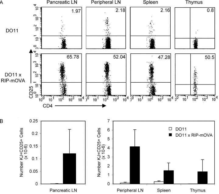
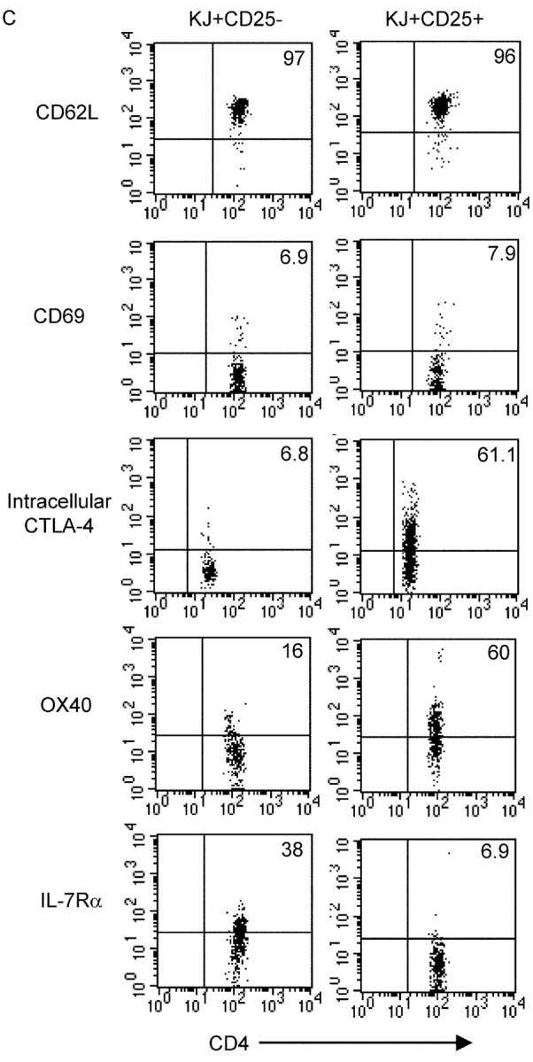
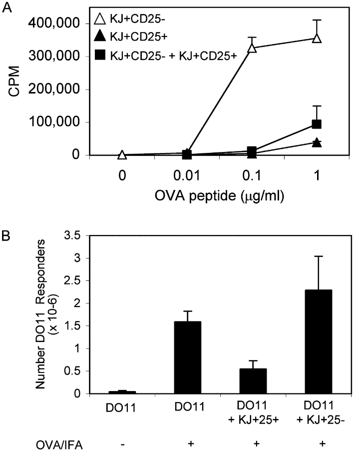
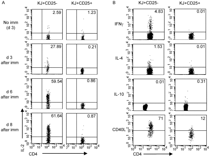
The failure of CD25+ regulatory T cells (Tregs) to proliferate after T cell receptor (TCR) stimulation in vitro has lead to their classification as naturally anergic. Here we use Tregs expressing a transgenic TCR to show that despite anergy in vitro, Tregs proliferate in response to immunization in vivo. Tregs also proliferate and accumulate locally in response to transgenically expressed tissue antigen whereas their CD25- counterparts are depleted at such sites. Collectively, these data suggest that the anergic state that characterizes CD25+ Tregs in vitro may not accurately reflect their responsiveness in vivo. These observations support a model in which Treg population dynamics are shaped by the local antigenic environment.


Figures

Figure 1.
Figure 1.
Abundant clonotype+ CD25+ cells in DO11 × RIP-mOVA double transgenic mice. (A) FACS® profiles of DO11 single positive versus DO11 × RIP-mOVA double transgenic littermates at 7 wk of age. Cells were isolated from the indicated lymphoid tissues and stained with CD4-PERCP, CD25-PE, and KJ-126-APC. Plots are gated on CD4+ KJ+ cells (except for thymus plots, which are gated on CD4+ CD8 KJ+ cells) and show the percentage of CD25+ cells. (B) Absolute number of CD4+ KJ+ CD25+ cells in the lymphoid tissues of DO11 single positive versus DO11 × RIP-mOVA double transgenic littermates. Data show mean and standard deviation from three mice of each genotype aged 6–7 wk. (C) Peripheral LN cells from DO11 × RIP-mOVA double transgenic mice were stained with various combinations of KJ-126-APC, CD62L-FITC, CD69PE, CD4-PERCP, OX40- biotin, streptavidin-PE, IL-7Rα-PE, and CD25-FITC/PE. For intracellular staining cells were surface stained and then permeabilized and stained with CTLA-4-PE. Expression profiles of each marker are shown for gated CD4+ KJ+ CD25 versus CD4+ KJ+ CD25+ cells.
Figure 1.
Figure 1.
Abundant clonotype+ CD25+ cells in DO11 × RIP-mOVA double transgenic mice. (A) FACS® profiles of DO11 single positive versus DO11 × RIP-mOVA double transgenic littermates at 7 wk of age. Cells were isolated from the indicated lymphoid tissues and stained with CD4-PERCP, CD25-PE, and KJ-126-APC. Plots are gated on CD4+ KJ+ cells (except for thymus plots, which are gated on CD4+ CD8 KJ+ cells) and show the percentage of CD25+ cells. (B) Absolute number of CD4+ KJ+ CD25+ cells in the lymphoid tissues of DO11 single positive versus DO11 × RIP-mOVA double transgenic littermates. Data show mean and standard deviation from three mice of each genotype aged 6–7 wk. (C) Peripheral LN cells from DO11 × RIP-mOVA double transgenic mice were stained with various combinations of KJ-126-APC, CD62L-FITC, CD69PE, CD4-PERCP, OX40- biotin, streptavidin-PE, IL-7Rα-PE, and CD25-FITC/PE. For intracellular staining cells were surface stained and then permeabilized and stained with CTLA-4-PE. Expression profiles of each marker are shown for gated CD4+ KJ+ CD25 versus CD4+ KJ+ CD25+ cells.
Figure 2.
Figure 2.
The generation of KJ+ CD25+ cells does not require endogenous TCRα chains. (A) Frequency of TCR Vα2 usage and expression levels of the transgenic TCR on peripheral LN cells from DO11 × RIP-mOVA mice. Histogram labels depict the median fluorescence channel. (B) Proportion of CD4+ KJ+ cells expressing CD25 in DO11 versus DO11 × RIP-mOVA mice on a RAG−/− background. Cells were isolated and stained as described in Fig. 1.
Figure 3.
Figure 3.
KJ+ CD25+ cells are suppressive in vitro and in vivo. (A) KJ+ CD25 and KJ+ CD25+ populations were purified from pooled peripheral LNs of DO11 × RIP-mOVA double transgenic mice by high speed cell sorting and stimulated in 96-well plates, either alone or in combination at a 1:1 ratio, with BALB/c splenocytes and the indicated concentration of OVA323–339 peptide. Data show one experiment that is representative of three. (B) 2 × 10⁶ DO11 T cells (from a single positive DO11 mouse) were injected into BALB/c recipients either alone or in combination with 106 CFSE-labeled KJ+ CD25+ cells or 106 CFSE-labeled KJ+ CD25 cells. Recipient mice were immunized s.c. with 200 μg OVA/IFA where indicated. Draining LNs were isolated at day 3 and the absolute number of responder DO11 cells (CFSE) is shown. Data show one experiment (two mice per group) and are representative of two separate experiments.
Figure 4.
Figure 4.
KJ+ CD25+ cells proliferate in vivo in response to immunization. 0.5 × 10⁶ CFSE-labeled KJ+ CD25 or KJ+ CD25+ cells were adoptively transferred into BALB/c recipients that were immunized s.c. 24 h later with 200 μg OVA/IFA where indicated. Draining and nondraining LNs were isolated at the indicating time points after immunization and a fraction were stained with KJ-126-APC, CD4-PERCP, and CD25-PE whereas the rest were reserved for restimulation. (A) CFSE profile of gated CD4+ KJ+ cells harvested at the indicated time points after immunization. (B) Absolute number of CD4+ KJ+ cells at day 3 after immunization (symbols depict separate experiments). (C) Draining LN cells from two recipients of KJ+ CD25 cells and two recipients of KJ+ CD25+ cells were isolated 3 d after immunization and restimulated in vitro with OVA323–339 peptide. Proliferation was assessed 72 h later. Similar data were obtained in three independent experiments. (D) KJ+ CD25+ cells were sorted from conventional DO11 mice, CFSE labeled, and 106 cells were adoptively transferred into BALB/c recipients that were immunized where indicated 24 h later as described above. CFSE profiles of gated CD4+ KJ+ cells 3 d after immunization are shown.
Figure 5.
Figure 5.
Cytokine profiles of KJ+ CD25+ cells responding to immunization. 0.5 × 10⁶ CFSE-labeled KJ+ CD25 or KJ+ CD25+ cells were adoptively transferred into BALB/c recipients that were immunized s.c. 24 h later with 200 μg OVA/IFA. Draining LNs were isolated at the indicated time point after immunization, restimulated for 4 h with 1 μg/ml OVA323–339 peptide, fixed, permeabilized, and stained for intracellular proteins as described in Materials and Methods. Plots are gated on CD4+ KJ+ cells. (A) Kinetic analysis of IL-2 induction in response to immunization. (B) Analysis of IFNγ, IL-4, IL-10, and CD40L at day 3 after immunization. For IL-10 detection, CD4 was detected with PERCP rather than FITC accounting for the slightly lower fluorescence observed. Data are representative of at least three independent experiments.
Figure 6.
Figure 6.
Proliferative response of KJ+ CD25 and KJ+ CD25+ cells to tissue-expressed antigen. 106 CFSE-labeled KJ+ CD25 or KJ+ CD25+ cells were adoptively transferred into RIP-mOVA recipients. 6 d later pancreatic LNs and inguinal LNs were isolated from recipient mice and stained with KJ-126-APC, CD4-PERCP, and CD25-PE. (A) CFSE profiles of gated CD4+ KJ+ cells in one experiment. MFI shows the median fluorescence channel for the CFSE low fraction. (B) Percentage of CD4+ KJ+ cells that were CFSE low in four separate experiments.
Figure 7.
Figure 7.
Differential regulation of KJ+ CD25 and KJ+ CD25+ cell numbers in response to self-antigen. 0.5 × 10⁶ KJ+ CD25, KJ+ CD25 RAG−/−, KJ+ CD25+, or KJ+ CD25+ RAG−/− cells were adoptively transferred into RIP-mOVA recipients or their transgene-negative littermates. 8 d later recipient mice were killed and pancreatic LN cells and inguinal LN cells were stained with KJ-126-APC, CD4-PERCP, and CD25-PE. (A) Absolute number of CD4+ KJ+ cells in the pancreatic LNs of RIP-mOVA or transgene-negative recipients. (B) Percentage of KJ+ cells (within the CD4+ population) in the pancreatic LNs and inguinal LNs of RIP-mOVA recipients. Lines join data points from the same experiment.

References

    1. Takahashi, T., Y. Kuniyasu, M. Toda, N. Sakaguchi, M. Itoh, M. Iwata, J. Shimizu, and S. Sakaguchi. 1998. Immunologic self-tolerance maintained by CD25+ CD4+ naturally anergic and suppressive T cells: induction of autoimmune disease by breaking their anergic/suppressive state. Int. Immunol. 12:1969–1980. - PubMed
    1. Thornton, A.M., and E.M. Shevach. 1998. CD4+ CD25+ immunoregulatory T cells suppress polyclonal T cell activation in vitro by inhibiting interleukin 2 production. J. Exp. Med. 188:287–296. - PMC - PubMed
    1. Read, S., S. Mauze, C. Asseman, A. Bean, R. Coffman, and F. Powrie. 1998. CD38+ CD45RB(low) CD4+ T cells: a population of T cells with immune regulatory activities in vitro. Eur. J. Immunol. 28:3435–3447. - PubMed
    1. Suri-Payer, E., A.Z. Amar, A.M. Thornton, and E.M. Shevach. 1998. CD4+ CD25+ T cells inhibit both the induction and effector function of autoreactive T cells and represent a unique lineage of immunoregulatory cells. J. Immunol. 160:1212–1218. - PubMed
    1. Read, S., V. Malmstrom, and F. Powrie. 2000. Cytotoxic T lymphocyte–associated antigen 4 plays an essential role in the function of CD25+ CD4+ regulatory cells that control intestinal inflammation. J. Exp. Med. 192:295–302. - PMC - PubMed

Publication types

MeSH terms