. 2003 Jul 21;198(2):281-91.
doi: 10.1084/jem.20030645.

Dendritic cells are responsible for the capacity of CpG oligodeoxynucleotides to act as an adjuvant for protective vaccine immunity against Leishmania major in mice

Affiliations

Dendritic cells are responsible for the capacity of CpG oligodeoxynucleotides to act as an adjuvant for protective vaccine immunity against Leishmania major in mice

Javeed A Shah et al. J Exp Med. .

Abstract

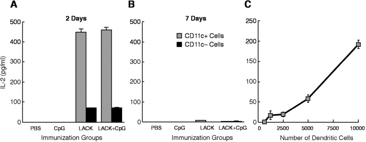
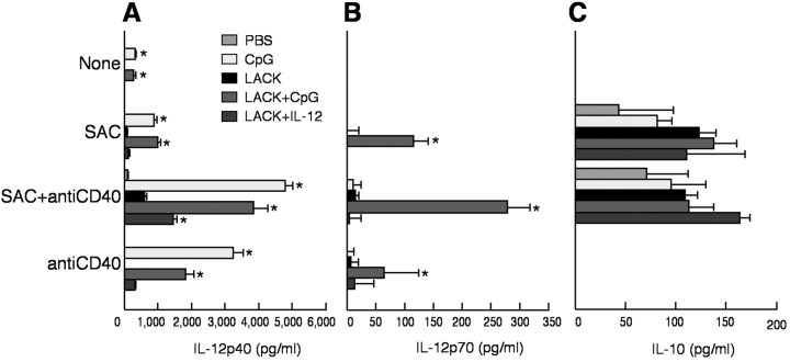
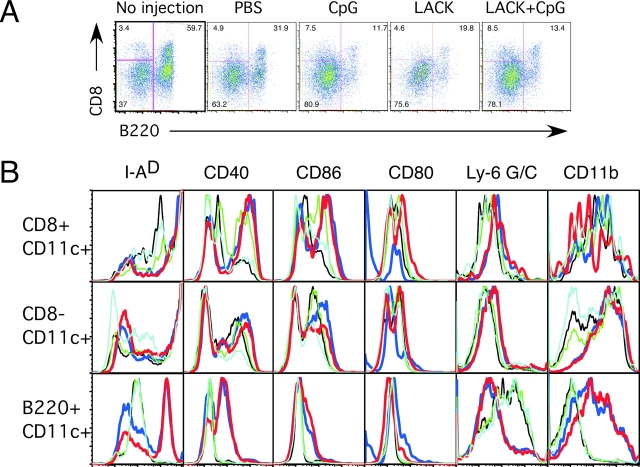
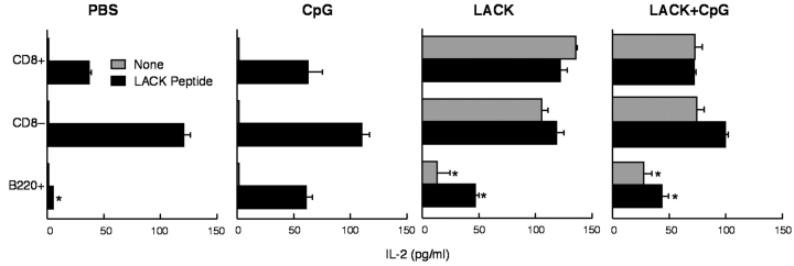
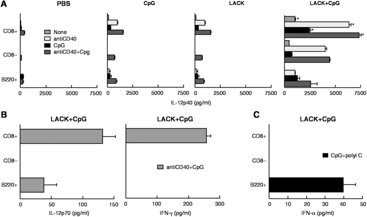
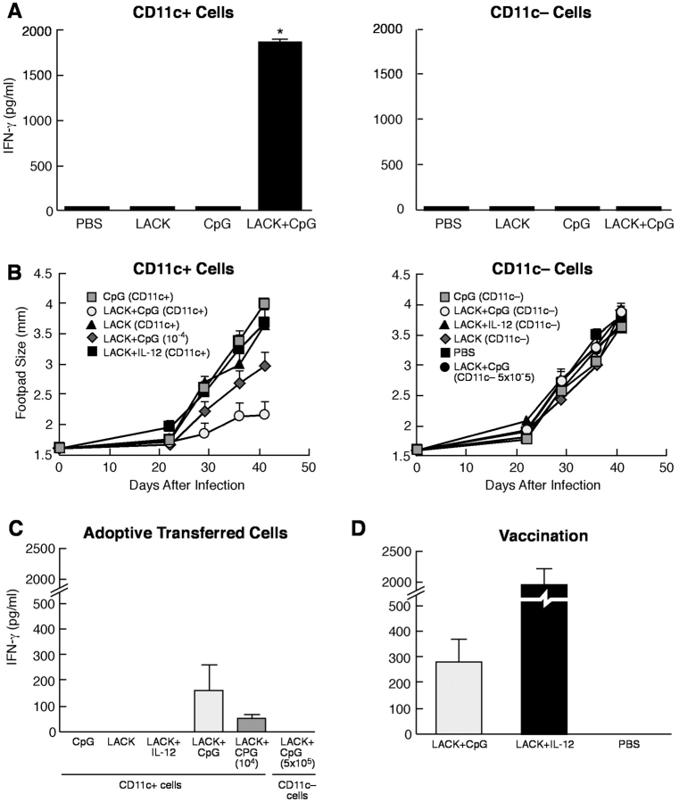
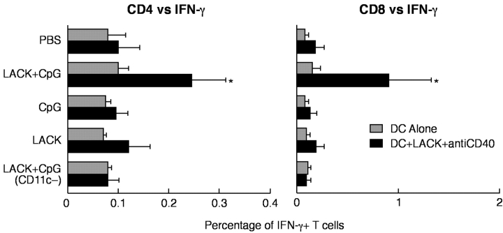
Vaccination with leishmanial Ag and CpG oligodeoxynucleotides (ODN) confers sustained cellular immunity and protection to infectious challenge up to 6 mo after immunization. To define the cellular mechanism by which CpG ODN mediate their adjuvant effects in vivo, the functional capacity of distinct dendritic cell (DC) subsets was assessed in the lymph nodes (LNs) of BALB/c mice, 36 h after immunization with the leishmanial antigen (LACK) and CpG ODN. After this immunization, there was a striking decrease in the frequency of the CD11c+B220+ plasmacytoid DCs with a proportionate increase in CD11c+CD8-B220- cells. CD11c+CD8+B220- cells were the most potent producers of interleukin (IL)-12 p70 and interferon (IFN)-gamma, while plasmacytoid DCs were the only subset capable of secreting IFN-alpha. In terms of antigen presenting capacity, plasmacytoid DCs were far less efficient compared with the other DC subsets. To certify that DCs were responsible for effective vaccination, we isolated CD11c+ and CD11c- cells 36 h after immunization and used such cells to elicit protective immunity after adoptive transfer in naive, Leishmania major susceptible BALB/c mice. CD11c+ cells but not 10-fold higher numbers of CD11c- cells from such immunized mice mediated protection. Therefore, the combination of LACK antigen and CpG ODN adjuvant leads to the presence of CD11c+ DCs in the draining LN that are capable of vaccinating naive mice in the absence of further antigen or adjuvant.


Figures

Figure 1.
Figure 1.
LACK Ag-presenting capacity of CD11c+ and CD11c cells derived in vivo after immunization. BALB/c mice (20 mice per group) were injected s.c. into four separate sites with PBS, CpG ODN, LACK, or LACK + CpG ODN. CD11c+ and CD11ccells were isolated 2 d (A) or 7 d (B) later from draining LN. CD11c+ (104; A and B) or CD11c- (105; A and B) were cocultured with LMR 7.5 hybridoma cells (105) for 24 h. IL-2 content was assessed from the supernatants from triplicate wells. (C) In a separate experiment, varying numbers of CD11c+ cells from mice immunized with LACK + CpG ODN were added to LMR 7.5 cells and IL-2 content assessed. Results are expressed as geometric mean ± SEM and are representative of four individual experiments.
Figure 2.
Figure 2.
IL-12 and IL-10 production from CD11c+ and CD11c cells after immunization. CD11c+ (104) and CD11c (104) cells from BALB/c mice immunized with PBS, CpG, or LACK + CpG ODN, or IL-12 were isolated from draining LN 2 d after immunization and cocultured with nothing, SAC, anti-CD40, or both for 24 h. IL-12 p40 (A), IL-12 p70 (B), and IL-10 (C) were measured by ELISA. Results are expressed as geometric mean ± SEM and are representative of four individual experiments. * indicates P < 0.05 for mice immunized with LACK ± CpG ODN as compared with PBS-immunized or LACK protein-immunized mice.
Figure 3.
Figure 3.
Frequency of DC subsets and their expression of cell surface markers from LN after immunization. (A) CD11c+ DCs from draining LNs of naive (no injection) and or mice immunized with PBS, CpG ODN, or LACK protein ± CpG ODN mice were enriched as described in the methods and stained with antibodies against CD11c, CD8 and B220. Cells were gated on CD11c+ cells and the percentage of CD11c+CD8+B220, CD11c+CD8B220, and CD11c+B220+ is shown. (B) Expression of cell surface markers was analyzed on subsets of CD11c+ DCs based on expression of CD8 and B220. Light blue, no injection; black, PBS; blue, CpG ODN; green, LACK; red, LACK + CpG ODN. Data are representative of three independent experiments.
Figure 4.
Figure 4.
LACK Ag-presenting capacity of CD11c+ DC subsets isolated directly from LNs after immunization. Sorted CD11c+CD8+, CD11c+CD8B220, and CD11c+CD8B220+ (104 cells) were cultured with LMR 7.5 cells (105) for 24 h with or without LACK peptide (10 μg/ml) and IL-2 content assessed. Results are expressed as geometric mean ± SEM. * indicates P < 0.05 from CD11c+CD8+ and CD8 as compared with that from CD11c+B220+ DCs.
Figure 5.
Figure 5.
Production of IL-12, IFN-γ, and IFN-α from CD11c+ DC subsets after immunization. Sorted CD11c+CD8+B220, CD11c+CD8B220, and CD11c+B220+ (5 × 10³) cells were isolated from draining LN 2 d after immunization with PBS, CPG ODN, or LACK ± CpG ODN and cocultured with nothing, anti-CD40 (1 μg/ml), CpG ODN (5 μg/ml), or both. (A) IL-12 p40 was measured from all immunized groups. IL-12 p70 (B) and IFN-γ (B) were only detected from mice immunized with LACK + CpG ODN. (C) IFN-α was measured by ELISA from CD11c+B220+ cells in response to stimulation with CpG ODN 1585 and Poly I:C. Results are expressed as geometric mean ± SEM and are representative of three independent experiments.
Figure 6.
Figure 6.
Immunogenicity and protection induced by CD11c+ DCs from immunized mice. CD11c+ and CD11c cells (105 cells) from immunized mice were transferred by s.c. injection into naive BALB/c mice (A). 4 d later, draining LN were removed, restimulated with LACK protein, and production of IFN-γ assessed from supernatants by ELISA. Results are expressed as geometric mean ± SEM and are representative of three independent experiments. * indicates P < 0.05 for cells of mice that received CD11c+ cells from LACK + CpG ODN-immunized mice compared with all other groups. (B) Varying numbers (5 × 10⁴ cells for all groups except where noted) of CD11c+ or CD11c cells from mice immunized with CpG ODN or LACK + CpG ODN or IL-12 protein were adoptively transferred s.c. into the footpad of naive BALB/c mice. Mice were boosted 2 wk later with the same regimen and then challenged with 105 L. major metacyclic promastigotes in the opposite footpad from which they received the cells from the immunized mice. Control of infection was assessed by measuring the diameter of the footpad lesions. Results are representative of three separate experiments. To assess immune responses post infection, splenocytes (2 × 10⁵ cells/200 μl) from infected mice that received CD11c+ or CD11c cells from immunized mice (C) or from naive BALB/c mice vaccinated with LACK + IL-12 or CpG ODN (D) were stimulated with LACK protein (10 μg/ml) for 48 h and production of IFN-γ assessed from supernatants. Results are the geometric mean of three replicate wells. Error bars represent the SEM. * indicates P < 0.05 for cells of mice that received CD11c+ cells from LACK + CpG ODN-immunized mice compared with all other groups.
Figure 7.
Figure 7.
Frequency of LACK-specific CD4+ and CD8+ IFN-γ–producing cells from BALB/c mice after transfer of CD11c+ DCs and infection. Pooled spleen cells from BALB/c mice (n = 3) that received two immunizations with sorted CD11c+ DCs from BALB/c mice immunized with LACK ± CpG ODN were harvested 3 wk after infection with L. major. Spleen cells were cultured (6 × 10⁶) with bone marrow–derived DCs from BALB/c mice (1.2 × 10⁶) with or without exposure to LACK protein, anti-CD40, and CpG ODN stimulation. Cells were processed and stained as described to enumerate the frequency of LACK-specific CD4+ and CD8+ T cell IFN-γ responses. Numbers represent the percentage of CD4+ or CD8+ T cells positive for IFN-γ. * indicates P < 0.05 for cells of mice that received CD11c+ cells from LACK + CpG ODN-immunized mice compared with all other groups.

References

    1. Seder, R.A., and A.V. Hill. 2000. Vaccines against intracellular infections requiring cellular immunity. Nature. 406:793–798. - PubMed
    1. O'Garra, A. 1998. Cytokines induce the development of functionally heterogeneous T helper cell subsets. Immunity. 8:275–283. - PubMed
    1. Murphy, K.M., W. Ouyang, J.D. Farrar, J. Yang, S. Ranganath, H. Asnagli, M. Afkarian, and T.L. Murphy. 2000. Signaling and transcription in T helper development. Annu. Rev. Immunol. 18:451–494. - PubMed
    1. Krieg, A.M. 2002. CpG motifs in bacterial DNA and their immune effects. Annu. Rev. Immunol. 20:709–760. - PubMed
    1. Barton, G.M., and R. Medzhitov. 2002. Control of adaptive immune responses by Toll-like receptors. Curr. Opin. Immunol. 14:380–383. - PubMed

MeSH terms