. 2003 Jul 21;198(2):305-13.
doi: 10.1084/jem.20030323.

Flt3 ligand regulates dendritic cell development from Flt3+ lymphoid and myeloid-committed progenitors to Flt3+ dendritic cells in vivo

Affiliations

Flt3 ligand regulates dendritic cell development from Flt3+ lymphoid and myeloid-committed progenitors to Flt3+ dendritic cells in vivo

Holger Karsunky et al. J Exp Med. .

Abstract

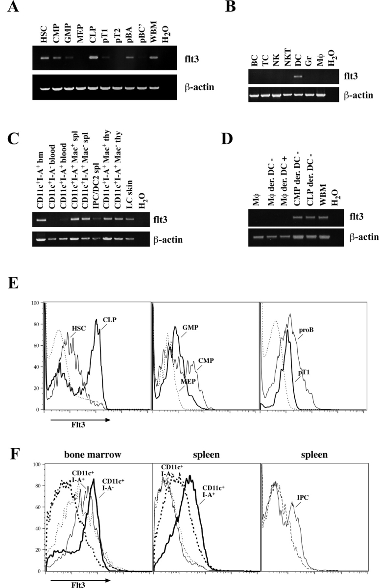
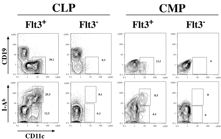
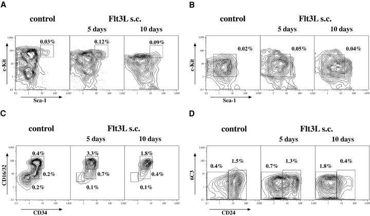
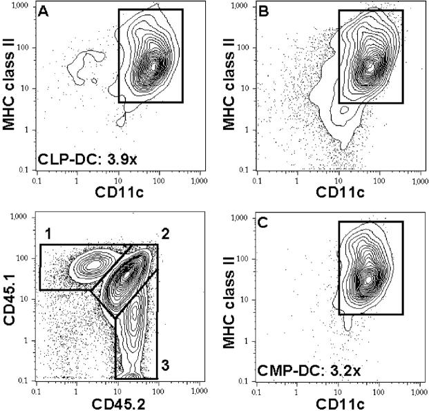
Stimulation of Flt3 receptor tyrosine kinase through its cognate ligand expands early hematopoietic progenitor and dendritic cells (DCs) in humans and mice. The exact developmental stages at which hematopoietic progenitors express Flt3, are responsive to its ligand, and subsequently develop to DCs, are not known. Here we show that common lymphoid and common myeloid progenitors, as well as steady state DCs in thymus, spleen, and epidermis, express Flt3. The receptor is down-regulated once definitive B cell, T cell, and megakaryocyte/erythrocyte commitment occurs, and Flt3 is not detectable on other steady state hematopoietic cell populations. Upon in vivo Flt3 ligand (Flt3L) administration, Flt3+ progenitor cells and their progeny DCs are expanded, whereas Flt3- downstream progenitors are not, or are only slightly increased. Transplantation of common lymphoid and common myeloid progenitors and subsequent Flt3L injection increases progeny DCs of both precursor populations. These findings provide a definitive map of Flt3 expression in the hematopoietic hierarchy and directly demonstrate that Flt3L can drive DC development along both the lymphoid and myeloid developmental pathways from Flt3+ progenitors to Flt3+ DCs.


Figures

Figure 1.
Figure 1.
Flt3 expression on defined hematopoietic progenitor cells, steady state DCs, and other mature hematopoietic cells. Expression of Flt3 was examined by nonquantitative RT-PCR on (A) hematopoietic progenitor populations from bone marrow and thymus, (B) on mature hematopoietic cells from spleen (BC, B cells; TC, T cells; NK, NK cells; NKT, NKT cells; DC, dendritic cells; Gr, granulocytes; Mφ, monocytes/macrophages), and (C) on steady state DC populations as indicated from bone marrow (bm), blood, spleen (spl), thymus (thy), and Langerhans cells (LC) from the epidermis. (D) In vitro–derived DCs (CD11c+ I-Ab+) from either splenic monocytes or common myeloid and lymphoid progenitors were sorted, and Flt3 expression was analyzed by RT-PCR. (E) FACS® analysis of Flt3 surface expression is shown on the left panel. thin line, HSCs; bold line, CLPs; dashed line, whole bone marrow for comparison. In the middle panel: thin line, CMPs; bold line, GMPs; dashed line, MEPs. On the right panel: thin line, pro-B cells; bold line, pro-T1 cells from thymus; dashed line, whole thymus. (F) Flt3 expression on CD11c+ I-Ab+ cells (thin line) and CD11c+ I-Ab− cells (bold line) from bone marrow (left) and spleen (middle). Right panel shows FACS® analysis of IPCs in the spleen. Dashed lines in F always show staining with the according isotype control antibody.
Figure 2.
Figure 2.
Only Flt3+ CLPs and CMPs generate DCs. Flt3+ and Flt3 subpopulations of CLPs and CMPs were sorted and 2,000 cells each were cultured in vitro on AC6 stroma cells in presence of IL-7/SLF/Flt3L for CLPs or SLF/Flt3L for CMPs. After 6 d these cultures were analyzed by FACS® and the percentages of the DC populations are given. Plots showing CMP-derived cells are gated on SSClow/intermediate cells to exclude macrophages with characteristically high autofluorescence. Data shown are representative for three independent experiments.
Figure 3.
Figure 3.
Flt3L expands hematopoietic progenitors. FACS® analysis of hematopoietic progenitor populations in the bone marrow after 5 and 10 d of s.c. Flt3L injections. Mice injected for 5 and 10 d (not depicted) with PBS were used as a control. (A) HSCs (electronically gated on Lin−/lo Thy1.1lo cells). (B) CLPs (gated on Lin−/lo Thy1.1 IL-7Rα+). (C) CMPs (CD34+ CD16/32lo), GMPs (CD34+ CD16/32hi), and MEPs (CD34 CD16/32lo; gated on Lin−/lo Sca-1 c-Kit+ cells). (D) Pro-B cells (gated on IgM NK1.1 B220+ CD43+ cells): pro B (A) are CD24−/lo 6C3 and pro-B (BC) are CD24+. Numbers delineate relative percentages of the according population in total bone marrow.
Figure 4.
Figure 4.
Flt3L expands CLP- and CMP-derived DCs in vivo. 104 CD45.1+ CLPs and 2 × 10⁴ CD45.2+ CMPs were competitively transplanted into sublethally irradiated CD45.1/CD45.2 F1 mice. Flt3L or control PBS was injected over 10 d, starting on day 2 after transplantation, and mice were evaluated 1 d after the last Flt3L injection. Lower left contour plot shows CD11c-enriched splenic progeny of CLPs (1), host cells (2), and CMPs (3). CD11c and MHC II expression of cells in gates 1, 2, and 3 are shown in contour plots A, B, and C, respectively. Average expansions of CLP- and CMP-derived DCs are indicated. Plots show DC read out of a Flt3L-injected animal and are representative of three experiments involving two to four mice in the Flt3L-treated group and two animals as controls each. For relative and total numbers of CLP- and CMP-derived DCs see Table II.

Similar articles

Cited by

References

    1. Matthews, W., C.T. Jordan, G.W. Wiegand, D. Pardoll, and I.R. Lemischka. 1991. A receptor tyrosine kinase specific to hematopoietic stem and progenitor cell-enriched populations. Cell. 65:1143–1152. - PubMed
    1. Small, D., M. Levenstein, E. Kim, C. Carow, S. Amin, P. Rockwell, L. Witte, C. Burrow, M.Z. Ratajczak, A.M. Gewirtz, et al. 1994. STK-1, the human homolog of Flk-2/Flt-3, is selectively expressed in CD34+ human bone marrow cells and is involved in the proliferation of early progenitor/stem cells. Proc. Natl. Acad. Sci. USA. 91:459–463. - PMC - PubMed
    1. Lyman, S.D., and S.E. Jacobsen. 1998. c-kit ligand and Flt3 ligand: stem/progenitor cell factors with overlapping yet distinct activities. Blood. 91:1101–1134. - PubMed
    1. Mackarehtschian, K., J.D. Hardin, K.A. Moore, S. Boast, S.P. Goff, and I.R. Lemischka. 1995. Targeted disruption of the flk2/flt3 gene leads to deficiencies in primitive hematopoietic progenitors. Immunity. 3:147–161. - PubMed
    1. McKenna, H.J., K.L. Stocking, R.E. Miller, K. Brasel, T. De Smedt, E. Maraskovsky, C.R. Maliszewski, D.H. Lynch, J. Smith, B. Pulendran, et al. 2000. Mice lacking flt3 ligand have deficient hematopoiesis affecting hematopoietic progenitor cells, dendritic cells, and natural killer cells. Blood. 95:3489–3497. - PubMed

Publication types

MeSH terms