. 2003 Jul 21;198(2):341-7.
doi: 10.1084/jem.20021279.

XIAP-mediated caspase inhibition in Hodgkin's lymphoma-derived B cells

Affiliations

XIAP-mediated caspase inhibition in Hodgkin's lymphoma-derived B cells

Hamid Kashkar et al. J Exp Med. .

Abstract

The malignant Hodgkin and Reed-Sternberg cells of Hodgkin's lymphoma (HL) and HL-derived B cell lines were previously shown to be resistant to different apoptotic stimuli. We show here that cytochrome c fails to stimulate caspases-9 and -3 activation in cytosolic extracts of HL-derived B cells, which is due to high level expression of X-linked inhibitor of apoptosis (XIAP). Coimmunoprecipitation studies revealed that XIAP, apoptosis protease-activating factor-1, and caspase-3 are complexed in HL-derived B cell lysates. Even after stimulation with exogenous cytochrome c and dATP, XIAP impairs the proteolytic processing and activation of caspase-3. In cytosolic extracts, inhibition of XIAP by the second mitochondria-derived activator of caspases (Smac)/DIABLO, or immunodepletion of XIAP restores cytochrome c-triggered processing and activation of caspase-3. Smac or a Smac-derived agonistic peptide also sensitized intact HL-derived B cells for the apoptotic action of staurosporine. Finally, Hodgkin and Reed-Sternberg cells of primary tumor HL tissues also constitutively and abundantly express XIAP. The results of this paper suggest that high level XIAP expression is a hallmark of HL, which may play a crucial role in resistance to apoptosis.


Figures

Figure 1.
Figure 1.
Failure of cytochrome c to induce caspase activation in cytosolic extracts of HL-derived B cell lines. Cytosolic extracts of L1236, L591, L428, and KMH2 cells, and of control B cell L1309 were prepared and equal amounts of protein were incubated with or without cytochrome c/dATP for 1 h at 30°C. Cytosolic extracts were resolved by SDS-PAGE and subjected to Western blot analysis. (A) Procaspase-9 (p46) and its fragments (p37, p35) were detected by polyclonal rabbit anti–caspase-9 antibody. (B) Procaspase-3 (p32) and its fragments (p20, p17) were detected by polyclonal anti–caspase-3 antibody. (C) Measurement of relative caspase activity using DEVD-AFC. Samples were normalized for total cytosolic protein content. Asterisk indicates nonspecific bands recognized by polyclonal antibodies.
Figure 2.
Figure 2.
Expression of caspase activators and inhibitors in HL-derived B cell lines. (A) Equal amounts of proteins from total cell lysate of L1236, L591, L428, and KMH2 cells and of control B cells L1309 were subjected to SDS-PAGE and Western blot analysis. Proteins were detected using antibodies against Apaf-1 and XIAP. (B) XIAP expression in primary Hodgkin's lymphoma (HL) tissues. XIAP positivity in H-RS cells: + = very weak; ++ = weak; +++ = moderate; ++++ = strong staining. (C) Paraffin section immunohistochemistry of cases 3, 5, 6, 7, 10, and 11 of classical HL cases listed in Fig. 2 B. XIAP was stained using anti-XIAP specific mAb. There are strong (Nos. 5, 6, 7, and 11) or moderate (Nos. 3 and 10) granular intracytoplasmic staining for XIAP in essentially all morphologically recognizable Hodgkin or Reed-Sternberg cells. Background lymphocytes are negative for XIAP staining.
Figure 2.
Figure 2.
Expression of caspase activators and inhibitors in HL-derived B cell lines. (A) Equal amounts of proteins from total cell lysate of L1236, L591, L428, and KMH2 cells and of control B cells L1309 were subjected to SDS-PAGE and Western blot analysis. Proteins were detected using antibodies against Apaf-1 and XIAP. (B) XIAP expression in primary Hodgkin's lymphoma (HL) tissues. XIAP positivity in H-RS cells: + = very weak; ++ = weak; +++ = moderate; ++++ = strong staining. (C) Paraffin section immunohistochemistry of cases 3, 5, 6, 7, 10, and 11 of classical HL cases listed in Fig. 2 B. XIAP was stained using anti-XIAP specific mAb. There are strong (Nos. 5, 6, 7, and 11) or moderate (Nos. 3 and 10) granular intracytoplasmic staining for XIAP in essentially all morphologically recognizable Hodgkin or Reed-Sternberg cells. Background lymphocytes are negative for XIAP staining.
Figure 3.
Figure 3.
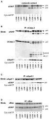
XIAP coimmunoprecipitates with activated caspase-3. Cytosolic extracts of L1236, L591, L428, and KMH2 cells and of control B cells L1309 were prepared, and equal amounts of protein were incubated with or without cytochrome c/dATP for 1 h at 30°C. (A) 100 μg samples were resolved by SDS-PAGE and subjected to Western blot analysis. XIAP was detected by mouse anti-XIAP mAb. (B–D) Caspase-3, Apaf-1, and Bax were immunoprecipitated by mouse anti–caspase-3, rat anti–Apaf-1, and mouse anti-Bax mAb in 600-μg cytosolic extracts and subjected to SDS-PAGE and Western blotting. XIAP, caspase-3, Apaf-1, and Bax were detected by mouse anti-XIAP mAb, polyclonal rabbit anti–caspase-3, polyclonal rabbit anti–Apaf-1, and polyclonal rabbit anti-Bax antibodies. Asterisk indicates mouse IgG.
Figure 4.
Figure 4.
Caspase-3 activation by caspase-8 and granzyme-B. Cytosolic extracts of L1236, L591, L428, and KMH2 cells and of control B cells L1309 were prepared, and equal amounts of protein were left untreated or incubated for 1 h with recombinant active caspase-8 (A) and granzyme-B (B) at 30°C. Cytosolic extracts were resolved by SDS-PAGE and subjected to Western blot analysis. Caspase-3 and XIAP were detected by polyclonal rabbit anti–caspase-3 antibody and mouse anti-XIAP mAb. Relative caspase activity was measured using DEVD-AFC after incubation of cytosolic extracts with recombinant active caspase-8 (A)and granzyme-B (B). Samples were normalized for total cytosolic protein content.
Figure 5.
Figure 5.
Depletion of XIAP restores caspase-3 processing and activity. Cytosolic extracts of L1236 and KMH2 cells and of control B cell L1309 were prepared, and equal amounts of protein were incubated with or without cytochrome c/dATP in the absence and presence of Smac protein for 1 h at 30°C. (A) Cytosolic extracts were resolved by SDS-PAGE and subjected to Western blot analysis. Caspase-3 was detected by polyclonal rabbit anti–caspase-3 antibody. (B) Relative caspase activity was measured by hydrolysis of DEVD-AFC. Samples were normalized for total cytosolic protein content. (C) XIAP was immunodepleted by mouse anti-XIAP mAb. Cytosolic extracts of KMH2 cells with or without XIAP were incubated with or without cytochrome c/dATP for 1 h at 30°C. Samples were normalized for total cytosolic protein content and relative caspase-3 activity was measured by DEVDase activity. (D) Cells were transfected with vehicle, β-galactosidase, Smac N7 peptide, or Smac protein and treated for 24 h with 1 μM staurosporine. Cell death was determined by trypan blue exclusion. Each value represents the average of results from two independent experiments.
Figure 5.
Figure 5.
Depletion of XIAP restores caspase-3 processing and activity. Cytosolic extracts of L1236 and KMH2 cells and of control B cell L1309 were prepared, and equal amounts of protein were incubated with or without cytochrome c/dATP in the absence and presence of Smac protein for 1 h at 30°C. (A) Cytosolic extracts were resolved by SDS-PAGE and subjected to Western blot analysis. Caspase-3 was detected by polyclonal rabbit anti–caspase-3 antibody. (B) Relative caspase activity was measured by hydrolysis of DEVD-AFC. Samples were normalized for total cytosolic protein content. (C) XIAP was immunodepleted by mouse anti-XIAP mAb. Cytosolic extracts of KMH2 cells with or without XIAP were incubated with or without cytochrome c/dATP for 1 h at 30°C. Samples were normalized for total cytosolic protein content and relative caspase-3 activity was measured by DEVDase activity. (D) Cells were transfected with vehicle, β-galactosidase, Smac N7 peptide, or Smac protein and treated for 24 h with 1 μM staurosporine. Cell death was determined by trypan blue exclusion. Each value represents the average of results from two independent experiments.

References

    1. Scaffidi, C., S. Fulda, A. Srinivasan, C. Friesen, F. Li, K.J. Tomaselli, K.M. Debatin, P.H. Krammer, and M.E. Peter. 1998. Two CD95 (APO-1/Fas) signaling pathways. EMBO J. 17:1675–1687. - PMC - PubMed
    1. Jürgensmeier, J.M., Z. Xie, Q.L. Deveraux, L.M. Ellerby, D.E. Bredesen, and J.C. Reed. 1998. Bax directly induces release of cytochrome c from isolated mitochondria. Proc. Natl. Acad. Sci. USA. 95:4997–5002. - PMC - PubMed
    1. Du, C., M. Fang, Y. Li, L. Li, and X. Wang. 2000. Smac, a mitochondrial protein that promotes cytochrome c-dependent caspase activation by eliminating IAP inhibition. Cell. 102:33–42. - PubMed
    1. Li, P., D. Nijhawan, I. Budihardjo, S.M. Srinivasula, M. Ahmad, E.S. Alnemri, and X.D. Wang. 1997. Cytochrome c and dATP-dependent formation of Apaf-1/caspase-9 complex initiates an apoptotic protease cascade. Cell. 91:479–489. - PubMed
    1. Renatus, M., H.R. Stennicke, F.L. Scott, R.C. Liddington, and G.S. Salvesen. 2001. Dimer formation drives the activation of the cell death protease caspase-9. Proc. Natl. Acad. Sci. USA. 98:14250–14255. - PMC - PubMed

Publication types