. 2003 Aug;71(8):4463-71.
doi: 10.1128/IAI.71.8.4463-4471.2003.

Identification of the agr locus of Listeria monocytogenes: role in bacterial virulence

Affiliations

Identification of the agr locus of Listeria monocytogenes: role in bacterial virulence

Nicolas Autret et al. Infect Immun. 2003 Aug.

Abstract

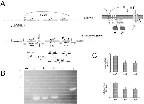
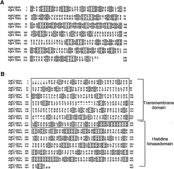
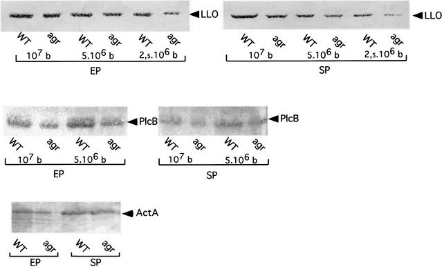
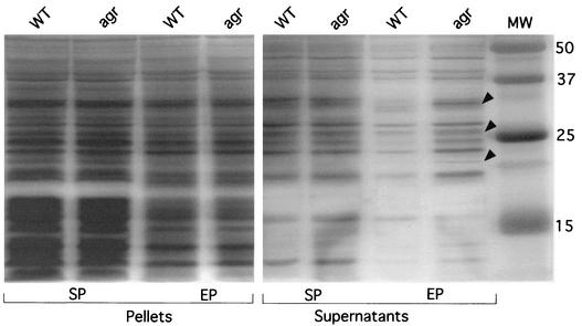
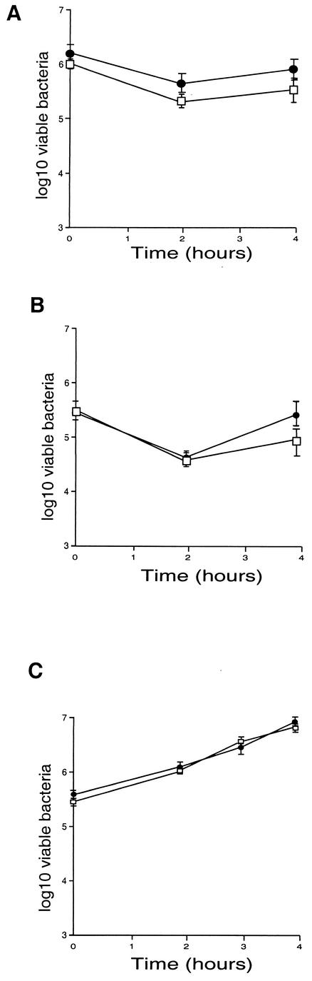
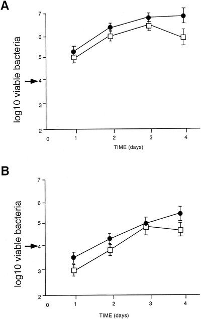
Listeria monocytogenes is a gram-positive facultative intracellular food-borne pathogen that can cause severe infections in humans and animals. We have recently adapted signature-tagged transposon mutagenesis (STM) to identify genes involved in the virulence of L. monocytogenes. A new round of STM allowed us to identify a new locus encoding a protein homologous to AgrA, the well-studied response regulator of Staphylococcus aureus and part of a two-component system involved in bacterial virulence. The production of several secreted proteins was modified in the agrA mutant of L. monocytogenes grown in broth, indicating that the agr locus influenced protein secretion. Inactivation of agrA did not affect the ability of the pathogen to invade and multiply in cells in vitro. However, the virulence of the agrA mutant was attenuated in the mouse (a 10-fold increase in the 50% lethal dose by the intravenous route), demonstrating for the first time a role for the agr locus in the virulence of L. monocytogenes.


Figures

FIG. 1.
FIG. 1.
Genetic organization of the agr locus and transcriptional analysis. (A) Genetic organization of the agr locus. (Upper panel) agr locus of S. aureus. The grey arrows indicate the orientations and approximate sizes of the different genes. The dotted lines ending in arrows indicate the two main divergent transcripts RNAII and RNAIII. To the right, a schematic representation of the various components of the agr locus is shown, with letters representing the proteins encoded A, AgrA; B, AgrB; C, AgrC; D, AgrD; asterisk, phosphorylated group; P2 and P3, promoters of RNAII and RNAIII, respectively. (Lower panel) agr locus of L. monocytogenes. The arrows indicate approximate sizes and orientations of the different genes. The predicted length of each protein is indicated below (in amino acids [aas]). The stop sign-shaped symbols indicate putative transcription terminators. The values in percentages below agrB, agrC, and agrA indicate the percentage of amino acid identity between the L. monocytogenes and S. aureus orthologues encoded by the genes of the agr loci. Numbers between parentheses indicate the sizes (in base pairs) of the intergenic regions. The site of Tn1545 insertion is represented by an inverted black triangle. (B) Transcriptional analysis by RT-PCR. The dotted lines enclosed by arrows in the lower half of panel A indicate the positions of the primers and PCR products used in the RT-PCR analysis. The amplified products, numbered 1 to 5, were subjected to Tris acetate-EDTA-agarose gel electrophoresis. The arrows preceded by numbers (in kilobases) to the left of the panel correspond to the molecular weight (MW) DNA ladder. (C) Expression of the agr genes in stationary (Stat) and exponential (Exp) phases. The expression was measured by real-time quantitative RT-PCR. The amount of agr mRNA relative to that of the normalizing gene gyr (upper panel) and the 16S gene (lower panel) was determined by real-time quantitative RT-PCR with bacteria grown in BHI in the exponential or stationary phase of growth. The amounts of gyr mRNA and 16S rRNA were constant under these conditions (data not shown). The values shown are means of results from three assays; the error bars indicate the standard deviations.
FIG. 2.
FIG. 2.
Multiple alignment of AgrA (A) and AgrC (B) from L. monocytogenes and S. aureus. Alignments were performed by using CLUSTALW and AlignP programs. Lismo, L. monocytogenes (EGD-e); Stau, S. aureus. Identical residues are boxed and shaded. (S. aureus AgrA is from strain N315, accession no. P13131; S. aureus AgrC is from strain KSI54, accession no. gi:1916243). In panel B, the transmembrane N-terminal domain (boxed) and the histidine kinase domain of AgrC are indicated.
FIG. 3.
FIG. 3.
Western blot analyses. Culture supernatants and membrane fractions from cells grown overnight at 37°C with agitation in BHI medium were tested in exponential phase (EP) and stationary phase (SP). Identical amounts of each culture supernatant were loaded onto SDS-10% polyacrylamide gels. Proteins were transferred electrophoretically onto nitrocellulose and detected with specific antibodies. (Upper panel) Anti-LLO MAb. Decreasing amounts of supernatant were loaded, corresponding to 107, 5 × 10⁶, and 2.5 × 10⁶ bacteria (b). The anti-LLO MAbs SE1 and SE2 were used at a final dilution of 1/1,000. (Middle panel) Anti-PC-PLC. Decreasing amounts of supernatant were loaded, corresponding to 107 and 5 × 10⁶ bacteria. The anti-PlcB polyclonal antibody was used at a final dilution of 1/200. (Lower panel) Anti-ActA. Membrane fractions corresponding to 107 bacteria were loaded. The anti-ActA polyclonal antibody was used at a final dilution of 1/1,000. WT, strain EGD-e; agr, agrA-Tn1545 insertion mutant.
FIG. 4.
FIG. 4.
Protein secretion in the agrA mutant. Culture supernatants and membrane fractions from cells grown overnight at 37°C with agitation in RPMI medium were tested in exponential phase (EP) and stationary phase (SP). The black arrowheads point to protein bands present in the supernatant of the agr mutant in the exponential phase of growth that are missing in that of the wild-type (WT) strain. Numbers to the right correspond to the molecular weight (MW) markers.
FIG.5.
FIG.5.
Invasiveness of the agrA mutant was evaluated in two different cell lines, Caco-2 cells (A) and HepG-2 cells (B), and in bone marrow macrophages (C). Cell monolayers were incubated for 1 h at 37°C with approximately 100 bacteria per cell. After washing, the cells were reincubated for 4 h in fresh culture medium. At 2, 3, and 4 h, the cells were washed again and lysed and viable bacteria were counted on BHI plates. Values and error bars represent the means and standard deviations of the numbers of bacteria per well (three wells per assay, two different assays). •, EGD-e; □, agrA mutant.
FIG. 6.
FIG. 6.
In vivo survival of the agrA mutant. The kinetics of bacterial growth were monitored in mice infected either with EGD-e or with the agrA mutant. Mice were inoculated with 2 × 10⁴ bacteria (indicated by an arrow to the left of the ordinate). Bacterial survival in the spleens (A) and livers (B) was monitored over a 4-day period. •, EGD-e; □, agrA mutant.

References

    1. Autret, N., I. Dubail, P. Trieu-Cuot, P. Berche, and A. Charbit. 2001. Identification of new genes involved in the virulence of Listeria monocytogenes by signature-tagged transposon mutagenesis. Infect. Immun. 69:2054-2065. - PMC - PubMed
    1. Berche, P. 1995. Bacteremia is required for invasion of the murine central nervous system by Listeria monocytogenes. Microb. Pathog. 18:323-336. - PubMed
    1. Celli, J., and P. Trieu-Cuot. 1998. Circularization of Tn916 is required for expression of the transposon-encoded transfer functions: characterization of long tetracycline-inducible transcripts reading through the attachment site. Mol. Microbiol. 28:103-117. - PubMed
    1. Cossart, P., and M. Lecuit. 1998. Interactions of Listeria monocytogenes with mammalian cells during entry and actin-based movement: bacterial factors, cellular ligands and signaling. EMBO J. 17:3797-3806. - PMC - PubMed
    1. Cotter, P. D., N. Emerson, C. G. Gahan, and C. Hill. 1999. Identification and disruption of lisRK, a genetic locus encoding a two-component signal transduction system involved in stress tolerance and virulence in Listeria monocytogenes. J. Bacteriol. 181:6840-6843. - PMC - PubMed

Publication types

MeSH terms

LinkOut - more resources