. 2003 Aug;71(8):4623-32.
doi: 10.1128/IAI.71.8.4623-4632.2003.

The C terminus of YopT is crucial for activity and the N terminus is crucial for substrate binding

Affiliations

The C terminus of YopT is crucial for activity and the N terminus is crucial for substrate binding

Isabel Sorg et al. Infect Immun. 2003 Aug.

Abstract

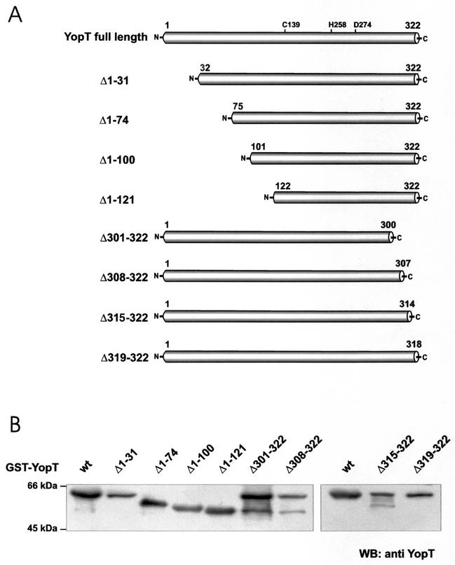
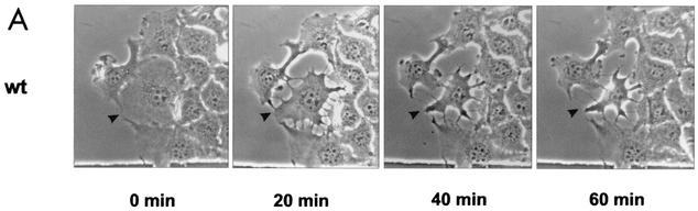
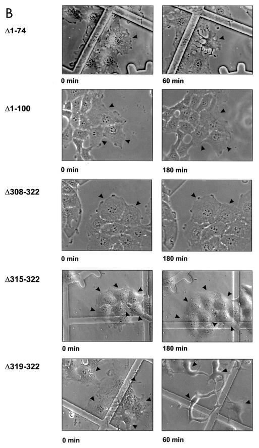
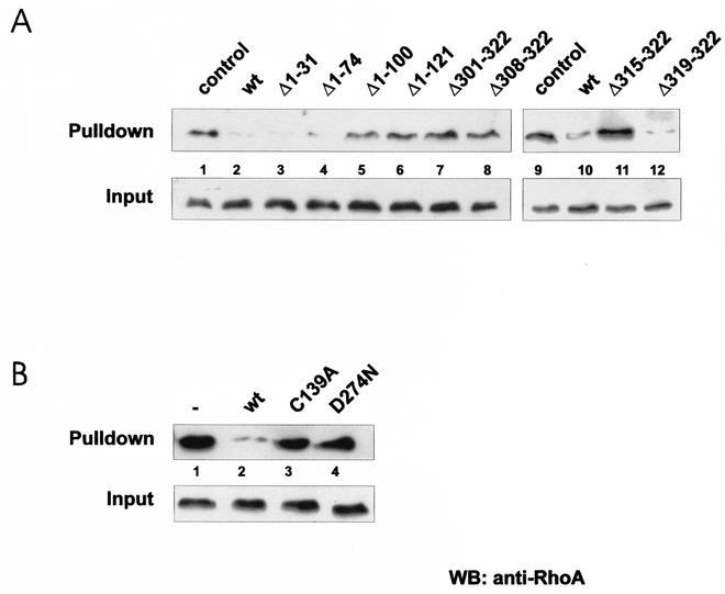
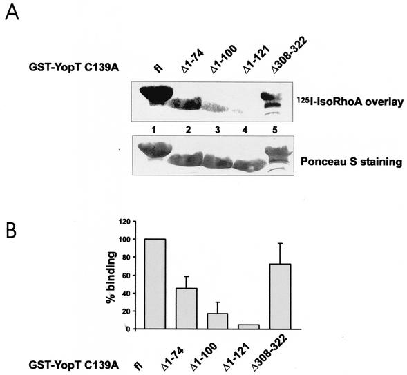
Recently, it was shown that Yersinia outer protein T (YopT) belongs to a new family of cysteine proteases containing invariant C, H, and D residues that are crucial for its activity. YopT cleaves RhoA, Rac, and Cdc42 at their C termini, thereby releasing them from the membrane. Moreover, YopT inhibits the Rho-rhotekin and Rho-guanine nucleotide dissociation inhibitor interactions. To characterize the active domain of YopT, we constructed N- and C-terminal truncations and expressed them as glutathione S-transferase fusion proteins in Escherichia coli. The toxin fragments were tested for stability by trypsin digestion. The activity of the proteins was studied by membrane release assay, rhotekin pulldown experiments, and microinjection. Whereas deletion of the first 74 N-terminal amino acids did not influence the activity of YopT, deletion of 8 amino acids from the C terminus led to complete loss of activity. N-terminal deletion of 100 amino acids led to an inactive protein, although it still contained the amino acids C139, H258, and D274, which are essential for catalysis. Loss of activity of the N-terminal deletions corresponded to the block of interaction with RhoA, indicating that residues 75 to 100 of YopT are essential for binding to the GTPase. By contrast, when up to 15 amino acids of the C terminus were deleted, the protein had no activity but was still able to interact with RhoA, suggesting a role for the C terminus in the enzyme activity of YopT.


Figures

FIG. 1.
FIG. 1.
(A) YopT fragments. The first and last amino acids of each fragment are indicated. N-terminal truncations were named Δ1-31, Δ1-74, Δ1-100, Δ1-121, and Δ301-322, whereas C-terminally truncated fragments were designated Δ301-322, Δ308-322, Δ315-322, and Δ319-322. All fragments contain the amino acids cysteine 139, histidine 258, and aspartate 274, which are crucial for the activity of full-length YopT. (B) Immunoblot of GST-YopT fragments. All of the fragments used in this study were expressed as GST fusion proteins in E. coli TG1. Expression of the different GST fusion proteins was checked by immunoblot analysis with a YopT antibody. wt, wild type; WB, Western blot.
FIG. 2.
FIG. 2.
Microinjection of YopT into EBL cells. (A) Time course of EBL cells after microinjection of GST-YopT. (B) N-terminal (Δ1-31, Δ1-74, Δ1-100, and Δ1-121) and C-terminal (Δ301-322, Δ308-322, Δ315-322, and Δ319-322) GST-YopT fragments were microinjected into EBL cells. Shown are pictures of crucial GST-YopT fragments, which were taken 60 and 180 min after microinjection, respectively. Injected cells are indicated by arrowheads. The experiment was repeated at least three times with similar results. wt, wild type.
FIG. 2.
FIG. 2.
Microinjection of YopT into EBL cells. (A) Time course of EBL cells after microinjection of GST-YopT. (B) N-terminal (Δ1-31, Δ1-74, Δ1-100, and Δ1-121) and C-terminal (Δ301-322, Δ308-322, Δ315-322, and Δ319-322) GST-YopT fragments were microinjected into EBL cells. Shown are pictures of crucial GST-YopT fragments, which were taken 60 and 180 min after microinjection, respectively. Injected cells are indicated by arrowheads. The experiment was repeated at least three times with similar results. wt, wild type.
FIG. 3.
FIG. 3.
Membrane release of RhoA. Isolated rat brain membranes were incubated with fragments (A) or mutant forms (B) of GST-YopT, as indicated. After separation of membranes and corresponding supernatants by ultracentrifugation, the samples were analyzed for RhoA by Western blotting (WB). Shown is a typical result of at least four independent experiments. wt, wild type.
FIG. 4.
FIG. 4.
Rhotekin pulldown assay. Lysates of CNF-1-treated HeLa cells were incubated as indicated with wild-type (wt) GST-YopT and fragments (A) or mutant forms (B) thereof for 30 min at 37°C. After incubation with GST-YopT, 1/15 of the volume was taken as input. The rest of the lysates were incubated with GST-rhotekin beads for 60 min at 4°C. The beads were then washed, and the rhotekin-bound RhoA and the input control were separated by SDS-PAGE and detected by Western blotting (WB) with a RhoA-specific antibody. Shown is a typical result of at least four independent experiments.
FIG. 5.
FIG. 5.
Influence of lovastatin on the effect of YopT. Subconfluent HeLa cells were treated with 30 μM lovastatin for 16 h and then incubated for 2 h with CNF-1 (400 ng/ml) or only treated with CNF-1. Cytosols were prepared and incubated with or without GST-YopT for 30 min at 37°C. After incubation with GST-YopT, 1/15 of the probes was taken as input while the rest was incubated with either GST-rhotekin or GST-GDI beads for 60 min at 4°C. The beads were then washed. Bound RhoA and the input control were separated by SDS-PAGE and detected by Western blotting (WB) with a RhoA-specific antibody. Shown is a typical result of at least four independent experiments.
FIG. 6.
FIG. 6.
Binding of YopT fragments to RhoA. The indicated YopT C139A fragments were expressed as GST fusion proteins, and then equal amounts of the proteins were separated by SDS-PAGE and transferred onto PVDF membrane. The membrane was subsequently used for an overlay assay with isoprenylated, 125I-labeled RhoA that had previously been activated with GTPγS. After incubation with RhoA, the membrane was washed extensively and the bound RhoA was analyzed by phosphorimaging. Equal amounts of the GST-YopT proteins were checked by Ponceau S staining (bottom of panel A). In panel A, a typical experiment is shown. Panel B shows the quantification of three independent experiments as means plus standard deviations. fl, full length.

Similar articles

Cited by

References

    1. Aktories, K., G. Schmidt, and F. Hofmann. 2000. GTPases targeted by bacterial toxins, p. 311-331. In A. Hall (ed.), GTPases. Oxford University Press, Oxford, England.
    1. Barz, C., T. N. Abahji, K. Trülzsch, and J. Heesemann. 2000. The Yersinia Ser/Thr protein kinase YpkA/YopO directly interacts with the small GTPases RhoA and Rac-1. FEBS Lett. 482:139-143. - PubMed
    1. Bishop, A. L., and A. Hall. 2000. Rho GTPases and their effector proteins. Biochem. J. 348:241-255. - PMC - PubMed
    1. Cornelis, G. R. 2000. Type III secretion: a bacterial device for close combat with cells of their eukaryotic host. Philos. Trans. R. Soc. Lond. B Biol. 355:681-693. - PMC - PubMed
    1. Cornelis, G. R., A. Boland, A. P. Boyd, C. Geuijen, M. Iriarte, C. Neyt, M.-P. Sory, and I. Stainier. 1998. The virulence plasmid of Yersinia, an antihost genome. Microbiol. Mol. Biol. Rev. 62:1315-1352. - PMC - PubMed

Publication types

MeSH terms

LinkOut - more resources