. 2003 Aug 5;100(16):9116-21.
doi: 10.1073/pnas.1632821100. Epub 2003 Jul 21.

A chemical approach for identifying O-GlcNAc-modified proteins in cells

Affiliations

A chemical approach for identifying O-GlcNAc-modified proteins in cells

David J Vocadlo et al. Proc Natl Acad Sci U S A. .

Abstract

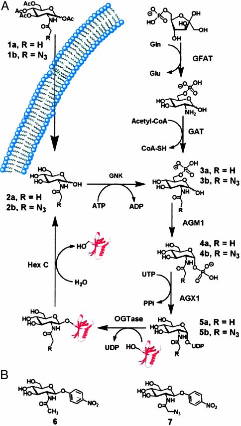
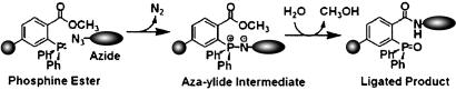
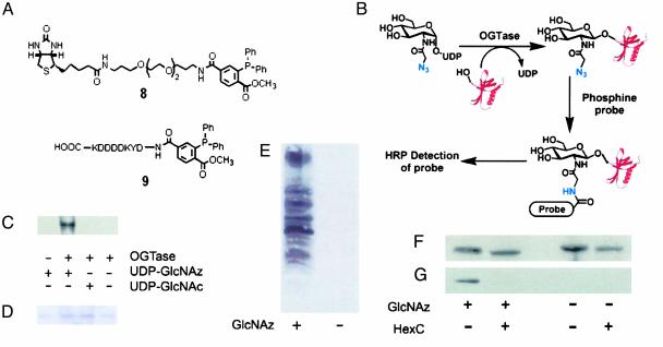
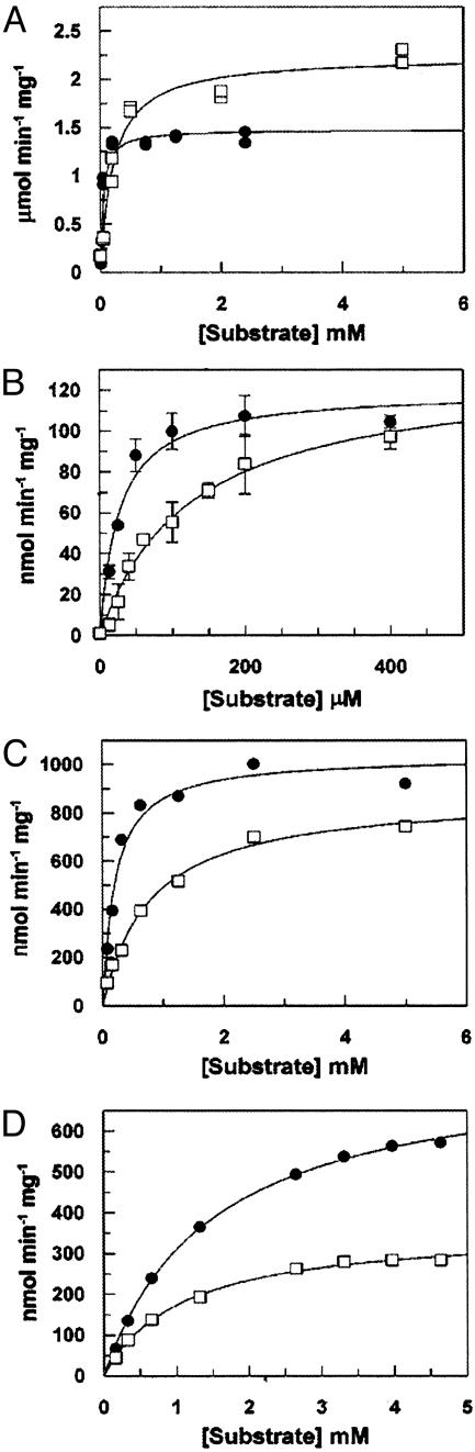
The glycosylation of serine and threonine residues with a single GlcNAc moiety is a dynamic posttranslational modification of many nuclear and cytoplasmic proteins. We describe a chemical strategy directed toward identifying O-GlcNAc-modified proteins from living cells or proteins modified in vitro. We demonstrate, in vitro, that each enzyme in the hexosamine salvage pathway, and the enzymes that affect this dynamic modification (UDP-GlcNAc:polypeptidtyltransferase and O-GlcNAcase), tolerate analogues of their natural substrates in which the N-acyl side chain has been modified to bear a bio-orthogonal azide moiety. Accordingly, treatment of cells with N-azidoacetylglucosamine results in the metabolic incorporation of the azido sugar into nuclear and cytoplasmic proteins. These O-azidoacetylglucosamine-modified proteins can be covalently derivatized with various biochemical probes at the site of protein glycosylation by using the Staudinger ligation. The approach was validated by metabolic labeling of nuclear pore protein p62, which is known to be posttranslationally modified with O-GlcNAc. This strategy will prove useful for both the identification of O-GlcNAc-modified proteins and the elucidation of the specific residues that bear this saccharide.


Figures

Fig. 1.
Fig. 1.
(A) The de novo hexosamine biosynthetic pathway, salvage pathway, and dynamic glycosylation of nucleocytoplasmic proteins. O-GlcNAc-modified proteins are biosynthesized in a series of enzymatic steps. The first committed step in the de novo process is the conversion of fructose-6-phosphate to glucosamine-6-phosphate through the action of the enzyme glutamine:fructose-6-phosphate amidotransferase (GFAT). Glucosamine-6-phosphate is then converted to the common intermediate N-acetylglucosamine-6-phosphate (3a) through the action of acetyl-CoA:d-glucosamine-6-phosphate N-acetyltransferase (GAT). The salvage pathway bypasses the de novo pathway through the action of GlcNAc kinase (GNK) and also generates GlcNAc-6-phosphate (3a). This intermediate is converted to GlcNAc-1-phosphate (4a) through the action of AGM1. N-acetylglucosamine-1-phosphate is converted to the end product of the hexosamine biosynthetic pathway, UDP-GlcNAc (5a), through the action of UDP-GlcNAc pyrophosphorylase (AGX1). OGTase transfers the saccharide moiety of UDP-GlcNAc to protein substrates within the cell. Hexosaminidase C (O-GlcNAcase) acts to cleave the glycosidic linkage of posttranslationally modified proteins to liberate the protein and GlcNAc (2a). Exogenously added Ac4GlcNAz (1b) diffuses into the cell and is deacylated through the action of intracellular esterases, and then enters into the salvage pathway. (B) Chromogenic substrates for O-GlcNAcase. Shown are para-nitrophenyl 2-acetamido-2-deoxy-β-d-glucopyranoside (6) and para-nitrophenyl 2-azidoacetamido-2-deoxy-β-d-glucopyranoside (7).
Fig. 2.
Fig. 2.
The Staudinger ligation. An alkyl azide moiety appended to a molecule of interest can be covalently derivatized with a triarylphosphine ester conjugated to a variety of probes. The reaction proceeds via an aza-ylide intermediate, which undergoes rearrangement to yield the final product.
Fig. 4.
Fig. 4.
UDP-GlcNAz is a substrate for OGTase both in vitro and in living cells. (A) Phosphine probes used in this study. Compound 8, biotin-phosphine; compound 9, FLAG-phosphine. (B) Schematic showing the labeling approach and Staudinger ligation to install the desired probe. The modification of proteins may be carried out either in vitro using recombinant OGTase or through metabolic processes within cultured cells. Chemoselective ligation of the glycosylated protein results in the attachment of a probe at the site of glycosylation that is detected by using a probe-specific HRP-antibody conjugate. (C) Western blot showing the in vitro enzymatic glycosylation of nuclear pore protein p62 using OGTase and UDP-GlcNAz. After enzymatic reaction the samples were labeled with phosphine 8 and then analyzed by Western blot using streptavidin-HRP. (D) SDS/PAGE gel of the same reactions as in C stained with Coomassie blue. (E) Western blot of purified nuclear extracts obtained from Jurkat cells cultured in the presence or absence of Ac4GlcNAz after reaction with FLAG-phosphine. The blot was probed with mouse anti-FLAG-HRP conjugate. (F) Western blot of p62 immunoprecipitated from Jurkat cells cultured in the presence or absence of Ac4GlcNAz with mouse anti-p62 antibody (mAb 414). Samples either were digested overnight with O-GlcNAcase before the ligation reaction or were untreated. The blot is the same as shown in G and was reprobed, after being stripped, using mAb 414 followed by donkey anti-mouse-HRP antibody conjugate. (G) Western blot of p62 immunoprecipitated from Jurkat cells cultured in the presence or absence of Ac4GlcNAz and digested overnight with O-GlcNAcase before the ligation reaction or untreated. All samples were subsequently reacted with FLAG-phosphine. The blot was probed by using the anti-FLAG-HRP conjugate.
Fig. 3.
Fig. 3.
Substrate specificity of the enzymes in the GlcNAc salvage pathway and O-GlcNAcase. (A) N-acetylglucosamine kinase. (B) AGM1. (C) UDP-GlcNAc pyrophosphorylase. (D) O-GlcNAcase. Each enzyme was incubated with the appropriate natural substrate (•) or corresponding azido substrate analogue (□) and assayed as described in Supporting Materials and Methods. Error bars (SD of three replicates) are shown only for the kinetic evaluation of AGM1 as random errors in the assay were significant compared with the assays of the other enzymes.

References

    1. Torres, C. R. & Hart, G. W. (1984) J. Biol. Chem. 259, 3308–3317. - PubMed
    1. Wells, L., Vosseller, K. & Hart, G. W. (2001) Science 291, 2376–2378. - PubMed
    1. Hanover, J. A. (2001) FASEB J. 15, 1865–1876. - PubMed
    1. Zachara, N. E. & Hart, G. W. (2002) Chem. Rev. 102, 431–438. - PubMed
    1. Jackson, S. P. & Tjian, R. (1988) Cell 55, 125–133. - PubMed

Publication types

MeSH terms

LinkOut - more resources