. 2003 Aug;163(2):465-76.
doi: 10.1016/s0002-9440(10)63676-0.

Tumor necrosis factor-alpha acts as a complete mitogen for primary rat hepatocytes

Affiliations

Tumor necrosis factor-alpha acts as a complete mitogen for primary rat hepatocytes

Heather A Iocca et al. Am J Pathol. 2003 Aug.

Abstract

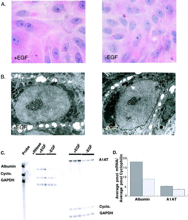
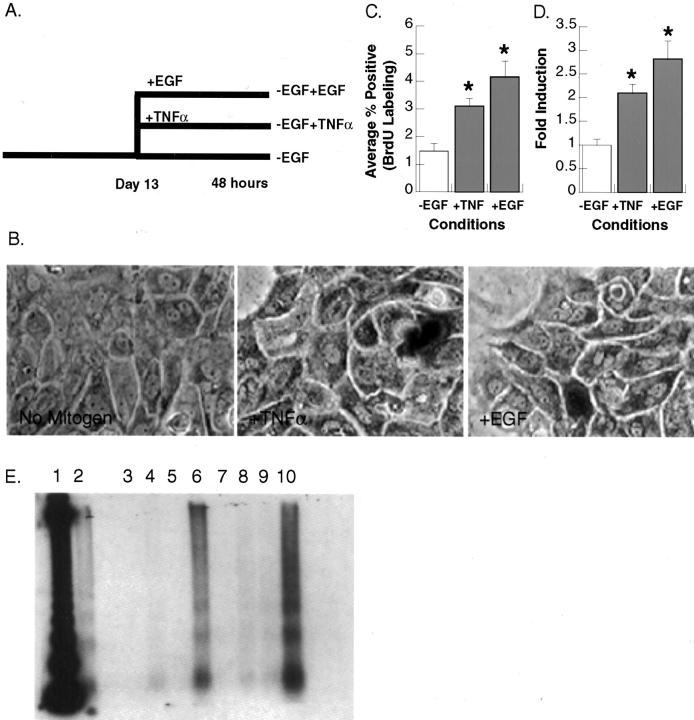
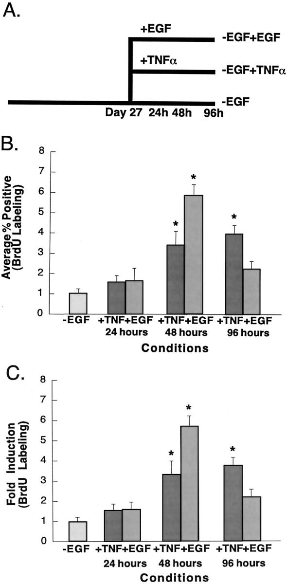
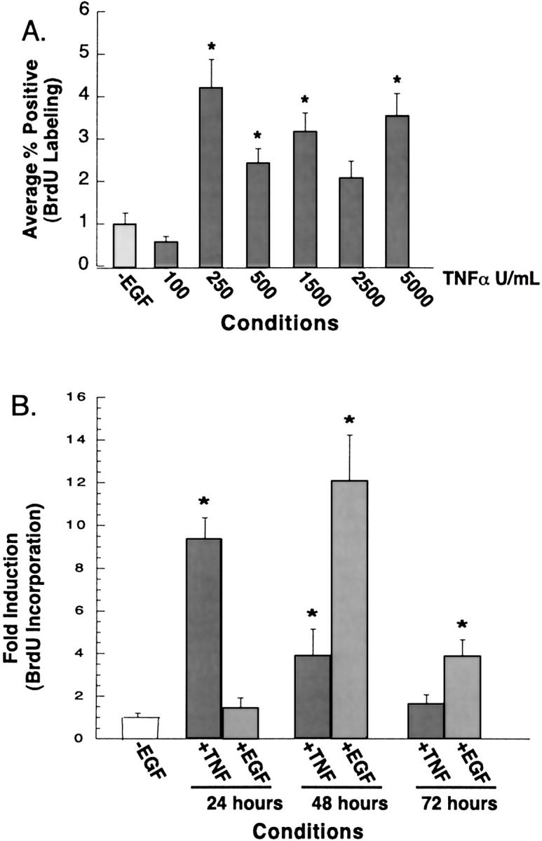
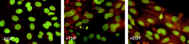
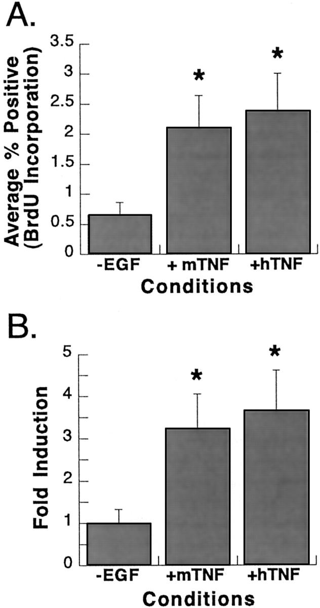
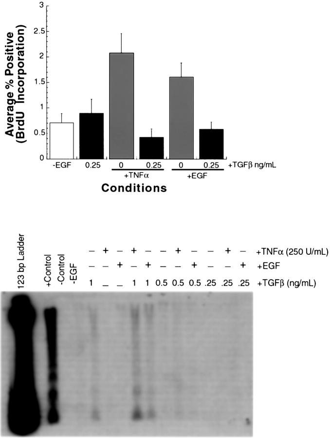
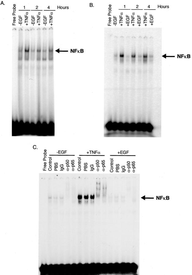
The cytokine tumor necrosis factor (TNF)-alpha has previously been shown to prime hepatocytes to a state of replicative competence, but has not been shown to act as a complete mitogen for these cells. In the present study we have altered our previously described long-term dimethyl sulfoxide culture system to exclude all known hepatocyte mitogens from the culture media and enable us to directly examine the effects of TNF-alpha on primary rat hepatocytes. We have shown that cells maintained under these culture conditions retain the biochemical and morphological features of well-differentiated hepatocytes. Treatment with TNF-alpha induced DNA synthesis relative to control, to a level not significantly different from that induced by the known hepatocyte mitogen, epidermal growth factor (EGF). Maximal DNA synthesis was induced by treatment with 250 U/ml TNF-alpha for 24 hours. Mitotic figures were observed in cultures treated with TNF-alpha or EGF but not in untreated controls. Treatment of cultures with TNF-alpha, but not EGF, induced activation of both nuclear factor-kappaB p50 homodimers and p50/p65 heterodimers. DNA synthesis induced by TNF-alpha was inhibited by treatment with transforming growth factor-beta. Based on the results of our studies, we conclude that TNF-alpha acts as a complete mitogen for rat hepatocytes.


Figures

Figure 1.
Figure 1.
Primary rat hepatocytes cultured in the absence of EGF retain the morphological and biochemical markers of well-differentiated hepatocytes. A: H&E stain of primary rat hepatocytes cultured in the presence (+EGF) or absence (−EGF) of EGF. B: Electron micrographs of primary rat hepatocytes cultured in the presence or absence of EGF. Arrow indicates presence of peroxisomes. C: Phosphorimager scan of RPA for rat albumin, A1AT, cyclophilin, and GAPDH. Left: RPA for albumin, cyclophilin, and GAPDH. Lane 1, Probe, no RNase (positive control); lane 2, probe, +RNase (negative control); lanes 3 to 5, +EGF cultures of hepatocytes; lanes 6 to 8, −EGF cultures of hepatocytes. Right: RPA for A1AT, cyclophilin, and GAPDH. Lanes 1 to 3, +EGF cultures of hepatocytes; lanes 4 to 6, −EGF cultures of hepatocytes. D: Graphical representation of RPA results after ImageQuant analysis. Liver-specific gene expression was normalized to cyclophilin expression to control for equal RNA loading. Original magnifications: ×400 (A); ×11,500 (B).
Figure 2.
Figure 2.
TNF-α and EGF act as primary mitogens for rat hepatocytes. A: Protocol for the induction of DNA synthesis by TNF-α and EGF. Hepatocytes were maintained for 13 days in the absence of EGF and for an additional 48 hours in control media (−EGF), media containing TNF-α (2500 U/ml) (−EGF +TNF-α), or media containing EGF (25 ng/ml) (−EGF +EGF). BrdU was added to the media of all cultures for the final 24 hours of the treatment period. B: BrdU labeling of primary rat hepatocytes. Cells were fixed and stained for BrdU incorporation. C and D: DNA synthesis in TNF-α- or EGF-treated hepatocytes. Percentages of BrdU-labeled nuclei were calculated for each condition. Fold induction in BrdU incorporation was calculated by normalizing data to control cultures. Error bars indicate the SE for individual conditions. *, Values indicate that results are significantly increased relative to −EGF controls (one-tailed t-test, P < 0.05). E: Induction of apoptosis in primary rat hepatocytes. Samples in lanes 3to 6 were extracted from +EGF cultures and samples in lanes 7 to 10 from −EGF cultures. Lane 1, 123-bp ladder; lane 2, +TNF-α +cycloheximide (5 μmol/L) (positive control); lanes 3 and 7, untreated control; lanes 4 and 8, +cycloheximide (5 μmol/L); lanes 5 and 9, +TNF-α (2500 U/ml); lanes 6 and 10, +cycloheximide (5 μmol/L) +TNF-α. Original magnification, ×200 (B).
Figure 3.
Figure 3.
Time course of TNF-α- and EGF-induced DNA synthesis. A: Protocol for the induction of DNA synthesis by EGF and TNF-α. Hepatocytes were maintained for 27 days in the absence of EGF. Cultures were either maintained in this media during the experiment to serve as controls (−EGF); treated with TNF-α (5000 U/ml) for 24, 48, or 96 hours (−EGF +TNF-α); or treated with EGF (25 ng/ml) for 24, 48, or 96 hours (−EGF +EGF). BrdU was added to the media of all cultures for the final 24 hours of treatment. B and C: DNA synthesis in TNF-α- and EGF-treated hepatocytes. Percentages of BrdU-labeled nuclei were calculated for the conditions described previously. Fold induction in BrdU incorporation was calculated by normalizing data to control cultures. Error bars indicate the SE for individual conditions. *, Values indicate results that were significantly increased relative to −EGF controls (one-tailed t-test, P < 0.05).
Figure 4.
Figure 4.
Concentration curve of TNF-α-induced DNA synthesis. Hepatocytes were maintained for 16 days in the absence of EGF. Cultures were either maintained in this media during the 48-hour experiment to serve as controls (−EGF) or treated with TNF-α (100, 250, 500, 1500, 2500, or 5000 U/ml). BrdU was added to the media of all cultures 24 hours after treatment. A: DNA synthesis in TNF-α-treated hepatocytes. Percentages of BrdU-labeled nuclei were calculated for each condition. B: Time courses of TNF-α- and EGF-induced DNA synthesis. Hepatocytes were maintained for 13 days in the absence of EGF. Cultures were either maintained in this media during the experiment to serve as controls (−EGF), treated with TNF-α (250 U/ml) or EGF (25 ng/ml) for 24, 48, or 72 hours. BrdU was added to the media of all cultures 24 hours before harvest. Percentages of BrdU-labeled nuclei were calculated for each condition. Fold induction in BrdU incorporation was calculated by normalizing data to control cultures. Error bars indicate the SE for individual conditions. *, Values indicate results that were significantly increased relative to −EGF controls (one-tailed t-test, P < 0.05).
Figure 5.
Figure 5.
Acridine orange staining of primary rat hepatocytes after TNF-α or EGF treatment. Cells were maintained for 13 days after plating in the absence of EGF. Cultures were then either maintained under control conditions for the duration of the experiment (−EGF), treated with TNF-α (250 U/ml) (+TNF) or EGF (25 ng/ml) (+EGF) for 48, 72, or 96 hours. Cells were fixed and stained with acridine orange. Original magnification, ×200.
Figure 6.
Figure 6.
BrdU labeling of primary rat hepatocytes after treatment with mouse or human TNF-α. Cultures were maintained in the absence of EGF for 13 days before the start of the experiment. Cells were then maintained under the following conditions for an additional 48 hours: DMEM-F12 lacking EGF (−EGF), DMEM-F12 containing mouse TNF-α (250 U/ml) (+mTNF), or DMEM-F12 containing human TNF-α (250 U/ml) (+hTNF). BrdU was added to the media for the last 24 hours of the treatment period. A: Percentages of BrdU-labeled nuclei were calculated for each condition. B: Fold induction in BrdU incorporation was calculated by normalizing data to control cultures. Error bars indicate the SE for individual conditions. *, Values indicate results that are significantly increased relative to −EGF controls (one-tailed t-test, P < 0.05).
Figure 7.
Figure 7.
TGF-β abrogates the induction of DNA synthesis by TNF-α and EGF in primary rat hepatocytes. Hepatocytes were maintained for 16 days in the absence of EGF. A: Hepatocytes were either maintained under these conditions for the duration of the experiment (−EGF) treated with TGF-β alone (0.25 ng/ml), TNF-α alone (250 U/ml), EGF alone (25 ng/ml), TNF-α in combination with TGF-β, or EGF in combination with TGF-β. Cultures were treated for 24 hours and BrdU was added to the culture media at the time of treatment. Percentages of BrdU-labeled nuclei were calculated for each condition. Error bars indicate the SE for individual conditions. B: DNA fragmentation after treatment with TNF-α and TGF-β. Hepatocytes were either maintained in media lacking EGF for the duration of the experiment (−EGF) and treated with TGF-β alone (0.25, 0.5, or 1.0 ng/ml), TNF-α alone (250 U/ml), EGF alone (25 ng/ml), TNF-α in combination with TGF-β, or EGF in combination with TGF-β for 24 hours. Untreated primary rat hepatocytes or hepatocytes treated with cycloheximide (5 μmol/L) and TNF-α (2500 U/ml) in combination were used as negative and positive controls, respectively. Low-molecular weight DNA was isolated and analyzed for internucleosomal cleavage.
Figure 8.
Figure 8.
NF-κB is activated after TNF-α treatment. Hepatocytes were maintained for 16 days in the absence of EGF. Arrows indicate the position of the NF-κB complex. A: Cells were either maintained in −EGF media (−EGF) or treated with TNF-α (250 U/ml) (+TNF-α) for an additional 1, 2, or 4 hours. Nuclear protein was incubated with 32P-labeled oligonucleotides corresponding to the consensus binding sequence for NF-κB. B: Cells were maintained in the absence of EGF for 15 days before the start of the experiment. Cells were maintained in this media for the duration of the experiment (−EGF) or treated with TNF-α (+TNF-α) or EGF (+EGF) for 1, 2, or 4 hours. Nuclear protein was incubated with 32P-labeled oligonucleotides corresponding to the consensus binding site for NF-κB. C: Supershift analysis of NF-κB activity. Extracts were incubated as described for gel shifts and subsequently incubated an additional half hour with no addition (control), addition of PBS, normal rabbit IgG (IgG), anti-p50 antibody (α-p50), or anti-p65 antibody (α-p65).

Similar articles

Cited by

References

    1. Loffreda S, Rai R, Yang SQ, Lin HZ, Diehl AM: Bile ducts and portal and central veins are major producers of tumor necrosis factor α in regenerating liver. Gastroenterology 1997, 112:2089-2098 - PubMed
    1. Akerman P, Cote P, Yang SQ, McClain C, Nelson S, Bagby GJ, Diehl AM: Antibodies to tumor necrosis factor-α inhibit liver regeneration after partial hepatectomy. Am J Physiol 1992, 263:G579-G585 - PubMed
    1. Webber EM, Bruix J, Pierce RH, Fausto N: Tumor necrosis factor primes hepatocytes for DNA replication in the rat. Hepatology 1998, 28:1226-1234 - PubMed
    1. Yamada Y, Kirillova I, Peschon JJ, Fausto N: Initiation of liver growth by tumor necrosis factor: deficient liver regeneration in mice lacking type I tumor necrosis factor receptor. Proc Natl Acad Sci USA 1997, 94:1141-1146 - PMC - PubMed
    1. Yamada Y, Webber EM, Kirillova I, Peschon JJ, Fausto N: Analysis of liver regeneration in mice lacking type I or type II TNF receptor: requirement for type I but not type II receptor. Hepatology 1998, 28:906-913 - PubMed

Publication types