Identification of genes up-regulated in urothelial tumors: the 67-kd laminin receptor and tumor-associated trypsin inhibitor
- PMID: 12875970
- PMCID: PMC1868207
- DOI: 10.1016/S0002-9440(10)63678-4
Identification of genes up-regulated in urothelial tumors: the 67-kd laminin receptor and tumor-associated trypsin inhibitor
Abstract
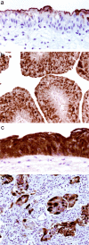
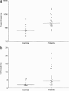
Studies investigating changes in gene expression in urothelial carcinoma have generally compared tumors of different stages and grades but comparisons between low-grade, noninvasive tumors and normal urothelium are needed to identify genes involved in early tumor development. We isolated the urothelium from a low-grade tumor and corresponding normal mucosa by laser capture microdissection on frozen sections. The RNA extracted was amplified to generate suppressive subtractive cDNA libraries. Random sequencing of cDNA clones identified approximately 100 unique species. Of these 83% were known genes, 15% had homology to genes with an unknown function in humans, and 2% did not show homology to any published gene sequence. Two of the known genes, the 67-kd laminin receptor (67LR) and tumor-associated trypsin inhibitor (TATI), had previously been associated with metastatic progression in many tumor types, although 67LR has not been investigated in urothelial tumors. Immunolabeling of the original tissue with antibodies against these two genes confirmed overexpression, validating our strategy: 67LR was not expressed in the normal urothelium but was present in the tumor, whereas TATI expression was confined to umbrella cells in the normal urothelium, but extended to all cell layers in the tumor. We investigated both markers further in a separate series of tumors of different stages and grades. TATI was more consistently overexpressed than 67LR in all tumor grades and stages. Levels of secreted TATI were significantly higher in urine samples from patients with tumors compared to controls. Our strategy, combining laser capture microdissection and cDNA library construction, has identified genes that may be involved in the early phases of urothelial tumor development rather than with disease progression, highlighting the importance of comparing tumor with normal rather than just tumors of different stages and grades.
Figures


Similar articles
-
Differential gene expression profiling in aggressive bladder transitional cell carcinoma compared to the adjacent microscopically normal urothelium by microdissection-SMART cDNA PCR-SSH.Cancer Biol Ther. 2006 Jan;5(1):104-10. doi: 10.4161/cbt.5.1.2348. Epub 2006 Jan 22. Cancer Biol Ther. 2006. PMID: 16357518
-
Association of tumor-associated trypsin inhibitor (TATI) expression with molecular markers, pathologic features and clinical outcomes of urothelial carcinoma of the urinary bladder.World J Urol. 2012 Dec;30(6):785-94. doi: 10.1007/s00345-011-0727-7. Epub 2011 Jul 8. World J Urol. 2012. PMID: 21739120
-
Chromosomal imbalances in successive moments of human bladder urothelial carcinoma.Urol Oncol. 2013 Aug;31(6):827-35. doi: 10.1016/j.urolonc.2011.05.015. Epub 2011 Jul 16. Urol Oncol. 2013. PMID: 21763161
-
When urothelial differentiation pathways go wrong: implications for bladder cancer development and progression.Urol Oncol. 2013 Aug;31(6):802-11. doi: 10.1016/j.urolonc.2011.07.017. Epub 2011 Sep 15. Urol Oncol. 2013. PMID: 21924649 Free PMC article. Review.
-
Autocrine induction of invasion and metastasis by tumor-associated trypsin inhibitor in human colon cancer cells.Oncogene. 2008 Jul 3;27(29):4024-33. doi: 10.1038/onc.2008.42. Epub 2008 Mar 3. Oncogene. 2008. PMID: 18317448 Review.
Cited by
-
Urothelial carcinoma: stem cells on the edge.Cancer Metastasis Rev. 2009 Dec;28(3-4):291-304. doi: 10.1007/s10555-009-9187-6. Cancer Metastasis Rev. 2009. PMID: 20012172 Free PMC article. Review.
-
Hypoxia promotes metastasis in human gastric cancer by up-regulating the 67-kDa laminin receptor.Cancer Sci. 2010 Jul;101(7):1653-60. doi: 10.1111/j.1349-7006.2010.01592.x. Epub 2010 Apr 19. Cancer Sci. 2010. PMID: 20491781 Free PMC article.
-
Differentiation of a highly tumorigenic basal cell compartment in urothelial carcinoma.Stem Cells. 2009 Jul;27(7):1487-95. doi: 10.1002/stem.92. Stem Cells. 2009. PMID: 19544456 Free PMC article.
References
-
- Harnden P, Parkinson MC: Transitional cell carcinoma of the bladder: diagnosis and prognosis. Curr Diagnostic Pathol 1996, 3:109-121
-
- Ross JS, Cohen MB: Biomarkers for the detection of bladder cancer. Adv Anat Pathol 2001, 8:37-45 - PubMed
-
- Gutierrez Banos JL, Henar Rebollo RM, Antolin Juarez FM, Garcia BM: Usefulness of the BTA STAT test for the diagnosis of bladder cancer. Urology 2001, 57:685-689 - PubMed
-
- Gromova I, Gromov P, Celis JE: bc10: a novel human bladder cancer-associated protein with a conserved genomic structure downregulated in invasive cancer. Int J Cancer 2002, 98:539-546 - PubMed
-
- Gromova I, Gromov P, Celis JE: A novel member of the glycosyltransferase family, beta 3 Gn-T2, highly downregulated in invasive human bladder transitional cell carcinomas. Mol Carcinog 2001, 32:61-72 - PubMed
Publication types
MeSH terms
Substances
LinkOut - more resources
Full Text Sources
Other Literature Sources
Medical

