. 2003 Aug;163(2):517-31.
doi: 10.1016/S0002-9440(10)63680-2.

The fibrinolytic system in dissemination and matrix protein deposition during a mycobacterium infection

Affiliations

The fibrinolytic system in dissemination and matrix protein deposition during a mycobacterium infection

Jun Sato et al. Am J Pathol. 2003 Aug.

Abstract

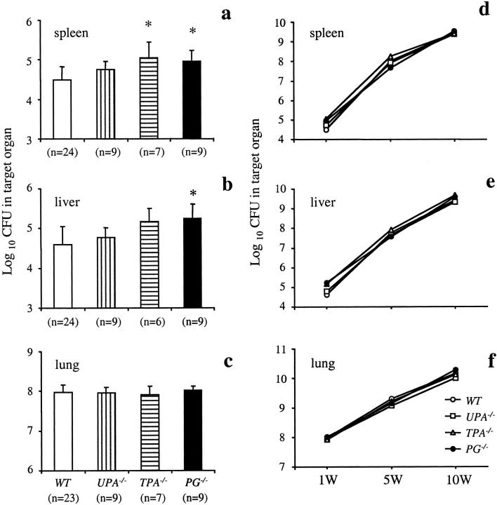
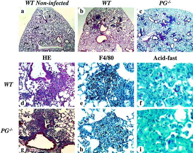
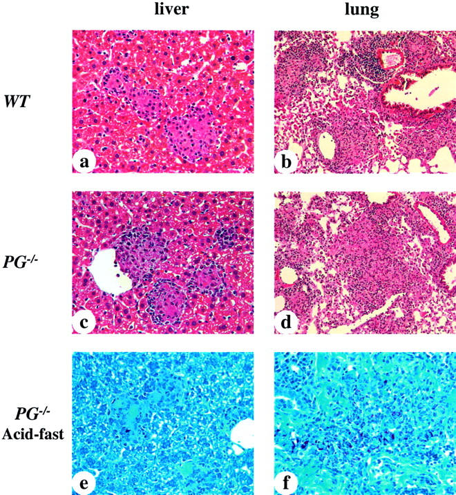
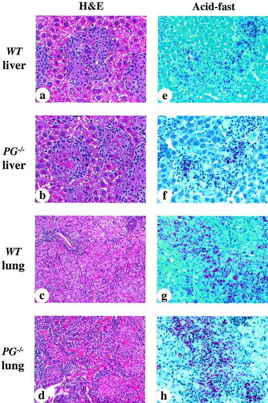
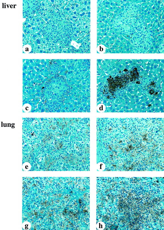
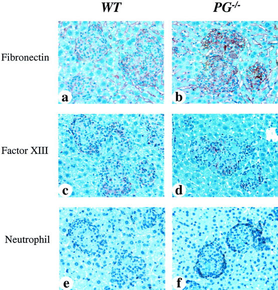
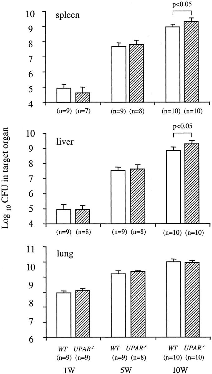
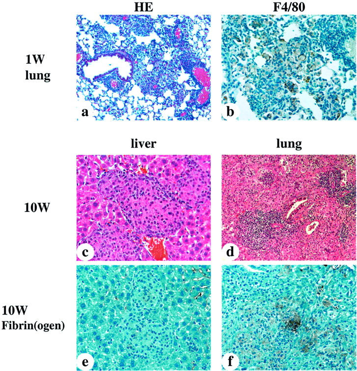
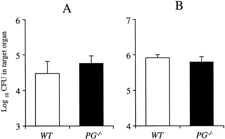
The fibrinolytic system is known to play an important role in the inflammatory response to bacterial infections. In the present study, relationships between protein components of the fibrinolytic system and infectivity by Mycobacterium avium were analyzed. Infections were initiated through noninvasive intratracheal administration of M. avium 724 in mice individually deficient for plasminogen, tissue-type plasminogen activator, urokinase-type plasminogen activator, and urokinase-type plasminogen activator receptor, along with wild-type control mice. There were no differences in lung colony counts among all mouse genotypes throughout a 10-week infection. However, in tissue-type plasminogen activator and plasminogen-deficient mice an earlier dissemination of M. avium to other organs was observed. Nevertheless, the M. avium growth rates in the liver, spleen, and lung did not differ between the various mouse populations throughout a 10-week infection. Histochemical and immunohistochemical analyses at 5 and 10 weeks after infection demonstrated that plasminogen-deficient mice, compared to wild-type mice, had enhanced fibrin and fibronectin deposition, as well as increased neutrophil infiltration within liver granulomas. These results suggest that plasmin(ogen) plays a role in the turnover of extracellular matrix proteins within granulomas and has a limited effect in the early dissemination of M. avium from lungs. Thus, plasmin(ogen) functions in limiting progressive fibrosis in the granuloma during a chronic mycobacterial infection.


Figures

Figure 1.
Figure 1.
The course of M. avium 724 infection in WT, UPA−/−, TPA−/−, and PG−/− mice. Mice were intratracheally infected with 1 × 10⁸ CFU of M. avium 724. Total colony counts were determined in the spleens (CFU/organ) (a), livers (CFU/g weight) (b), and lungs (CFU/organ) (c) at 1 week after infection. d–f: Plots of the colony counts at 1, 5, and 10 weeks after infection in each genotype. The data represent the mean ± SD of CFU in the organs of gene-inactivated mice and WT mice. *, P < 0.05.
Figure 2.
Figure 2.
Photomicrographs of representative livers and lungs from WT and PG−/− mice 1 week after infection with 1 × 10⁸ CFU of M. avium 724. a to d and g were stained with H&E, e and h were stained with anti-mouse F4/80 antibody, and f and i were stained for acid-fast bacilli. a: PBS-treated WT mouse lung showing normal alveolar structure and no cell infiltration. b and c: WT and PG−/− mouse lungs showing patchy distributions of lesions with normal alveolar space. d: WT mouse lung displaying focal cell infiltration of macrophages and lymphocytes in the alveolar space around bronchioles and arteries. e: In the focal cell-infiltrated area of the WT mouse lung, macrophages are tightly packed and aggregated with lymphocytes. f: WT mouse lung showing acid-fast bacilli in the cytoplasma of macrophages. g and h: A PG−/− mouse lung showing similar findings to those observed in WT mice. i: In a PG−/− mouse lung, macrophages with acid-fast bacilli were similar to WT mice. Original magnifications: ×40 (a–c); ×200 (d, g); ×400 (e, h); ×1000 (f, i).
Figure 3.
Figure 3.
Photomicrographs of representative livers and lungs from WT and PG−/− mice 5 weeks after infection with 1 × 10⁸ CFU of M. avium 724. a to d were stained with H&E and e and f were stained for acid-fast bacilli. a and c: WT and PG−/− mouse livers showing small and mature granulomas. The sizes and distributions of granuloma formations were similar in both groups. b and d: WT and PG−/− mouse lungs showing much more cell infiltration than that observed at 1 week after infection, as well as a decrease of alveolar space, and thickening of alveolar walls. e and f: PG−/− murine liver and lungs showing large macrophages parasitized by acid-fast bacilli. These findings were similar to those of WT mice. Original magnifications: ×400 (a, c, e, f); ×200 (b, d).
Figure 4.
Figure 4.
Photomicrographs of representative liver and lungs from WT and PG−/− mice at 10 weeks after infection. a to d were stained with H&E; e to h were stained for acid-fast bacilli. a: WT mouse liver showing mature and fused granulomas. The numbers and sizes of the granulomas were increased as compared to those at 5 weeks after infection. b: PG−/− mouse liver showing similar granuloma formations to those of WT livers, containing some fibrin deposition in granulomas. c: WT mouse lung showing extensive cell infiltration occupying most of the alveolar space. d: PG−/− murine lung showing similar findings to those of WT lungs, and more extensive fibrin deposition. e and f: WT and PG−/− mouse livers showing heavily parasitized macrophages within granulomas. g and h: WT and PG−/− mouse lungs showing diffuse distribution of heavily parasitized macrophages. Original magnifications: ×400 (a, b, e–h); ×200 (c, d).
Figure 5.
Figure 5.
Anti-fibrin(ogen) staining in liver and lungs from infected WT and PG−/− mice 5 weeks after infection. a: WT mouse liver showing no fibrin deposition in granulomas. b: PG−/− mouse liver showing dense fibrin deposition in granulomas. c: WT murine lung showing a small amount of diffuse fibrin deposition. d: PG−/− mouse lung showing dense and highly extensive fibrin deposition. Original magnifications, ×400.
Figure 6.
Figure 6.
Anti-fibrin(ogen) staining in liver and lungs from infected WT and gene-inactivated mice 10 weeks after infection. a–c: WT, UPA−/−, and TPA−/− mouse livers, respectively, showing no fibrin deposition in granulomas. d: PG−/− mouse liver showing denser and more extensive fibrin deposition in granulomas, as compared with genotypically similar mice at 5 weeks after infection. e: WT mouse lung showing a small amount of diffuse fibrin deposition. f and g: Murine UPA−/− and TPA−/− lungs displaying increased fibrin deposition as compared to WT murine lungs. h: PG−/− mouse lung showing dense and more extensive fibrin deposition.
Figure 7.
Figure 7.
Immunohistochemical analyses for fibronectin, Factor XIII, and neutrophils in livers from WT and PG−/− mice at 10 weeks after infection. a and b were stained with anti-fibronectin antibody, c and d were stained with anti-Factor XIII subunit-A antibody, and e and f were stained with anti-neutrophil antibody. a: WT mouse liver showing a small amount of diffuse fibronectin deposition in granulomas. b: PG−/− mouse liver showing a more dense and extensive fibronectin deposition in granulomas, as compared with WT mice. c and d: WT and PG−/− mouse livers, respectively, showing a small amount of Factor XIII deposition in granulomas. e: WT mouse had a few neutrophils at the peripheral areas of the granulomas. f: PG−/− mouse had much stronger neutrophil infiltration around the granulomas. The distribution of fibronectin deposition was similar to fibrin in granulomas. Original magnifications, ×400.
Figure 8.
Figure 8.
The time course of M. avium 724 infection in WT and UPAR−/− mice. Mice were infected with 1 × 10⁸ CFU of M. avium 724. Total colony counts were determined in the spleens (CFU/organ), livers (CFU/g weight), and lungs (CFU/organ) of both genotypes at 1, 5, and 10 weeks after infection. The data represent the mean ± SD of CFU in the organs of WT and UPAR−/− mice. *, P < 0.05 versus WT mice.
Figure 9.
Figure 9.
Photomicrographs of representative livers and lungs from UPAR−/− mice at 1 and 10 weeks after infection. a, c, and d were stained with H&E, b was stained with anti-mouse F4/80 antibody, and e and f were stained with anti-fibrin(ogen) antibody. a: UPAR−/− mouse lung at 1 week after infection showing focal cell infiltration of macrophages and lymphocytes in the alveolar space around bronchioles and arteries. These findings were similar to those of WT lung (Figure 2, a and b) ▶ . b: Anti-F4/80 antibody staining showing macrophages aggregated with lymphocytes in UPAR−/− mouse lung at 1 week after infection. These findings were similar to those of infected WT lungs (Figure 2, a to e) ▶ . c: UPAR−/− mouse liver at 10 weeks after infection showing mature and fused granulomas. d: UPAR−/− mouse lung at 10 weeks after infection displaying extensive cell infiltration that occupies most of the alveolar space. e and f: UPAR−/− mouse liver and lung showing a minimal amount of fibrin deposition. Original magnifications: ×200 (a); ×400 (b–f).
Figure 10.
Figure 10.
M. bovis BCG infection in WT and PG−/− mice at 5 weeks after infection. Mice were intratracheally infected with 5 × 10⁷ CFU of M. bovis BCG. Total colony counts (CFU/organ) were determined in the spleens (A) and lungs (B) at 5 weeks after infection. Total colony counts in livers were undetectable in both WT and PG−/− mice. The data represent the mean ± SD of five mice per group.

Similar articles

Cited by

References

    1. Chackerian AA, Alt JM, Perera TV, Dascher CC, Behar SM: Dissemination of Mycobacterium tuberculosis is influenced by host factors and precedes the initiation of T-cell immunity. Infect Immun 2002, 70:4501-4509 - PMC - PubMed
    1. Saunders BM, Cooper AM: Restraining mycobacteria: role of granulomas in mycobacterial infections. Immunol Cell Biol 2000, 78:334-341 - PubMed
    1. Ehlers S: Immunity to tuberculosis: a delicate balance between protection and pathology. FEMS Immunol Med Microbiol 1999, 23:49-58 - PubMed
    1. Castellino FJ, Ploplis VA: Plasminogen and streptokinase Bachmann F eds. Handbook of Experimental Pharmacology, Fibrinolytics and Antifibrinolytics 2000, vol 146:pp 25-56 Springer-Verlag, New York
    1. Lijnen HR: Plasmin and matrix metalloproteinases in vascular remodeling. Thromb Haemost 2001, 86:324-333 - PubMed

Publication types

MeSH terms

LinkOut - more resources