. 2003 Aug;163(2):553-62.
doi: 10.1016/S0002-9440(10)63683-8.

Bone-marrow-derived cells contribute to glomerular endothelial repair in experimental glomerulonephritis

Affiliations

Bone-marrow-derived cells contribute to glomerular endothelial repair in experimental glomerulonephritis

Maarten B Rookmaaker et al. Am J Pathol. 2003 Aug.

Abstract

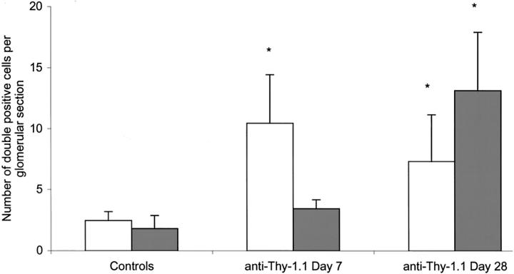
Glomerular endothelial injury plays an important role in the pathogenesis of renal diseases and is centrally involved in renal disease progression. Glomerular endothelial repair may help maintain renal function. We examined whether bone-marrow (BM)-derived cells contribute to glomerular repair. A rat allogenic BM transplant model was used to allow tracing of BM-derived cells using a donor major histocompatibility complex class-I specific mAb. In glomeruli of chimeric rats we identified a small number of donor-BM-derived endothelial and mesangial cells, which increased in a time-dependent manner. Induction of anti-Thy-1.1-glomerulonephritis (transient mesangial and secondary glomerular endothelial injury) caused a significant, more than fourfold increase in the number of BM-derived glomerular endothelial cells at day 7 after anti-Thy-1.1 injection compared to chimeric rats without glomerular injury. The level of BM-derived endothelial cells remained high at day 28. We also observed a more than sevenfold increase in the number of BM-derived mesangial cells at day 28. BM-derived endothelial and mesangial cells were fully integrated in the glomerular structure. Our data show that BM-derived cells participate in glomerular endothelial and mesangial cell turnover and contribute to microvascular repair. These findings provide novel insights into the pathogenesis of renal disease and suggest a potential role for stem cell therapy.


Figures

Figure 1.
Figure 1.
Histological changes in anti-Thy-1.1 nephritis in WRBM→BN rats. A: Renal morphology in control BN rats. B: Renal morphology in control WRBM→BN rats. C: in WRBM→BN rats at 6 months after bone marrow transplantation no significant changes in renal morphology have occurred. D: At day 7 after injection of anti-Thy-1.1 there is marked loss of mesangial cells with dissolution of the mesangial matrix and formation of microaneurysms. E: At day 28 after anti-Thy-1.1 injection glomerular morphology has partially recovered. (PAS, ×400).
Figure 2.
Figure 2.
A: Immunoperoxidase staining of 5-μm cryostat kidney sections of a control BN rat (no irradiation/no bone marrow transplantation/no anti-Thy-1.1 nephritis), negative staining with U9F4. B: Donor bone-marrow-derived U9F4-positive cells (brown) are present in both glomeruli (arrows) as well as tubulo-interstitium of control WRBM→BN rat kidney, 2 months after irradiation and bone marrow reconstitution (no anti-Thy-1.1 injection) (×200).
Figure 3.
Figure 3.
Anti-Thy-1.1 injection causes extensive loss of mesangial cells followed by regeneration. Immunofluorescent double-staining at day 7 with U9F4 (green) and OX-7 (red) shows a few mesangial cells, which occasionally form small clusters located at the vascular pole of the glomerulus (arrow). BM-derived mesangial cells (yellow) are often located in these small cell clusters near the vascular pole. (×630).
Figure 4.
Figure 4.
Immunofluorescent double-staining of glomerular sections from WRBM→BN rats at 28 days after induction of anti-Thy-1.1 nephritis. Confocal laser-scanning microscopy (×630, pinhole 2.5). A: Staining for BM donor (WR) MHC-I (U9F4, green). B: Staining for endothelial cells (RECA-1, red). C: U9F4/RECA-1 double-staining identifies bone-marrow-derived endothelial cells, randomly integrated in the glomerular endothelium (U9F4/ED-1 double-positive, yellow). D: Staining for BM donor (WR) MHC-I (U9F4, green). E: Staining for mesangial cells (OX-7, red). F: U9F4/OX-7 double-staining showing donor bone-marrow-derived mesangial cells (U9F4/OX-7 double-positive, yellow) integrated in the glomerulus. G: Staining for BM donor (WR) MHC-I (U9F4, red). H: Staining for monocytes/macrophages (ED-1, green). I: U9F4/ED-1 double-staining shows a BM donor-derived (U9F4/ED-1 double positive, yellow) monocyte/macrophage. ▵▵, U9F4/OX-7, U9F4/RECA-1, U9F4/ED-1 double-positive cells (yellow), ie, donor-derived mesangial and endothelial cells and a donor-derived monocyte/macrophage. ∧∧, single-U9F4 staining.
Figure 5.
Figure 5.
The average number of BM-derived endothelial cells (RECA-1/U9F4 double-positive cells, white bars) and bone-marrow-derived mesangial cells (OX-7/U9F4 double-positive cells, shaded bars) per glomerular section in control WRBM→BN rats and in rats at 7 and 28 days after anti-Thy-1.1 injection. Values are expressed as mean ± SD per glomerular section. *, P < 0.05 versus controls.
Figure 6.
Figure 6.
Controls of the immunofluorescent double-staining as shown in Figure 4 ▶ . All pictures were taken with the same settings as used for Figure 4 ▶ and were processed by the same procedure. Confocal laser-scanning microscopy (×630, pinhole 2.5). The following controls were included: substitution of the first primary antibody with a different biotinylated control antibody and normal second primary and substitution of the second primary antibody with a different biotinylated control antibody. A: ED-1+/U9F4 control. B: U9F4 control/RECA-1+. C: U9F4 control/OX-7+. D: ED-1 control/U9F4+. E: U9F4+/RECA-1 control. F: U9F4 control/RECA-1 control. A to F: All controls were negative, indicating that the biotin-blocking steps were complete and that no cross-reactivity occurred.

References

    1. Kang DH, Kanellis J, Hugo C, Truong L, Anderson S, Kerjaschki D, Schreiner GF, Johnson RJ: Role of the microvascular endothelium in progressive renal disease. J Am Soc Nephrol 2002, 13:806-816 - PubMed
    1. Yamanaka N, Shimizu A: Role of glomerular endothelial damage in progressive renal disease. Kidney Blood Press Res 1999, 22:13-20 - PubMed
    1. Abouna GM, Al Adnani MS, Kremer GD, Kumar SA, Daddah SK, Kusma G: Reversal of diabetic nephropathy in human cadaveric kidneys after transplantation into non-diabetic recipients. Lancet 1983, 2:1274-1276 - PubMed
    1. Clark G, White RH, Glasgow EF, Chantler C, Cameron JS, Gill D, Comley LA: Poststreptococcal glomerulonephritis in children: clinicopathological correlations and long-term prognosis. Pediatr Nephrol 1988, 2:381-388 - PubMed
    1. Iruela-Arispe L, Gordon K, Hugo C, Duijvestijn AM, Claffey KP, Reilly M, Couser WG, Alpers CE, Johnson RJ: Participation of glomerular endothelial cells in the capillary repair of glomerulonephritis. Am J Pathol 1995, 147:1715-1727 - PMC - PubMed

Publication types

MeSH terms

LinkOut - more resources