. 2003 Aug;163(2):571-81.
doi: 10.1016/S0002-9440(10)63685-1.

Persistent down-regulation of Fli1, a suppressor of collagen transcription, in fibrotic scleroderma skin

Affiliations

Persistent down-regulation of Fli1, a suppressor of collagen transcription, in fibrotic scleroderma skin

Masahide Kubo et al. Am J Pathol. 2003 Aug.

Abstract

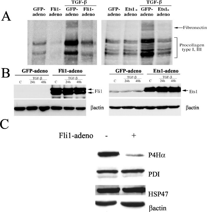
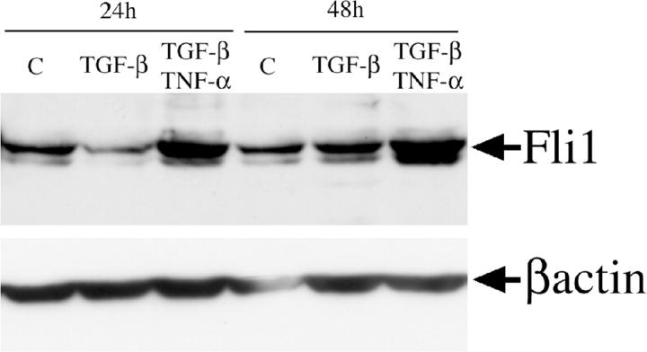
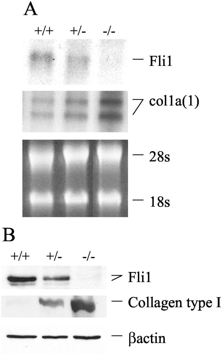
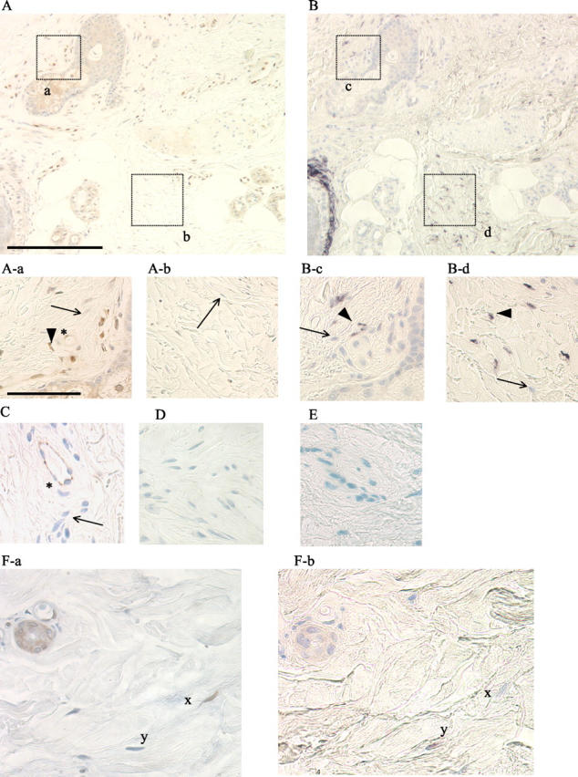
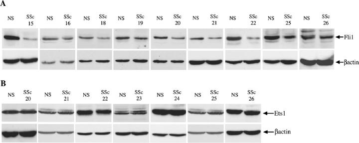
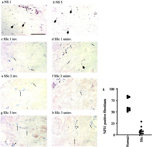
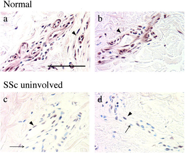
The molecular and cellular mechanisms that maintain proper collagen homeostasis in healthy human skin and are responsible for the dysregulated collagen synthesis in scleroderma remain primarily unknown. This study demonstrates that Fli1 is a physiological negative regulator of collagen gene expression in dermal fibroblasts in vitro and in human skin in vivo. This conclusion is supported by the analyses of mouse embryonic fibroblasts from Fli1(-/-), Fli1(+/-), and Fli1(+/+) mice. In cultured human and mouse fibroblasts Fli1 expression levels are inversely correlated with the collagen type I expression levels. These in vitro observations were validated in vivo. In healthy human skin Fli1 protein is expressed in fibroblasts and endothelial cells. Significantly, absence of Fli1 expression in individual fibroblasts correlates with elevated collagen synthesis. In contrast to healthy skin, Fli1 protein is consistently absent from fibroblasts and significantly reduced in endothelial cells in clinically involved scleroderma skin, which correlates with enhanced collagen synthesis in systemic sclerosis skin. This study supports the role of Fli1 as a suppressor of collagen transcription in human skin in vivo. Persistent down-regulation of Fli1 in scleroderma fibroblasts in vivo may directly contribute to uncontrolled matrix deposition in scleroderma skin.


Figures

Figure 1.
Figure 1.
Fli1 inhibits basal and TGF-β-induced synthesis of collagenous proteins. A: Newly synthesized collagenous proteins in control (GFP) and either Fli1- or Ets1-infected fibroblasts were measured in a [3H]proline incorporation assay after 30 hours of TGF-β stimulation. Aliquots of conditioned media normalized for cell number were analyzed for collagenous protein content via SDS-polyacrylamide gel electrophoresis and fluorography. B: Fli1 and Ets1 levels in GFP- and either Fli1 or Ets1 adenovirus-transduced fibroblasts (multiplicity of infection = 100) before and after TGF-β treatment. Blots were reprobed with β-actin antibody. C: Fli1 down-regulates synthesis of prolyl 4-hydroxylase α in dermal fibroblasts. Protein levels of prolyl 4-hydroxylase α (P4Hα), PDI, and HSP 47 in control and Fli1-infected fibroblasts (multiplicity of infection = 100) were determined using specific antibodies. The blots were probed with anti-p4Hα antibody, anti-PDI antibody, anti-HSP 47 antibody, and anti-β-actin antibody.
Figure 2.
Figure 2.
Fli1 protein level is modulated by cytokines. Serum-starved, confluent fibroblasts were treated with TGF-β (2 ng/ml) alone or a combination of TGF-β and TNF-α (10 ng/ml) added simultaneously for indicated periods of time. Fli1 protein was determined in 100 μg of total cell extract by Western blot. The blots were reprobed with anti-β actin antibody.
Figure 3.
Figure 3.
Fli1 expression levels inversely correlate with collagen type I synthesis in Fli1+/+, Fli1+/−, and Fli1−/− MEFs. A: Representative Northern blot of Fli1 and collagen α1(I) mRNAs. B: Representative Western blot of Fli1 and collagen type I proteins. β-actin was used as loading control.
Figure 4.
Figure 4.
Fli1 and collagen expression in healthy human skin in vivo. Serial skin sections were used for Fli1 immunodetection (A) and COL1A1 in situ hybridization (B) (original magnifications, ×100; scale bar, 200 μm). Note: Fli1-positive fibroblasts represented by brown nuclear staining (thick arrow) and Fli-1-negative fibroblasts (blue counterstain, thin arrow) in A-a and A-b (original magnifications, ×400; scale bar, 50 μm). B-c and B-d represent collagen mRNA expression from the corresponding fields. Note: COL1A1-positive fibroblasts (thick arrowhead) and Fli1-negative fibroblasts (thin arrow). Endothelial origin of cells expressing Fli1 (marked by asterisk) in A-a was confirmed using anti-CD31 antibody (C). In D (negative control), primary antibody was omitted. In E, sense (negative control) COL1A1 probe was used. F-a and F-b (original magnifications, ×400) show serial sections of skin in which corresponding fibroblasts are marked x and y. Note that Fli1-positive fibroblast (x) does not express COL1A1 mRNA, while Fli1-negative fibroblast (y) is positive for collagen mRNA.
Figure 5.
Figure 5.
Fli1 protein is underexpressed in a subset of SSc fibroblasts. Dermal SSc and healthy control fibroblasts (NS) were analyzed for Fli1 and Ets1 protein expression levels. Nuclear extracts were used for Western blot with polyclonal Fli1 antibody or with polyclonal Ets1 antibody. Blots were reprobed with β-actin antibody. A: Representative data of Fli1 protein levels in SSc and NS fibroblasts B: Representative data of Ets1 protein levels in SSc and NS fibroblasts.
Figure 6.
Figure 6.
Immunodetection of Fli1 in skin from healthy individuals and uninvolved and involved skin from patients with SSc. Positively stained fibroblasts and endothelial cells are present in healthy skin (a, b) (original magnifications, ×400; scale bar, 100 μm). Thick arrow points to Fli1-positive fibroblasts, an asterisk indicates endothelial cell; c and d, e and f, and g and h involved and uninvolved skin sections from patients with SSc. Note absence of Fli1 in fibroblasts (thin arrow points to Fli1-negative fibroblast) and in some endothelial cells (asterisk) in SSc skin. k: Percentage of Fli-1-positive fibroblasts in SSc and healthy skin. At least 100 fibroblasts were counted for each specimen.
Figure 7.
Figure 7.
Immunodetection of Fli1 in endothelial cells in healthy skin (a, b) and in uninvolved skin from patients with SSc (c, d) (original magnifications, ×400; scale bar, 100 μm). Thick arrow points to Fli1-positive endothelial cells, thin arrow points to Fli1-negative endothelial cells.

References

    1. LeRoy EC: The connective tissue in scleroderma. Coll Relat Res 1981, 1:301-308 - PubMed
    1. Trojanowska M, LeRoy EC, Eckes B, Krieg T: Pathogenesis of fibrosis: type 1 collagen and the skin. J Mol Med 1998, 76:266-274 - PubMed
    1. Kahari VM: Activation of dermal connective tissue in scleroderma. Ann Med 1993, 25:511-518 - PubMed
    1. Fleischmajer R, Gay S, Meigel WN, Perlish JS: Collagen in the cellular and fibrotic stages of scleroderma. Arthritis Rheum 1978, 21:418-428 - PubMed
    1. Ohtsuka T, Koibuchi N, Sakai H, Yamakage A, Yamazaki S: Quantitative analysis of alpha 1(I) and alpha 1(III) procollagen mRNA expression in systemic sclerosis skin tissue—an in situ hybridization study. Arch Dermatol Res 1999, 291:575-582 - PubMed

Publication types

MeSH terms